. 2009 Dec 11:3:21.
doi: 10.3389/neuro.04.021.2009. eCollection 2009.

Optogenetic Dissection of Neuronal Circuits in Zebrafish using Viral Gene Transfer and the Tet System

Affiliations

Optogenetic Dissection of Neuronal Circuits in Zebrafish using Viral Gene Transfer and the Tet System

Peixin Zhu et al. Front Neural Circuits. .

Abstract

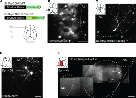
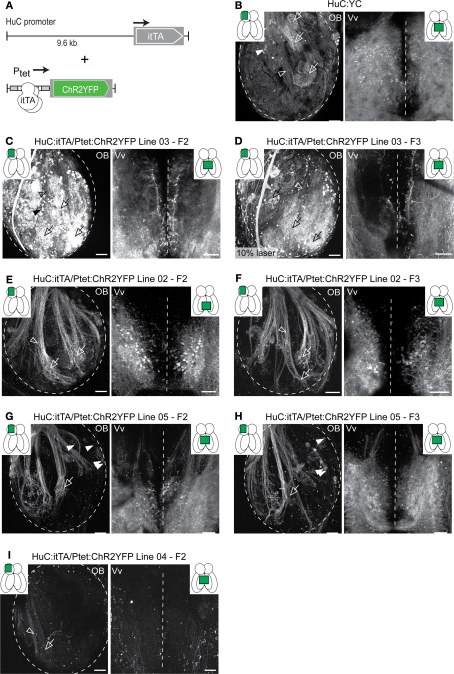
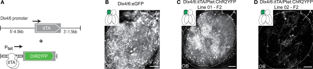
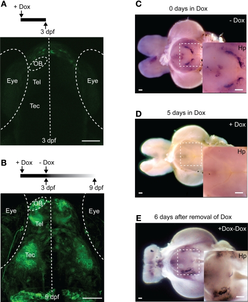
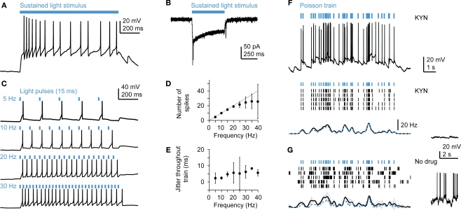
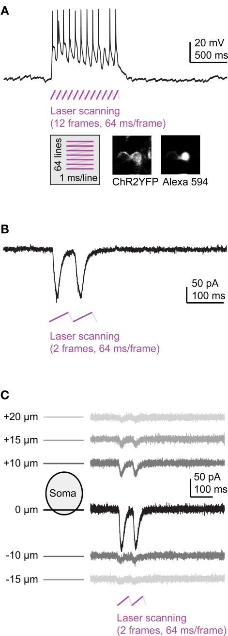
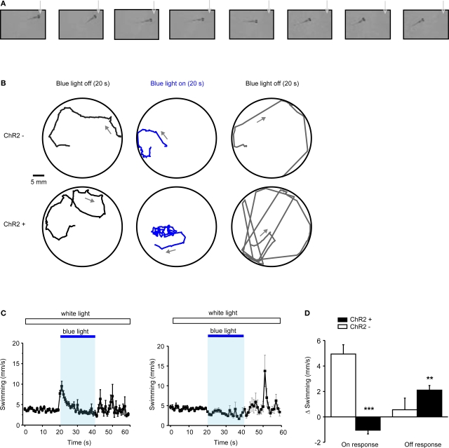
The conditional expression of transgenes at high levels in sparse and specific populations of neurons is important for high-resolution optogenetic analyses of neuronal circuits. We explored two complementary methods, viral gene delivery and the iTet-Off system, to express transgenes in the brain of zebrafish. High-level gene expression in neurons was achieved by Sindbis and Rabies viruses. The Tet system produced strong and specific gene expression that could be modulated conveniently by doxycycline. Moreover, transgenic lines showed expression in distinct, sparse and stable populations of neurons that appeared to be subsets of the neurons targeted by the promoter driving the Tet-activator. The Tet system therefore provides the opportunity to generate libraries of diverse expression patterns similar to gene trap approaches or the thy-1 promoter in mice, but with the additional possibility to pre-select cell types of interest. In transgenic lines expressing channelrhodopsin-2, action potential firing could be precisely controlled by two-photon stimulation at low laser power, presumably because the expression levels of the Tet-controlled genes were high even in adults. In channelrhodopsin-2-expressing larvae, optical stimulation with a single blue LED evoked distinct swimming behaviors including backward swimming. These approaches provide new opportunities for the optogenetic dissection of neuronal circuit structure and function.

Keywords: Tet system; channelrhodopsin; multiphoton; olfactory bulb; optogenetics; viral gene transfer; zebrafish.


Figures

Figure 1
Figure 1
Viral gene delivery. (A) Top: rSindbis constructs with ChR2YFP or AlstR:IRES:eGFP reporters. Bottom: schematic outline of the adult zebrafish forebrain (ventral view). (B,C) Fluorescence images of neurons expressing ChR2YFP (B) or AlstR:IRES:eGFP (C) 12–24 h after injection of Sindbis virus (2-photon microscopy; z-projections of stacks). Insets indicate injection sites and visualized areas. Titer of injected virus was high in (B) and low in (C). (D) All-or-none expression of mCherry in OB mitral cells after injection of Rabies virus in a target area (Dp), indicating retrograde tracing. (E) Visualization of an axonal trajectory from Vv to Dp in a HuC:YC transgenic fish. Background fluorescence is from YC. Scale bars: 50 μm.
Figure 2
Figure 2
Expression patterns in Tet system transgenics. (A) Constructs used to express the transactivator (itTA) under control of the HuC promoter and ChR2YFP under Ptet control. (B) Expression of the cytosolic fluorescent protein, YC, under the control of the HuC promoter in a transgenic fish generated by conventional techniques. Expression is shown in two forebrain regions of an adult fish (OB and Vv; ventral view; z-projections of small confocal stacks; regions are indicated in insets). Variation among different founder lines is minimal (not shown). In the OB, expression is restricted to afferent sensory axons (open arrowheads) terminating in glomeruli (open arrows) and mitral cells (filled arrowheads). In Vv, expression is seen in distributed neurons along the midline. (C–I) Expression patterns in Tet system transgenics. Genotype, line number and generation (F0: injected generation) are indicated. Unless noted otherwise, images were taken with the same settings to compare brightness. Expression patterns vary between founder lines but are stable between generations. (C,D) Offspring of HuC:itTA/Ptet:ChR2YFP Line 03 strongly express in sensory afferents (open arrowheads), glomeruli (open arrows), mitral cells (filled arrowheads) and Vv neurons. Expression in the OB was similar to HuC:YC transgenics (B). Laser power in (D) was 10 % of that used for other images. (E,F) Offspring of HuC:itTA/Ptet:ChR2YFP Line 02 express selectively in subsets of sensory afferents and in Vv neurons. (G,H) Offspring of HuC:itTA/Ptet:ChR2YFP Line 05 express in subsets of sensory afferents projecting to the lateral OB and to a ventral glomerulus (open arrow), in a small group of mitral cells in the medial OB (filled arrowheads), and in a dense population of neurons in posterior Vv. (I) Offspring of HuC:itTA/Ptet:ChR2YFP Line 04 express in a sparse set of sensory afferents projecting to the posterior-lateral OB. Scale bars: 50 μm.
Figure 3
Figure 3
Expression patterns in Tet system transgenics using a second promoter. (A) Constructs used to express the transactivator (itTA) under the control of the Dlx4/6 promoter and ChR2YFP under Ptet control. (B) GFP expression in the adult OB of Dlx4/6:eGFP transgenic fish generated by conventional methods. Expression is observed in interneurons in the intermediate layers. (C) Offspring of Dlx4/6:itTA/Ptet:ChR2YFP Line 01 express in many OB interneurons. (D) Offspring of Dlx4/6:itTA/Ptet:ChR2YFP Line 02 express in a sparse subset of OB interneurons. Scale bars: 50 μm.
Figure 4
Figure 4
Regulation of gene expression by Doxycycline. (A) Raising larvae in Dox (20 ng/ml) suppresses reporter expression in HuC:itTA/Ptet:ChR2YFP transgenics (Line 03) at 3 dpf. Weak fluorescence anterior to the OB is autofluorescence in the skin. (B) Strong expression resumed 6 days after Dox removal. (C) Detection of ChR2YFP mRNA by whole mount in situ hybridization in the brain of an adult zebrafish (HuC:itTA/Ptet:ChR2YFP Line 03; ventral view; OB removed). A strong signal was detected in the hypothalamus (Hp; enlarged in inset). (D) In situ signal after 5 days of Dox treatment (50 μg/ml). (E) In situ signal 6 days after removal of Dox. OB: olfactory bulb; Tel: telencephalon; Tec: optic tectum. Scale bars: 100 μm.
Figure 5
Figure 5
Optical manipulation of neuronal activity in Tet system transgenics expressing channelrhodopsin-2. (A) Response of a mitral cell to prolonged illumination with blue light in current clamp. (B) Response of a mitral cell to blue light stimulation in voltage clamp. (C) Responses of a mitral cell to trains of light pulses (15 ms) at constant frequencies. Other mitral cells showed very similar responses (n = 3). (D) Number of action potentials (mean ± SD) evoked by regular trains of light pulses as a function of frequency, averaged over n = 3 mitral cells. (E) Jitter of action potential timing (mean ± SD) as a function of frequency, averaged over n = 3 mitral cells. (F) Responses to Poisson trains of light pulses (15 ms). Top: example of an intracellular recording. Middle: action potentials (black ticks) evoked by the same stimulus train (blue ticks) in five repeated trials in the presence of kynurenic acid (KYN; 2 mM), which suppresses spontaneous activity (see membrane potential trace on the right). Bottom: Mean firing rate as a function of time. Spike trains were convolved with a Gaussian kernel (sigma: 100 ms) and averaged. Gray shading indicates SD; dashed blue trace shows event rate of the pulse train. Other mitral cells showed very similar responses (n = 3). (G) Responses of the same mitral cell as in (F) in the absence of kynurenic acid, which results in a background of high spontaneous activity (right).
Figure 6
Figure 6
Multiphoton activation of channelrhodopsin-2 in Tet system transgenics. (A) Frame scanning at low laser power evokes a strong depolarization and action potential firing (current clamp). (B) Frame scanning evokes inward currents (voltage clamp). (C) Currents evoked by frame scanning as a function of focus position.
Figure 7
Figure 7
Optical modulation of swimming behavior in larvae expressing channelrhodopsin-2 under Tet control. (A) Video frames taken immediately after onset of blue light (LED; inter-frame interval: 300 ms). Note that the larva (HuC:itTA/Ptet:ChR2YFP Line 03) swims backwards (see also Movie S1 in Supplementary Material). (B) Examples of swimming trajectories of individual larvae (ChR+: HuC:itTA/Ptet:ChR2YFP Line 03; ChR-: wild-type sibling) during 20-s periods before, during and after exposure to blue light. Arrows indicate direction of swimming. (C) Mean swimming speed of ChR- and ChR+ larvae before, during and after a 20-s exposure to blue light (n = 30 for each group). (D) Quantification of on-response (mean change in swimming speed during 2 s after light onset) and off-response (mean change in swimming speed during 20 s after light offset) in ChR- and ChR+ larvae (n = 30 for each group). **P < 0.01; ***P < 0.001 (paired t-test).

References

    1. Adamantidis A. R., Zhang F., Aravanis A. M., Deisseroth K., de Lecea L. (2007). Neural substrates of awakening probed with optogenetic control of hypocretin neurons. Nature 450, 420–42410.1038/nature06310 - DOI - PMC - PubMed
    1. Arrenberg A. B., Del Bene F., Baier H. (2009). Optical control of zebrafish behavior with halorhodopsin. Proc. Natl. Acad. Sci. U.S.A. 106, 17968–1797310.1073/pnas.0906252106 - DOI - PMC - PubMed
    1. Asakawa K., Kawakami K. (2008). Targeted gene expression by the Gal4-UAS system in zebrafish. Dev. Growth Differ. 50, 391–39910.1111/j.1440-169X.2008.01044.x - DOI - PubMed
    1. Baier H., Scott E. K. (2009). Genetic and optical targeting of neural circuits and behavior-zebrafish in the spotlight. Curr. Opin. Neurobiol., 19, 553–56010.1016/j.conb.2009.08.001 - DOI - PMC - PubMed
    1. Baron U., Gossen M., Bujard H. (1997). Tetracycline-controlled transcription in eukaryotes: novel transactivators with graded transactivation potential. Nucleic Acids Res. 25, 2723–272910.1093/nar/25.14.2723 - DOI - PMC - PubMed

LinkOut - more resources