. 2010 Jan 4:3:19.
doi: 10.3389/neuro.03.019.2009. eCollection 2010.

Cerebellar and extracerebellar involvement in mouse eyeblink conditioning: the ACDC model

Affiliations

Cerebellar and extracerebellar involvement in mouse eyeblink conditioning: the ACDC model

Henk-Jan Boele et al. Front Cell Neurosci. .

Abstract

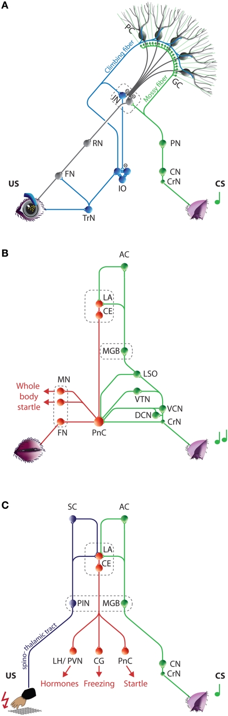
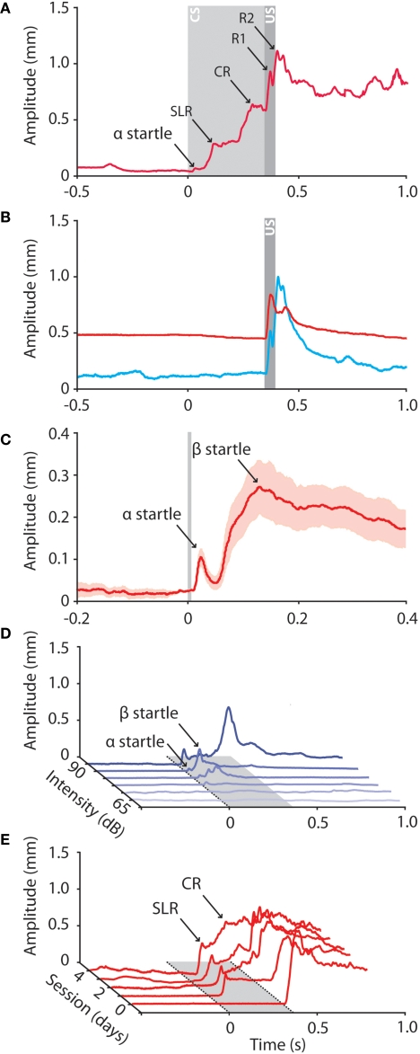
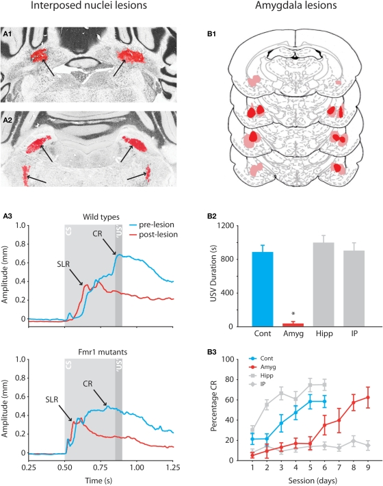
Over the past decade the advent of mouse transgenics has generated new perspectives on the study of cerebellar molecular mechanisms that are essential for eyeblink conditioning. However, it also appears that results from eyeblink conditioning experiments done in mice differ in some aspects from results previously obtained in other mammals. In this review article we will, based on studies using (cell-specific) mouse mutants and region-specific lesions, re-examine the general eyeblink behavior in mice and the neuro-anatomical circuits that might contribute to the different peaks in the conditioned eyeblink trace. We conclude that the learning process in mice has at least two stages: An early stage, which includes short-latency responses that are at least partly controlled by extracerebellar structures such as the amygdala, and a later stage, which is represented by well-timed conditioned responses that are mainly controlled by the pontocerebellar and olivocerebellar systems. We refer to this overall concept as the Amygdala-Cerebellum-Dynamic-Conditioning Model (ACDC model).

Keywords: ACDC model; amygdala; cerebellum; cued fear conditioning; eyeblink conditioning; mouse.


Figures

Figure 1
Figure 1
Neurocircuitries underlying eyeblink conditioning, auditory startle reflexes, and cued fear conditioning. (A) Neural circuits engaged during eyeblink conditioning. The mossy fiber CS-pathway (green) and climbing fiber US-pathway (blue) converge at the PCs in the cerebellar cortex and to a much lesser extent at the IN neurons. The CR-pathway (gray) is formed by the cerebellar output neurons and relayed via the RN to the FN and OMN, which innervate the eyelid muscles. (For simplicity only the eyelid innervation by the FN is depicted, see text for more details.) Conditioned induced plasticity at the PC and possibly also in the IN gradually leads to the establishment of an adequate CR. (B) Neural circuits engaged during auditory startle reflexes. The fastest route for transmission of acoustic input into motor output is from the CrN via the PnC to the motor neurons, including the FN. In addition, multiple afferent systems including the LSO, VTN, DCN, and VCN excitate the giant PnC neurons. Amygdala activity directly controls the expression of the startle reflex by its projections to the PnC. (C) Neural circuits engaged during cued fear conditioning. The tone (CS) and electric foot shock (US) are relayed to the LA from thalamic and cortical regions of the auditory (green) and somatosensory (purple) systems, respectively. The LA directly and indirectly projects to the CE, which efferents (red) control the expression of the different aspects of the fear reaction. One or two paired trials induces efficient plasticity in the LA resulting in typical fear CRs including freezing, tachycardia, tachypneu, and facial responses. AC, Auditory cortex; CE, Central amygdala; CG, Central gray; CN, Cochlear nucleus; CrN, Cochlear root nucleus; CS, Conditioned stimulus; DCN, Dorsal cochlear nucleus; FN, Facial nucleus; GC, Granule cell; IN, Interposed nuclei; IO, Inferior olive; LA, Lateral amygdala; LH, Lateral hypothalamus; LSO, Lateral superior olive; MGB, Medial geniculate body of the thalamus; MN, Motor neurons; OMN Oculomotor Nucleus, PC, Purkinje cell; PIN, Posterior intralaminar nucleus of the thalamus; PN, Pontine nuclei; PnC, Caudal pontine reticular nucleus; PVN, Paraventricular hypothalamic nucleus ; RN, Red nucleus; SC, Somatosensory cortex; TrN, Trigeminal nerve nucleus; US, Unconditioned stimulus; VCN, Ventral cochlear nucleus; VTN, Ventrolateral tegmental nucleus.
Figure 2
Figure 2
Raw data traces obtained from mice during eyeblink conditioning and auditory startle reflexes. (A) Different peaks can be distinguished in the conditioned eyeblink trace, including a small startle peak, a short-latency response (SLR), a conditioned response (CR), and two or more unconditioned response peaks (R1 and R2). In all panels: tone onset at t = 0; puff onset at t = 350; CS duration 380 ms; US duration 30 ms; ISI 350 ms; at amplitude = 0 mm the eyelid is maximum opened, at 1 mm the eyelid is fully closed. (B) Raw data traces of a reflexive eyeblink response of a mouse when awake behaving (blue) and during ‘quiet wakefulness’ (red). When behaving the eyelid is fully open and the oscillatory properties of the eyelid motor system are clearly visible in the eyeblink response. During quiet wakefulness the mouse sits very quiet, the eyelid is half closed, the baseline is completely flat, and there is a virtual absence of the normal oscillations of the eyelid response. (C) Mean (±SEM) of 20 raw data traces of eyelid startle reflexes in response to a loud auditory tone (90 dB, 10 kHz). Note that, when presented such a loud tone, two startle peaks (α and β) can be distinguished in the raw data traces. (D) The amplitude of the auditory startle reflex correlates with the intensity of the auditory stimulus. First small α responses emerge, whereas β responses appear when the tone intensity is increased. (E) Example raw data traces of a mouse over the consecutive training sessions. During habituation session (T-0) the behaviorally neutral CS does not elicit eyelid responses. During the first paired training sessions SLRs emerge whereas prolonged training results in well-timed CRs.
Figure 3
Figure 3
Effects of anterior interposed nucleus lesions in mice and amygdala lesions in rats on eyeblink conditioning. (A1) Example of bilateral lesion (arrows) of anterior interposed nucleus in Nissl-stained section of Fmr1 mutant. (A2) Example of degenerated axonal fibers (silver staining is indicated by arrows) in the superior cerebellar peduncle and ipsilateral descending tracts. (A3) Eyeblink traces showing the average amplitudes of the CRs in wild-type and Fmr1 mutants before (blue) and after (red) the lesions. In both mutants and wild-types lesions of the anterior interposed nucleus abolish well-timed cerebellar CRs, whereas startle reflexes and SLRs are still present in the eyeblink trace. Reproduced with permission from Koekkoek et al. (2005). (B1) Histological reconstructions of amygdala lesion sites in rats. Numbers indicate distance in millimeters posterior to Bregma. (B2) Mean ultrasonic vocalization (USV) during day 6 of the training sessions. The USV duration is a valid model of anxiety in rats (Sanchez, 2003), and a reduced USV duration behaviorally confirms the lesions of the amygdala. (B3) Mean percentage of CR (±SEM) during daily training sessions from control (n = 9), interposed nuclei lesioned (n = 9), amygdala lesioned (n = 9), and hippocampus lesioned (n = 10) rats. Lesions of the amygdala robustly slowed down the acquisition of eyeblink CRs indicating that the amygdala modulates the eyeblink conditioning process. Reproduced with permission from Lee and Kim (2004).
Figure 4
Figure 4
The Amygdala-Cerebellum-Dynamic-Conditioning Model. An integrated model to explain the different phases in the mouse eyeblink conditioning learning process and the different peaks in the individual eyeblink traces. The colors in the model eyeblink trace represent the anatomical afferents to the FN or PnC. During eyeblink conditioning the tone (CS) and corneal air puff (US) converge at least in the amygdala and cerebellum. They are relayed to the LA from thalamic and cortical regions of the auditory (green) and somatosensory (purple) systems. Pontocerebellar (green) and olivocerebellar (blue) systems mediate the convergence of CS and US on Purkinje cells in the cerebellar cortex and to a lesser extent on the IN neurons. Amygdala and cerebellum control the FN activity via PnC (red) and RN (gray), respectively. During the first learning phase in eyeblink conditioning very efficient plasticity in the LA results in mild conditioned fear responses, including rapid facial responses such as an eyelid closure. This is represented in the eyeblink trace by an SLR (red). A few CS-US pairings are enough to obtain clear SLRs in mice. Prolonged training will induce cerebellar learning, which behaviorally is represented by a perfectly timed eyelid closure (gray). In addition, direct projections from the amygdala to the PN might contribute directly to the CS input of the cerebellum. Thus, the ACDC model assumes that neuronal mechanism in different brain regions contribute to the establishment of an adequate CR. AC, Auditory cortex; CE, Central amygdala; CN, Cochlear nucleus; CrN, Cochlear root nucleus; CS, Conditioned stimulus; FN, Facial nucleus; GC, Granule cell; IN, Interposed nuclei; IO, Inferior olive; LA, Lateral amygdala; MGB, Medial geniculate body of the thalamus; MN, Motor neurons; PC, Purkinje cell; PIN, Posterior intralaminar nucleus of the thalamus; PN, Pontine nuclei; PnC, Caudal pontine reticular nucleus; RN, Red nucleus; SC, Somatosensory cortex; TrN, Trigeminal nerve nucleus; US, Unconditioned stimulus.

Similar articles

Cited by

References

    1. Aiba A., Kano M., Chen C., Stanton M. E., Fox G. D., Herrup K., Zwingman T. A., Tonegawa S. (1994). Deficient cerebellar long-term depression and impaired motor learning in mGluR1 mutant mice. Cell 79, 377–38810.1016/0092-8674(94)90205-4 - DOI - PubMed
    1. Aksenov D., Serdyukova N., Irwin K., Bracha V. (2004). GABA neurotransmission in the cerebellar interposed nuclei: involvement in classically conditioned eyeblinks and neuronal activity. J. Neurophysiol. 91, 719–72710.1152/jn.00859.2003 - DOI - PubMed
    1. Albus J. S. (1971). A theory of cerebellar function. Math. Biosci. 10, 25–6110.1016/0025-5564(71)90051-4 - DOI
    1. Aou S., Woody C. D., Birt D. (1992). Changes in the activity of units of the cat motor cortex with rapid conditioning and extinction of a compound eye blink movement. J. Neurosci. 12, 549–559 - PMC - PubMed
    1. Araki K., Meguro H., Kushiya E., Takayama C., Inoue Y., Mishina M. (1993). Selective expression of the glutamate receptor channel delta 2 subunit in cerebellar Purkinje cells. Biochem. Biophys. Res. Commun. 197, 1267–127610.1006/bbrc.1993.2614 - DOI - PubMed