. 2010 Feb 1;5(2):e8981.
doi: 10.1371/journal.pone.0008981.

Vertebrate Lrig3-ErbB interactions occur in vitro but are unlikely to play a role in Lrig3-dependent inner ear morphogenesis

Affiliations

Vertebrate Lrig3-ErbB interactions occur in vitro but are unlikely to play a role in Lrig3-dependent inner ear morphogenesis

Victoria E Abraira et al. PLoS One. .

Abstract

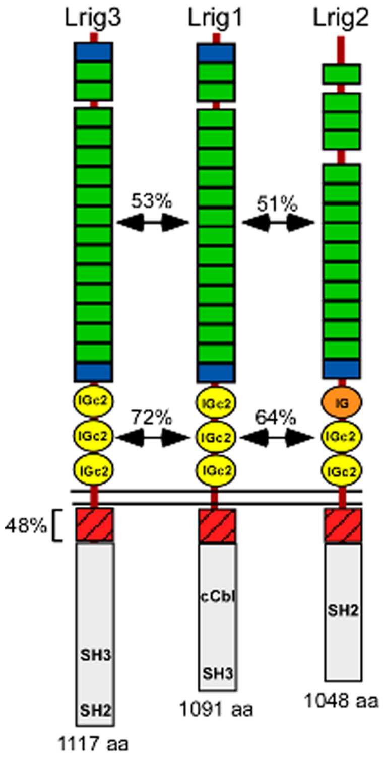
Background: The Lrig genes encode a family of transmembrane proteins that have been implicated in tumorigenesis, psoriasis, neural crest development, and complex tissue morphogenesis. Whether these diverse phenotypes reflect a single underlying cellular mechanism is not known. However, Lrig proteins contain evolutionarily conserved ectodomains harboring both leucine-rich repeats and immunoglobulin domains, suggesting an ability to bind to common partners. Previous studies revealed that Lrig1 binds to and inhibits members of the ErbB family of receptor tyrosine kinases by inducing receptor internalization and degradation. In addition, other receptor tyrosine kinase binding partners have been identified for both Lrig1 and Lrig3, leaving open the question of whether defective ErbB signaling is responsible for the observed mouse phenotypes.

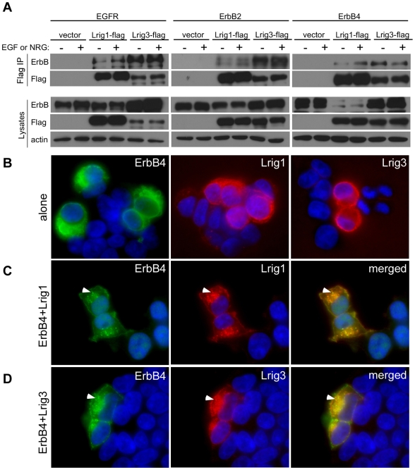
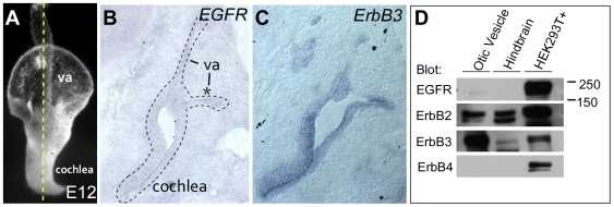
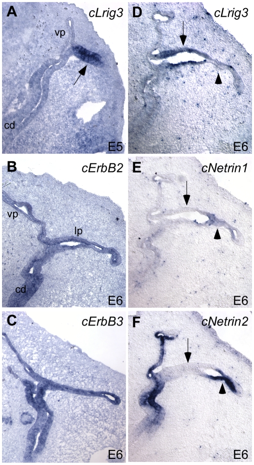
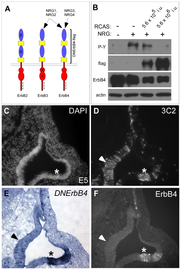
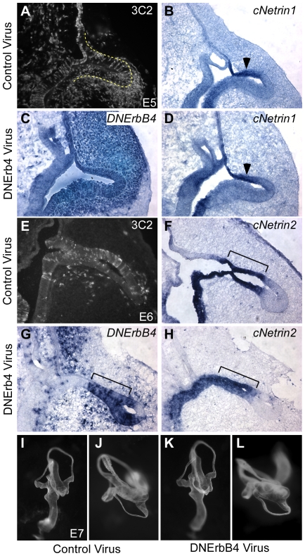
Methodology/principal findings: Here, we report that Lrig3, like Lrig1, is able to interact with ErbB receptors in vitro. We examined the in vivo significance of these interactions in the inner ear, where Lrig3 controls semicircular canal formation by determining the timing and extent of Netrin1 expression in the otic vesicle epithelium. We find that ErbB2 and ErbB3 are present in the early otic epithelium, and that Lrig3 acts cell-autonomously here, as would be predicted if Lrig3 regulates ErbB2/B3 activity. However, inhibition of ErbB activation in the chick otic vesicle has no detectable effect on Netrin gene expression or canal morphogenesis.

Conclusions/significance: Our results suggest that although both Lrig1 and Lrig3 can interact with ErbB receptors in vitro, modulation of Neuregulin signaling is unlikely to contribute to Lrig3-dependent processes of inner ear morphogenesis. These results highlight the similar binding properties of Lrig1 and Lrig3 and underscore the need to determine how these two family members bind to and regulate different receptors to affect diverse aspects of cell behavior in vivo.


Conflict of interest statement

Competing Interests: The authors have declared that no competing interests exist.

Figures

Figure 1
Figure 1. Lrig family protein structures.
Alignment of Lrig1, Lrig2, and Lrig3 shows that Lrig1 and Lrig3 share the same number and spacing of Leucine Rich Repeats (LRRs, green) and Immunoglobulin domains (Ig, yellow). In contrast, Lrig2 lacks an N' terminal LRR (blue) and has a different kind of Ig repeat, as reflected in the lower degree of identity with Lrig1. In the intracellular domains, all three family members share a highly conserved (48% identical) stretch of 48 amino acids (bracketed). Apart from this motif and some putative SH2 and/or SH3 domains, the cytoplasmic tails vary widely both in length and composition. Lrig1 is the only family member with a confirmed ability to bind c-Cbl. Arrows indicate degrees of identity between the LRR domain-containing regions and the Ig domain-containing regions. The amino acid (aa) length of each protein is also indicated. Domain structure was obtained with SMART protein (http://smart.embl-heidelberg.de/) and putative intracellular domains were defined by Scansite (http://scansite.mit.edu/).
Figure 2
Figure 2. Lrig1 and Lrig3 bind to and co-localize with ErbB receptor tyrosine kinases in vitro.
(A) Like Lrig1, Lrig3 can co-immunoprecipitate with multiple ErbB receptors in HEK293T cells. Lysates were immunoprecipitated with anti-flag antibodies and blotted with anti-ErbB (top) and anti-flag (bottom) antibodies. Both Lrig1-flag and Lrig3-flag can bind to EGFR (left), ErbB2 (middle), and ErbB4 (right) in the absence (−) or presence (+) of ligand (EGF or NRG). No ErbB receptors were precipitated in the absence of epitope-tagged Lrig (vector). Western blotting confirms the presence of each ErbB receptor (ErbB), and epitope-tagged Lrig family member (flag) in total lysates (bottom). Actin was used as a loading control. (B–D) Flag and ErbB4 immunocytochemistry on HEK293T cells transfected with ErbB4, Lrig1-flag, and/or Lrig3-flag alone (B) or in combinations (C,D). When expressed on their own, Lrig1, Lrig3 and ErbB4 are detected at the cell surface (B). When either Lrig1 (C) or Lrig3 (D) is expressed in combination with ErbB4, the proteins co-localize in intracellular compartments (arrowheads in C and D).
Figure 3
Figure 3. ErbB2 and ErbB3 are present in the developing mouse otic vesicle.
(A) Paintfill of an E12 mouse otic vesicle depicting plane of section shown in B and C (dashed line). The vestibular apparatus (va) develops dorsally and the cochlea extends ventrally. The lateral canal will develop from the lateral pouch, which is indicated by an asterisk. (B,C) In situ hybridization of EGFR (B) and ErbB3 (C) on adjacent transverse sections through an E12 mouse head. The otic epithelium is outlined and annotated as in A. Dorsal is up; lateral is right. EGFR message is not detectable in the otic vesicle, but is present in other tissues (not shown), confirming that the probe worked. ErbB3 is expressed throughout the otic vesicle epithelium. (D) Western blot of dissected otic vesicle tissue. ErbB2 and ErbB3 are present in abundance, with low levels of EGFR and no detectable ErbB4. HEK293T cells transfected with each ErbB receptor and hindbrain tissue served as controls. Only ErbB2 and ErbB3 are expressed in hindbrain at this stage.
Figure 4
Figure 4. Lrig3 acts within the otic epithelium to regulate canal morphogenesis.
(A,B) Lateral (A) and top-down (B) views of a paintfilled E14 Lrig3flox/flox inner ear reveal normal formation of all structures, including the posterior (pc) and lateral canals (lc). (C,D) Lateral (C) and top-down (D) views of a paintfilled E14 Lrig3 null inner ear show a truncation of the lateral canal (arrowhead). (E) X-gal staining of a transverse section through an E12 Pax2Cre;R26R embryo. βgalactosidase activity is restricted to the otic vesicle epithelium. (F,G) Paintfills of E14 inner ears derived from a Pax2Cre cross to the Lrig3flox allele. Pax2Cre;Lrig3flox/- embryos develop a lateral canal truncation (arrowhead) identical to Lrig3 nulls (D). Thus, loss of Lrig3 from Pax2-positive cells in the otic epithelium recapitulates the null phenotype.
Figure 5
Figure 5. Expression of cLrig3, cNetrin1, cNetrin2, cErbB2 and cErbB3 in chick otic epithelium.
(A–F) In situ hybridization of cLrig3 (A,D), cErbB2 (B), cErbB3 (C), cNetrin1 (E), and cNetrin2 (F) on transverse sections of E5 (A) or E6 (B–F) chicken heads. Dorsal is up; lateral is right. As in the mouse, chicken Lrig3 is initially present throughout the lateral pouch at E5 (arrows, A), and then sustained in the non-fusing epithelium (arrow, D) but not the fusion plate (arrowhead, D) at E6. Hybridization of adjacent sections confirms that chicken Netrin1 expression is complementary to Lrig3, with transcript detected in the fusing (arrowhead, E) but not non-fusing epithelium (arrow, E). In addition, cNetrin2, which does not exist in mice, is robustly expressed overlapping with cNetrin1 in the fusion plate (arrowhead, F). Both ErbB2 and ErbB3 are transcribed throughout the otic epithelium (B) including the vertical pouch (vp), lateral pouch (lp) and cochlear duct (cd).
Figure 6
Figure 6. RCAS(A)/DNErbB4-flag produces DNErbB4-flag protein in vitro and in vivo.
(A) Diagrams of ErbB2-4 receptors depicting their extracellular domains (blue and yellow), intracellular kinase modules (red), and tyrosine residues (Y). Four Neuregulin ligands (NRG1-4) bind to ErbB3 and ErbB4 as shown. Since ErbB2 cannot bind ligand and ErbB3 is kinase dead, ErbB2 and ErbB3 typically heterodimerize in order to bind and transduce Neuregulin signals. Dominant negative (DN) ErbB4 consists of the extracellular and transmembrane domains of the human ErbB4 receptor (bracketed), which binds to all four NRG ligands and prevents them from interacting with their endogenous receptors. Therefore, DNErbB4 effectively blocks Neuregulin signaling through all ErbB receptors. (B) Western blot analysis of lysates of DF1 chicken cells transiently transfected with ErbB4 and infected with RCAS(A)/DNErbB4-flag in the presence or absence of NRG, as indicated. Production of ErbB4 was confirmed by probing lysates with antibodies to ErbB4 (third row). Antibodies against the flag epitope reveal the relative amount of DNErbB4-flag in cells infected with 5×10⁵ or 5×10⁶ infectious units of RCAS(A)/DNErbB4-flag (second row). NRG stimulation of uninfected cells induces phosphorylation of ErbB4, as detected by blotting with anti-phosphotyrosine (P-Y) antibodies (top). This phosphorylation is reduced in the presence of increased levels of DNErbB4-flag. Actin served as a loading control for the P-Y blot (bottom). (C–F) Adjacent transverse sections through E5 chicken otic vesicles infected with RCAS(A)/DNErbB4-flag 48 hours earlier and processed for DAPI staining (C), anti-3C2 immunostaining (D), DNErbB4 in situ hybridization (E), or anti-ErbB4 immunostaining (F). The DNErbB4 in situ probe and the anti-ErbB4 antibody are specific to human ErbB4. 3C2-gag immunostaining reveals broad infection of the otic epithelium, including the lateral pouch (asterisk). Both DNErbB4 transcript (E) and DNErbB4 protein (F) are produced here (asterisks). In some areas, viral infection does not generate detectable amounts of DNErbB4-flag (arrowhead). Therefore, in all subsequent experiments, the extent of infection was monitored by in situ hybridization for DNErbB4.
Figure 7
Figure 7. Broad expression of dominant negative ErbB has no effect on Netrin gene expression or canal morphogenesis.
(A–H) Transverse sections through E5 (A–D) or E6 (E-H) chick heads infected with control (A,B,E,F) or DNErbB4 (C,D,G,H) virus. The lateral pouch is outlined in A. The extent of infection was assayed by 3C2-gag immunostaining for control virus (A,C) or by in situ hybridization for DNErbB4 (C,G). Adjacent sections were probed for cNetrin1 (B,D) or cNetrin2 (F,H). At both E5 and E6, cNetrin1 and cNetrin2 are expressed normally in the fusion plate epithelium (arrowheads, brackets) despite abundant expression of DNErbB4 here. (I–L) Lateral (I,K) and top-down (J,L) views of E7 paintfilled inner ears of control (I,J) and experimental (K,L) embryos. No change in the size or shape of the overall inner ear (I,L) or lateral canal (J,L) is evident.

Similar articles

Cited by

References

    1. Dolan J, Walshe K, Alsbury S, Hokamp K, O'Keeffe S, et al. The extracellular leucine-rich repeat superfamily; a comparative survey and analysis of evolutionary relationships and expression patterns. BMC Genomics. 2007;8:320. - PMC - PubMed
    1. Ghiglione C, Amundadottir L, Andresdottir M, Bilder D, Diamonti JA, et al. Mechanism of inhibition of the Drosophila and mammalian EGF receptors by the transmembrane protein Kekkon 1. Development. 2003;130:4483–4493. - PubMed
    1. Ghiglione C, Carraway KL, 3rd, Amundadottir LT, Boswell RE, Perrimon N, et al. The transmembrane molecule kekkon 1 acts in a feedback loop to negatively regulate the activity of the Drosophila EGF receptor during oogenesis. Cell. 1999;96:847–856. - PubMed
    1. Goldoni S, Iozzo RA, Kay P, Campbell S, McQuillan A, et al. A soluble ectodomain of LRIG1 inhibits cancer cell growth by attenuating basal and ligand-dependent EGFR activity. Oncogene. 2007;26:368–381. - PubMed
    1. Gur G, Rubin C, Katz M, Amit I, Citri A, et al. LRIG1 restricts growth factor signaling by enhancing receptor ubiquitylation and degradation. Embo J. 2004;23:3270–3281. - PMC - PubMed

Publication types

MeSH terms