. 2010 Feb 1;5(2):e8994.
doi: 10.1371/journal.pone.0008994.

The translation regulatory subunit eIF3f controls the kinase-dependent mTOR signaling required for muscle differentiation and hypertrophy in mouse

Affiliations

The translation regulatory subunit eIF3f controls the kinase-dependent mTOR signaling required for muscle differentiation and hypertrophy in mouse

Alfredo Csibi et al. PLoS One. .

Abstract

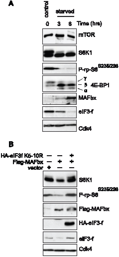
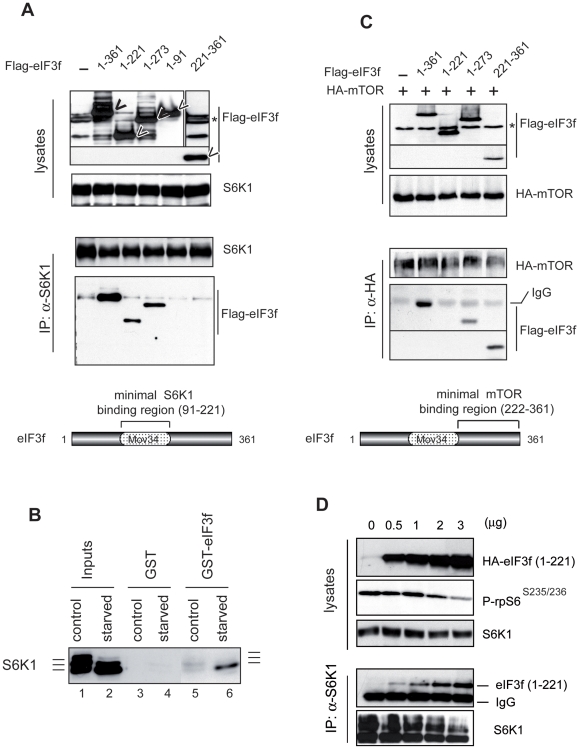
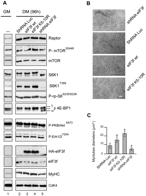
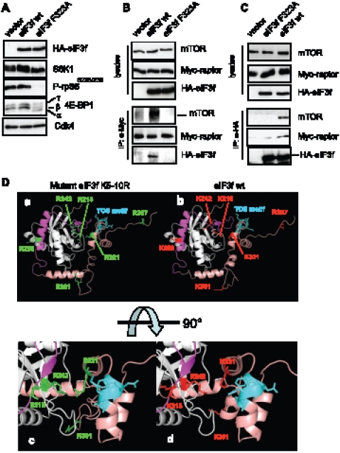
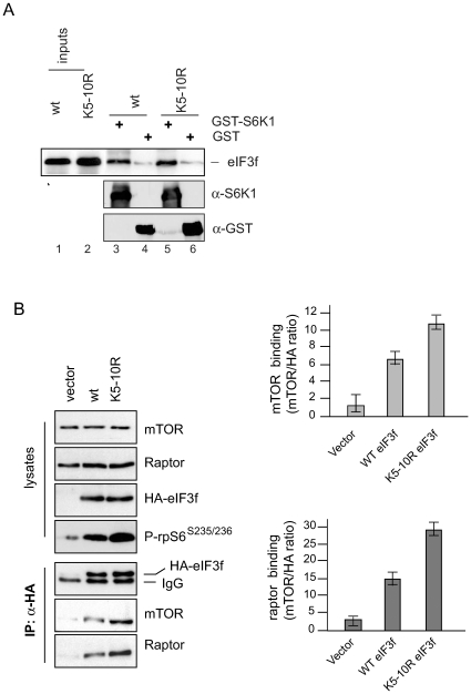
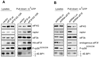
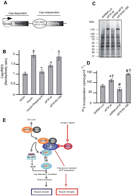
The mTORC1 pathway is required for both the terminal muscle differentiation and hypertrophy by controlling the mammalian translational machinery via phosphorylation of S6K1 and 4E-BP1. mTOR and S6K1 are connected by interacting with the eIF3 initiation complex. The regulatory subunit eIF3f plays a major role in muscle hypertrophy and is a key target that accounts for MAFbx function during atrophy. Here we present evidence that in MAFbx-induced atrophy the degradation of eIF3f suppresses S6K1 activation by mTOR, whereas an eIF3f mutant insensitive to MAFbx polyubiquitination maintained persistent phosphorylation of S6K1 and rpS6. During terminal muscle differentiation a conserved TOS motif in eIF3f connects mTOR/raptor complex, which phosphorylates S6K1 and regulates downstream effectors of mTOR and Cap-dependent translation initiation. Thus eIF3f plays a major role for proper activity of mTORC1 to regulate skeletal muscle size.


Conflict of interest statement

Competing Interests: The authors have declared that no competing interests exist.

Figures

Figure 1
Figure 1. regulation of mTORC1 activity is correlated to MAFbx mediated eIF3f degradation during skeletal muscle atrophy.
(A) Effects of starvation on components of the mTOR/S6K1 kinase pathway. Mouse primary cultured satellite cells myotubes at 4th day of differentiation were starved by removal of growth medium and incubated in PBS for the indicated times. Proteins were extracted and subjected to immunoblots analysis. (B) Primary cultures of satellite cells were transfected with expression vectors encoding Flag-tagged MAFbx and/or the HA-tagged mutant eIF3f K5–10R and cultured in differentiation medium for 4 days. Total cellular lysates were analyzed by immunoblotting.
Figure 2
Figure 2. mTOR and S6K1 physically interact with two different domains of eIF3f.
(A) Interaction of eIF3f mutants with S6K1. Mouse primary cultured satellite cells were transfected with expression vectors encoding Flag-tagged eIF3f wt and deletion mutants of eIF3f. Total cellular extracts were subjected to immunoprecipitation with anti-S6K1 followed by immunoblotting analysis with anti-Flag antibodies. (B) Phosphorylation of S6K1 regulates the interaction with eIF3f. Interaction of hyperphosphorylated (control) or hypophosphorylated S6K1 (starved) was tested for binding to eIF3-f. GST or GST-eIF3f beads were incubated with total cellular extracts (300 µg) of mouse primary culture of satellite myotubes in differentiation medium (control) or starved for 3h. Bound proteins were eluted, subjected to SDS-PAGE and analyzed by immunoblotting. (C) Interaction of eIF3f mutants with mTOR. Same as in (B) except that mouse primary cultured satellite cells were cotransfected with expression vector encoding HA-tagged mTOR. (D) An eIF3f mutant deleted of the mTOR-binding domain suppresses S6K1 phosphorylation. Mouse primary cultured satellite cells were transfected with increasing amounts of expression vectors encoding the deletion mutants eIF3f (aa1–221). Total cellular extracts were subjected to immunoprecipitation with anti-S6K1 followed by immunoblotting.
Figure 3
Figure 3. eIF3f regulates the mTORC1 pathway in differentiated myotubes.
(A) Mouse primary cultured satellite cells were transfected with expression vectors encoding HA-tagged eIF3f wt or the mutant K5–10R, or subjected to RNAi-mediated silencing of eIF3f. At 24h posttransfection cells were induced to differentiate. Total lysates from proliferating control cells (GM) or 4 days (DM 96h) differentiated myotubes were analyzed by immunoblotting using the indicated antibodies and phospho-specific antibodies. (B) Effects of the overexpression and/or the knockdown of eIF3f on myotubes size. Mouse primary cultured satellite cells were transfected with expression vectors as indicated in (A). Cells were cultured in differentiation medium for 4 days. Bright-field images of differentiated myotubes are shown. Scale bar, 20 µm. (C) Myotube mean diameter of experiments as in (A) was measured. Data represent the average ± s.e.m for three experiments #, P<0,05 compared to control. At least 150 myotubes for each condition were analyzed.
Figure 4
Figure 4. A TOS motif in eIF3f is necessary for binding to mTOR-raptor.
(A) Effects of TOS mutation on the activity of mTORC1. Mouse primary muscle cells were transfected with expression vectors encoding HA-tagged eIF3f wt, HA-tagged TOS motif mutant eIF3f F323A and the empty vector. At 24h post-transfection cells were induced to differentiate. Protein expression and phosphorylation levels were assayed from lysates of 4 days differentiated myotubes by immunoblotting. (B) Mouse primary muscle cells were co-transfected with expresion vector encoding Myc-raptor and either HA-eIF3f wt, the TOS motif mutant HA-eIF3f F323A and/or the empty vector. Transfected cells were induced to differentiate during 4 days and lysed in immunoprecipitation buffer without detergent. Total cellular extracts were subjected to immunoprecipitation with anti-Myc antibody, followed by immnoblotting. Asterisk indicates a non specific band . (C) Mouse primary muscle cells were transfected, differentiated and lysed as indicated in B. Total cellular extracts were subjected to immunoprecipitation with anti-HA antibody, followed byimmunoblotting. (D) Modeling of eIF3f shows that the functional TOS is accessible for eIF3f/raptor interaction. Models of the three-dimensional structures of wild type and mutant eIF3f K5-10R. The left-side images correspond to mutant eIF3f K5–10R (panels a and c), right-side to eIF3f wt (panels b and d). The mutated lysines are coloured red, and corresponding arginines are colored green. The C-terminal arm is colored salmon. The mTOR binding region is colored pink. The TOS motif is colored blue. The bottom panel is a zoom on the central region in a 90° rotation.
Figure 5
Figure 5. The hyperactive eIF3f mutant K5–10R shows increased affinity for binding to mTOR/raptor.
(A) Interaction of S6K1 with eIF3f wt and mutant K5–10R in vitro. GST or GST-S6K1 beads were incubated with in vitro translated HA-tagged eIF3f wt or mutant K5–10R. Bound proteins were eluted, subjected to SDS-PAGE and analyzed by immunoblotting. (B) Co-immunoprecipitation of endogenous mTOR/raptor with eIF3f wt and mutant eIF3f K5–10R. Mouse primary skeletal muscle cells were transfected with expression vectors encoding HA-tagged eIF3f wt or the mutant K5–10R. Cell extracts of 3 days differentiated myotubes were subjected to immunoprecipitation with anti-HA antibody. Immune complexes were subjected to SDS-PAGE and Western blotting. The bar graphs show the ratio of mTOR and raptor recovered relative to HA-tagged eIF3f wt or mutant K5–10R. Data represent the combined results from three different experiments.
Figure 6
Figure 6. eIF3f regulates the recruitment of translational proteins to the mRNA 7-methylguanosine cap structure.
(A) Binding of translational components to the 7-methylguanosine cap complex is enhanced by eIF3f. Mouse primary cultured satellite cells were transfected with expression vectors encoding HA-tagged eIF3f wt and/or the mutant eIF3f K5–10R and differentiated for 3 days. Cap-binding proteins in lysates were purified by 7-methyl GTP (m7GTP) affinity beads. Levels of proteins and phosphoproteins were analyzed by Western blotting. (B) eIF3f silencing leads to decreased recruitment of translational components to the m7-GTP cap complex. Mouse primary cultured satellite cells were subjected to RNAi-mediated silencing of eIF3f using specific small hairpin RNA. A nonspecific shRNAi was used as control. Cell lysates were purified by m7-GTP and analyzed as described in (A).
Figure 7
Figure 7. eIF3f regulates cap-dependent translation.
(A) Structure of the bicistronic reporter plasmid allowing cap-dependent expression of renilla luciferase and expression of firefly luciferase dependent on HCV IRES. (B) Overexpression of eIF3f modulates cap-dependent translation. Mouse primary cultured satellite cells were cotransfected with the bicistronic reporter vector and expression vectors encoding HA-tagged eIF3f wt and the mutant eIF3f K5–10R. Twenty-four hours posttransfection cells were grown for an additional 24h in 20% serum (control), stimulated with insulin or pretreated with rapamycin and stimulated with insulin for and additional 24 h. Cells transfected to express eIF3f wt or the mutant eIF3f K5–10R were grown in 20% serum. Luciferase activities were measured by a dual-luciferase assay. The ratio of Renilla (Cap-dependent) to Firefly (IRES-dependent) luciferase activity was calculated. Data are presented as the mean ± standard error from three independent experiments carried out in triplicate, *P<0,05 compared to control; # P<0,05 compared to Insulin + rapamycin. (C) Mouse primary cultured satellite cells were transfected with expression vectors as described in (B) and/or subjected to shRNAi-mediated silencing of eIF3f prior to labeling new protein synthesis with 35S methionine. Newly synthesized proteins were separated by SDS-PAGE, and visualized by autoradiography. (D) Newly synthesized proteins from three experiments as in (C) were quantified. *P = 0,002 and # P<0,001 compared to control; P>0,001 compared to shRNAi eIF3f. (E). Model depicting the central role of eIF3f in the signaling pathways controlling skeletal muscle mass.

Similar articles

Cited by

References

    1. Gingras AC, Raught B, Sonenberg N. Regulation of translation initiation by FRAP/mTOR. Genes Dev. 2001;15:807–826. - PubMed
    1. Richardson CJ, Schalm SS, Blenis J. PI3-kinase and TOR: PIKTORing cell growth. Semin Cell Dev Biol. 2004;15:147–159. - PubMed
    1. Schmelzle T, Hall MN. TOR, a central controller of cell growth. Cell. 2000;103:253–262. - PubMed
    1. Wullschleger S, Loewith R, Hall MN. TOR signaling in growth and metabolism. Cell. 2006;124:471–484. - PubMed
    1. Jacinto E, Loewith R, Schmidt A, Linn S, Rüegg MA, et al. Mammalian TOR complex 2 controls the actin cytoskeleton and is rapamycin insensitive. Nat Cell Biol. 2004;6:1122–1128. - PubMed

Publication types

MeSH terms