

Application of Fluoro-Jade C in acute and chronic neurodegeneration models: utilities and staining differences
- PMID: 20126570
- PMCID: PMC2808500
- DOI: 10.1267/ahc.09018
Application of Fluoro-Jade C in acute and chronic neurodegeneration models: utilities and staining differences
Abstract
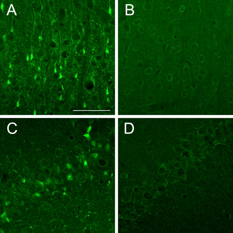
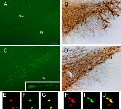
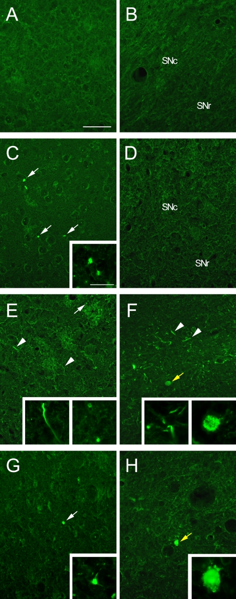
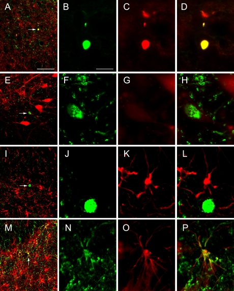
Recent neuropathological studies have shown that Fluoro-Jade C (FJC), an anionic fluorescent dye, is a good marker of degenerating neurons. However, those studies have mostly examined acute rather than chronic models of neurodegeneration. We therefore compared FJC staining using the intrastriatal 6-hydroxydopamine (6-OHDA)-injected rat as an acute model and the zitter rat as a chronic model, as both show dopaminergic (DA) neurodegeneration. In the 6-OHDA-injected rat, FJC-positive neurons were found in the substantia nigra pars compacta (SNc) before the loss of tyrosine hydroxylase (TH)-positive DA neurons. In the zitter rat, FJC-labeled fibers were first detected at 1 month old (1M) and were considerably increased in the striatum at 4M, whereas FJC-labeled cell bodies were found at 4M, but not at 1M in the SNc. Furthermore, FJC-labeled neurons of the zitter rat showed TH-immunoreactivity in fibers, but little in cell bodies, while those from the 6-OHDA-injected rat showed TH-immunoreactivity even in the cell bodies. These results demonstrate that FJC is a useful tool for detecting chronically degenerating neurons, and suggest that intracellular substances bound to FJC may accumulate in the cell bodies from fibers at a slower rate in the chronic model than in the acute model.
Keywords: 6-OHDA; Fluoro-Jade C; chronic neurodegenerative model; dopaminergic neurons; zitter rat.
Figures
Similar articles
-
Improvement in Double Staining With Fluoro-Jade C and Fluorescent Immunostaining: FJC Staining Is Not Specific to Degenerating Mature Neurons.J Histochem Cytochem. 2021 Sep;69(9):597-610. doi: 10.1369/00221554211043340. Epub 2021 Aug 31. J Histochem Cytochem. 2021. PMID: 34463186 Free PMC article.
-
Evaluation of Fluoro-Jade C as a marker of degenerating neurons in the rat retina and optic nerve.Exp Eye Res. 2009 Mar;88(3):426-37. doi: 10.1016/j.exer.2008.10.015. Epub 2008 Oct 29. Exp Eye Res. 2009. PMID: 19010324
-
Fluoro-Jade C can specifically stain the degenerative neurons in the substantia nigra of the 1-methyl-4-phenyl-1,2,3,6-tetrahydro pyridine-treated C57BL/6 mice.Brain Res. 2007 May 30;1150:55-61. doi: 10.1016/j.brainres.2007.02.078. Epub 2007 Mar 6. Brain Res. 2007. PMID: 17397812
-
Cyclosporin A attenuates the decrease in tyrosine hydroxylase immunoreactivity in nigrostriatal dopaminergic neurons and in striatal dopamine content in rats with intrastriatal injection of 6-hydroxydopamine.Exp Neurol. 1997 Aug;146(2):526-35. doi: 10.1006/exnr.1997.6575. Exp Neurol. 1997. PMID: 9270064
-
Development and application of novel histochemical tracers for localizing brain connectivity and pathology.Brain Res. 2016 Aug 15;1645:31-5. doi: 10.1016/j.brainres.2016.03.053. Epub 2016 May 4. Brain Res. 2016. PMID: 27155454 Review.
Cited by
-
Progesterone and vitamin D: Improvement after traumatic brain injury in middle-aged rats.Horm Behav. 2013 Aug;64(3):527-38. doi: 10.1016/j.yhbeh.2013.06.009. Epub 2013 Jul 27. Horm Behav. 2013. PMID: 23896206 Free PMC article.
-
Effects of Long-Term Vagus Nerve Electrical Stimulation Therapy on Acute Cerebral Infarction and Neurological Function Recovery in Post MCAO Mice.Oxid Med Cell Longev. 2022 Mar 29;2022:8131391. doi: 10.1155/2022/8131391. eCollection 2022. Oxid Med Cell Longev. 2022. PMID: 35391930 Free PMC article.
-
High-resolution fluorescence-guided transcranial ultrasound mapping in the live mouse brain.Sci Adv. 2021 Dec 10;7(50):eabi5464. doi: 10.1126/sciadv.abi5464. Epub 2021 Dec 8. Sci Adv. 2021. PMID: 34878843 Free PMC article.
-
Vdac1 Downregulation Causes Mitochondrial Disintegration Leading to Hippocampal Neurodegeneration in Scopolamine-Induced Amnesic Mice.Mol Neurobiol. 2019 Mar;56(3):1707-1718. doi: 10.1007/s12035-018-1164-z. Epub 2018 Jun 19. Mol Neurobiol. 2019. PMID: 29916145
-
Progesterone and low-dose vitamin D hormone treatment enhances sparing of memory following traumatic brain injury.Horm Behav. 2012 Apr;61(4):642-51. doi: 10.1016/j.yhbeh.2012.02.017. Horm Behav. 2012. PMID: 22570859 Free PMC article.
References
-
- Bian G. L., Wei L. C., Shi M., Wang Y. Q., Cao R., Chen L. W. Fluoro-Jade C can specifically stain the degenerative neurons in the substantia nigra of the 1-methyl-4-phenyl-1,2,3,6-tetrahydro pyridine-treated C57BL/6 mice. Brain Res. 2007;1150:55–61. - PubMed
-
- Brown D. A., Sawchenko P. E. Time course and distribution of inflammatory and neurodegenerative events suggest structural bases for the pathogenesis of experimental autoimmune encephalomyelitis. J. Comp. Neurol. 2007;502:236–260. - PubMed
-
- Chen L. W., Wang Y. Q., Bian G. L., Wei L. C., Yung K. L. Neurokinin-3 peptide instead of neurokinin-1 synergistically exacerbates kainic acid-inducing degeneration of neurons in the substantia nigra of mice. J. Neurochem. 2008;105:203–216. - PubMed
-
- Chidlow G., Wood J. P., Sarvestani G., Manavis J., Casson R. J. Evaluation of Fluoro-Jade C as a marker of degenerating neurons in the rat retina and optic nerve. Exp. Eye Res. 2009;88:426–437. - PubMed
-
- Cicchetti F., Brownell A. L., Williams K., Chen Y. I., Livni E., Isacson O. Neuroinflammation of the nigrostriatal pathway during progressive 6-OHDA dopamine degeneration in rats monitored by immunohistochemistry and PET imaging. Eur. J. Neurosci. 2002;15:991–998. - PubMed
LinkOut - more resources
Full Text Sources
Miscellaneous