

Immunohistochemical organization patterns of the follicular dendritic cells, myofibroblasts and macrophages in the human spleen--new considerations on the pathological diagnosis of splenectomy pieces
- PMID: 20126587
- PMCID: PMC2809999
Immunohistochemical organization patterns of the follicular dendritic cells, myofibroblasts and macrophages in the human spleen--new considerations on the pathological diagnosis of splenectomy pieces
Abstract
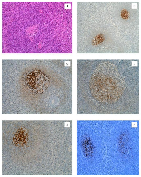
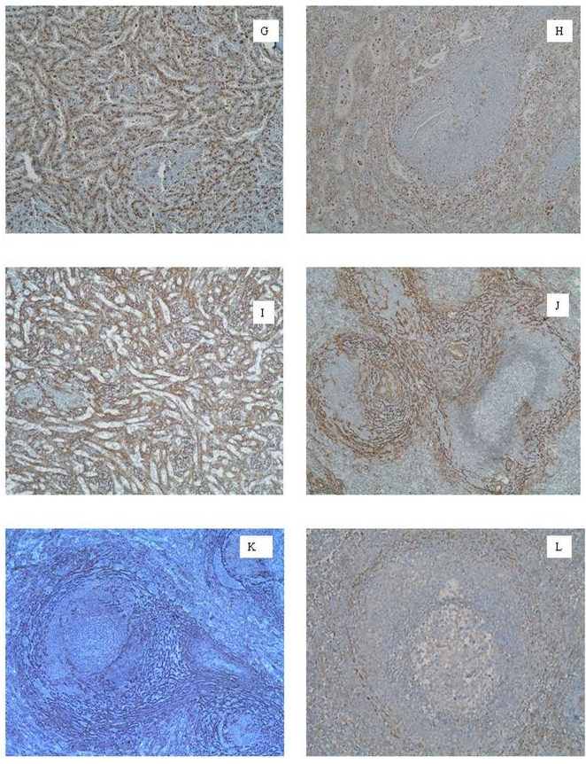
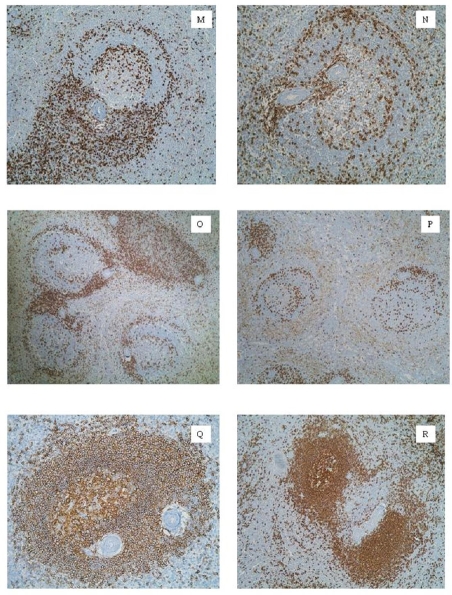
There is reliable information about how changes in spleen histology are influenced by the relationship among B and T lymphocytes, macrophages, dendritic cells and myofibroblasts. Moreover, if it can be applied in the day-by-day pathology laboratory. This work intends to elucidate morpho-functional aspects of relationships of these cells in the different spleen compartments, how they are influenced by pathological conditions and how basic immunohistochemical techniques could optimize the histopathological diagnosis. We analyzed the usefulness of the monoclonal antibodies CD45RO, CD20, CD21, CD35, CD68, caldesmon, the smooth muscle alpha-actin type 1 (SMA-1) in 91 specimens. CD21(+) CD35(+) follicular dendritic cells were organized into three patterns in agreement with the immune condition of the lymphoid follicle. Smooth muscle alpha-actin type 1(+)and caldesmon(+)myofibroblasts draw two double rings: marginal-perifollicular and germinal-marginal. The latter is closely related to T-cells. CD68(+)red pulp macrophages had clear and linear configuration. The interruption of this CD68(+) linear pattern in splenic marginal zone lymphoma cases could be a criterion to differentiate it from reactive hyperplasia. CD45RO, CD20, CD21, CD68 and SMA-1 provide a basic and quality immunohistochemical battery for a better comprehension of the human spleen and could improve its histopathological diagnosis.
Keywords: Follicular dendritic cells; histopathology; immune system; immunohistochemical; macrophages; myofibroblasts; spleen.
Figures
Similar articles
-
Inflammatory pseudotumor-like follicular dendritic cell sarcoma of the spleen: a report of six cases with increased IgG4-positive plasma cells.Pathol Int. 2013 May;63(5):245-51. doi: 10.1111/pin.12057. Pathol Int. 2013. PMID: 23714251
-
[Inflammatory pseudotumor-like follicular dendritic cell tumor of spleen].Zhonghua Bing Li Xue Za Zhi. 2008 Jan;37(1):40-4. Zhonghua Bing Li Xue Za Zhi. 2008. PMID: 18509984 Chinese.
-
Follicular dendritic cells in reactive and neoplastic lymphoid tissues: a reevaluation of staining patterns of CD21, CD23, and CD35 antibodies in paraffin sections after wet heat-induced epitope retrieval.Appl Immunohistochem Mol Morphol. 2001 Jun;9(2):117-24. doi: 10.1097/00129039-200106000-00003. Appl Immunohistochem Mol Morphol. 2001. PMID: 11396628
-
[Malignant lymphoma of the spleen. Histological and immunohistochemical studies of morphology and differential diagnosis].Veroff Pathol. 1991;136:1-265. Veroff Pathol. 1991. PMID: 1746166 Review. German.
-
Clinicopathologic characteristics of inflammatory pseudotumor-like follicular dendritic cell sarcoma.Int J Clin Exp Pathol. 2014 Apr 15;7(5):2421-9. eCollection 2014. Int J Clin Exp Pathol. 2014. PMID: 24966952 Free PMC article. Review.
Cited by
-
B lymphocyte compartments in the human splenic red pulp: capillary sheaths and periarteriolar regions.Histochem Cell Biol. 2014 May;141(5):507-18. doi: 10.1007/s00418-013-1172-z. Epub 2013 Dec 17. Histochem Cell Biol. 2014. PMID: 24337546
-
The human splenic microcirculation is entirely open as shown by 3D models in virtual reality.Sci Rep. 2022 Oct 1;12(1):16487. doi: 10.1038/s41598-022-19885-z. Sci Rep. 2022. PMID: 36182999 Free PMC article.
-
Assessment of immune cells and function of the residual spleen after subtotal splenectomy due to splenomegaly in cirrhotic patients.BMC Immunol. 2014 Oct 8;15:42. doi: 10.1186/s12865-014-0042-3. BMC Immunol. 2014. PMID: 25293512 Free PMC article.
-
Human spleen microanatomy: why mice do not suffice.Immunology. 2015 Jul;145(3):334-46. doi: 10.1111/imm.12469. Immunology. 2015. PMID: 25827019 Free PMC article. Review.
-
Heterogeneity of stromal cells in the human splenic white pulp. Fibroblastic reticulum cells, follicular dendritic cells and a third superficial stromal cell type.Immunology. 2014 Nov;143(3):462-77. doi: 10.1111/imm.12325. Immunology. 2014. PMID: 24890772 Free PMC article.
References
-
- Fujita T, Kashimura M, Adachi K. Scanning electron microscopy (SEM) studies of the spleen-normal and pathological. Scanning Electron Microsc. 1982;1:435–444. - PubMed
-
- Kashimura M, Fujita T. A scanning electron microscopy study of human spleen: relationship between microcirculation and functions. Scann Microsc. 1987;1:841–851. - PubMed
-
- van Krieken JHJM, Te Velde J. Normal Histology of the Human Spleen. Am J Surg Pathol. 1988;12:777–785. - PubMed
-
- van Krieken JHJM, Te Velde J, Hermans J, et al. The amount of white pulp in the spleen; a morphometrical study done in methylmethacry-late-embedded splenectomy specimens. Histopathology. 1983;7:167–182. - PubMed
-
- Steiniger B, Barth P. Berlin: Springer-Verlag; 2000. Microanatomy and function of the spleen. - PubMed
MeSH terms
LinkOut - more resources
Full Text Sources
Other Literature Sources