

Nuclear expression of KLF6 tumor suppressor factor is highly associated with overexpression of ERBB2 oncoprotein in ductal breast carcinomas
- PMID: 20126619
- PMCID: PMC2812494
- DOI: 10.1371/journal.pone.0008929
Nuclear expression of KLF6 tumor suppressor factor is highly associated with overexpression of ERBB2 oncoprotein in ductal breast carcinomas
Abstract
Background: Krüppel-like factor 6 (KLF6) is an evolutionarily conserved and ubiquitously expressed protein that belongs to the mammalian Sp1/KLF family of transcriptional regulators. Though KLF6 is a transcription factor and harbors a nuclear localization signal it is not systematically located in the nucleus but it was detected in the cytoplasm of several tissues and cell lines. Hence, it is still not fully settled whether the tumor suppressor function of KLF6 is directly associated with its ability to regulate target genes.
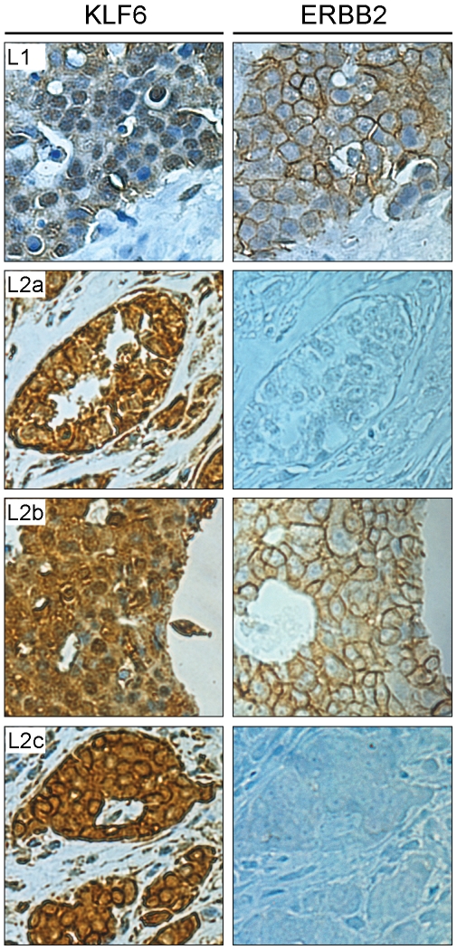
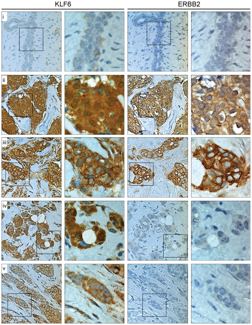
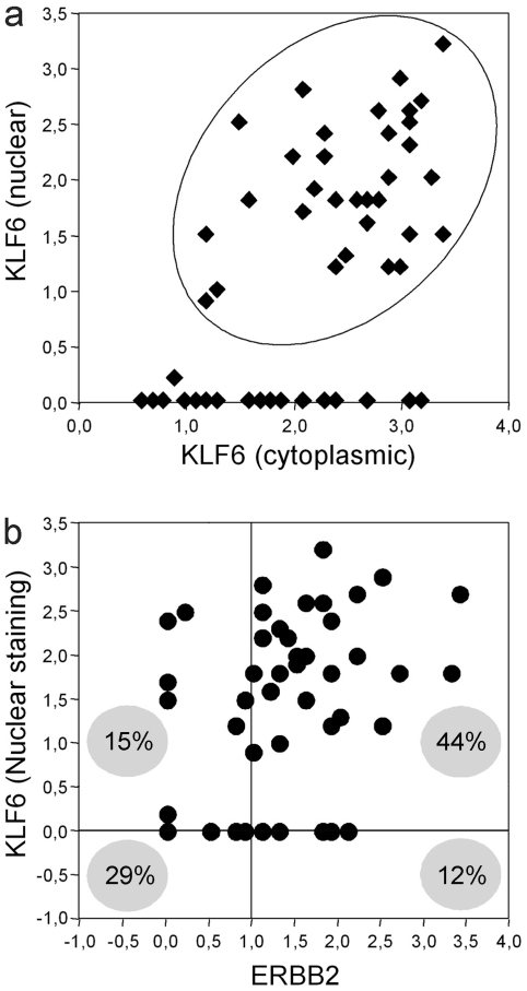
Methodology/principal findings: In this study we analyzed KLF6 expression and sub-cellular distribution by immunohistochemistry in several normal and tumor tissues in a microarray format representing fifteen human organs. Results indicate that while both nuclear and cytoplasmic distribution of KLF6 is detected in normal breast tissues, breast carcinomas express KLF6 mainly detected in the cytoplasm. Expression of KLF6 was further analyzed in breast cancer tissues overexpressing ERBB2 oncoprotein, which is associated with poor disease prognosis and patient's survival. The analysis of 48 ductal carcinomas revealed a significant population expressing KLF6 predominantly in the nuclear compartment (X(2)p = 0.005; Fisher p = 0.003). Moreover, this expression pattern correlates directly with early stage and small ductal breast tumors and linked to metastatic events in lymph nodes.
Conclusions/significance: Data are consistent with a preferential localization of KLF6 in the nuclear compartment of early stage and small HER2-ERBB2 overexpressing ductal breast tumor cells, also presenting lymph node metastatic events. Thus, KLF6 tumor suppressor could represent a new molecular marker candidate for tumor prognosis and/or a potential target for therapy strategies.
Conflict of interest statement
Figures
Similar articles
-
Krüppel-like factor 6 expression changes during trophoblast syncytialization and transactivates ßhCG and PSG placental genes.PLoS One. 2011;6(7):e22438. doi: 10.1371/journal.pone.0022438. Epub 2011 Jul 22. PLoS One. 2011. PMID: 21799854 Free PMC article.
-
Nucleo-cytoplasmic localization domains regulate Krüppel-like factor 6 (KLF6) protein stability and tumor suppressor function.PLoS One. 2010 Sep 9;5(9):e12639. doi: 10.1371/journal.pone.0012639. PLoS One. 2010. PMID: 20844588 Free PMC article.
-
KLF6-SV1 overexpression accelerates human and mouse prostate cancer progression and metastasis.J Clin Invest. 2008 Aug;118(8):2711-21. doi: 10.1172/JCI34780. J Clin Invest. 2008. PMID: 18596922 Free PMC article.
-
Biology of Krüppel-like factor 6 transcriptional regulator in cell life and death.IUBMB Life. 2010 Dec;62(12):896-905. doi: 10.1002/iub.396. IUBMB Life. 2010. PMID: 21154818 Review.
-
The role of KLF6 and its splice variants in cancer therapy.Drug Resist Updat. 2009 Feb-Apr;12(1-2):1-7. doi: 10.1016/j.drup.2008.11.001. Epub 2008 Dec 18. Drug Resist Updat. 2009. PMID: 19097929 Review.
Cited by
-
Identification of miRNAs that specifically target tumor suppressive KLF6-FL rather than oncogenic KLF6-SV1 isoform.RNA Biol. 2014;11(7):845-54. doi: 10.4161/rna.29356. Epub 2014 Jun 12. RNA Biol. 2014. PMID: 24921656 Free PMC article.
-
A novel KLF6-Rho GTPase axis regulates hepatocellular carcinoma cell migration and dissemination.Oncogene. 2016 Sep 1;35(35):4653-62. doi: 10.1038/onc.2016.2. Epub 2016 Feb 15. Oncogene. 2016. PMID: 26876204 Free PMC article.
-
Krüppel-like factor 6 expression changes during trophoblast syncytialization and transactivates ßhCG and PSG placental genes.PLoS One. 2011;6(7):e22438. doi: 10.1371/journal.pone.0022438. Epub 2011 Jul 22. PLoS One. 2011. PMID: 21799854 Free PMC article.
-
Oncogenic lncRNAs alter epigenetic memory at a fragile chromosomal site in human cancer cells.Sci Adv. 2022 Mar 4;8(9):eabl5621. doi: 10.1126/sciadv.abl5621. Epub 2022 Mar 2. Sci Adv. 2022. PMID: 35235361 Free PMC article.
-
KLF15 in breast cancer: a novel tumor suppressor?Cell Oncol (Dordr). 2015 Jun;38(3):227-35. doi: 10.1007/s13402-015-0226-8. Epub 2015 Apr 14. Cell Oncol (Dordr). 2015. PMID: 25869021
References
-
- Rowland BD, Peeper DS. KLF4, p21 and context-dependent opposing forces in cancer. Nat Rev Cancer. 2006;6:11–23. - PubMed
-
- Suske G, Bruford E, Philipsen S. Mammalian SP/KLF transcription factors: bring in the family. Genomics. 2005;85:551–556. - PubMed
-
- Gehrau RC, D'Astolfo DS, Prieto C, Bocco JL, Koritschoner NP. Genomic organization and functional analysis of the gene encoding the Kruppel-like transcription factor KLF6. Biochim Biophys Acta. 2005;1730:137–146. - PubMed
Publication types
MeSH terms
Substances
LinkOut - more resources
Full Text Sources
Medical
Research Materials
Miscellaneous
