. 2010 Jan 26;5(1):e8900.
doi: 10.1371/journal.pone.0008900.

An electrostatically preferred lateral orientation of SNARE complex suggests novel mechanisms for driving membrane fusion

Affiliations

An electrostatically preferred lateral orientation of SNARE complex suggests novel mechanisms for driving membrane fusion

Ting Guo et al. PLoS One. .

Abstract

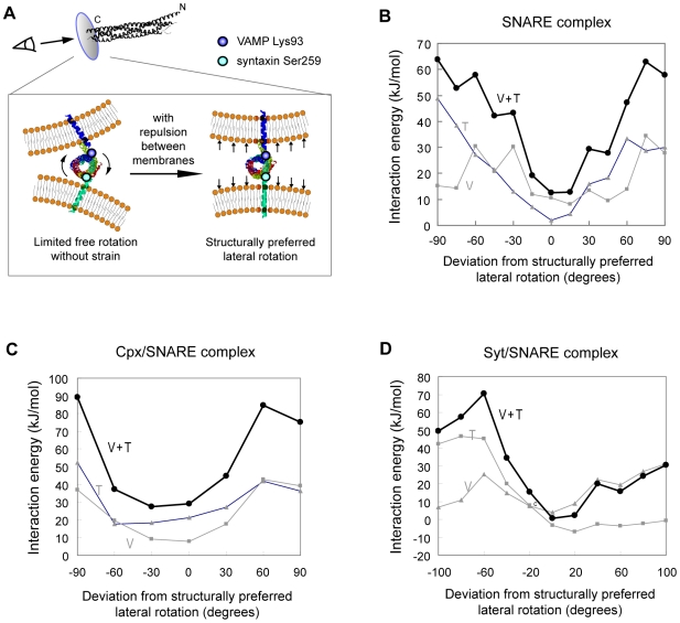
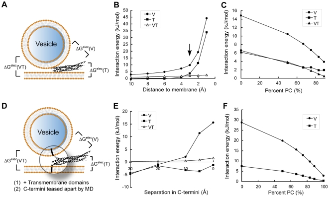
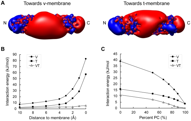
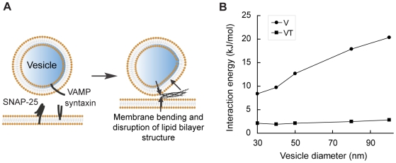
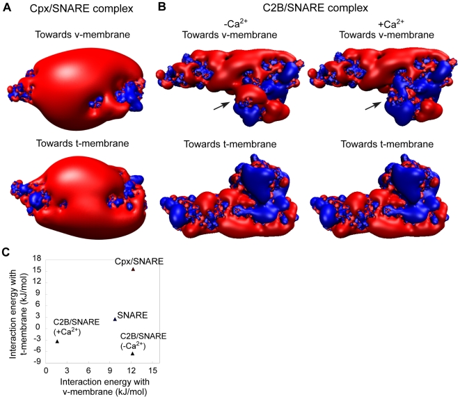
Biological membrane fusion is a basic cellular process catalyzed by SNARE proteins and additional auxiliary factors. Yet, the critical mechanistic details of SNARE-catalyzed membrane fusion are poorly understood, especially during rapid synaptic transmission. Here, we systematically assessed the electrostatic forces between SNARE complex, auxiliary proteins and fusing membranes by the nonlinear Poisson-Boltzmann equation using explicit models of membranes and proteins. We found that a previously unrecognized, structurally preferred and energetically highly favorable lateral orientation exists for the SNARE complex between fusing membranes. This preferred orientation immediately suggests a novel and simple synaptotagmin-dependent mechanistic trigger of membrane fusion. Moreover, electrostatic interactions between membranes, SNARE complex, and auxiliary proteins appear to orchestrate a series of membrane curvature events that set the stage for rapid synaptic vesicle fusion. Together, our electrostatic analyses of SNAREs and their regulatory factors suggest unexpected and potentially novel mechanisms for eukaryotic membrane fusion proteins.


Conflict of interest statement

Competing Interests: The authors have declared that no competing interests exist.

Figures

Figure 1
Figure 1. Electrostatic potential profile of the SNARE complex.
(A) and (B) Patterns of surface electrostatic potential of the neuronal SNARE complex colored to molecular surfaces at ±7 kT/e (top panels) or to ±1 kT/e isopotential contours at (bottom panels). Positive potential is colored in blue, and negative potential is in red. Asterisk: the bulk of negative charges facing the v-membrane. Arrows: positive charges enriched in C-terminus of the SNARE complex. Arrowheads: positive charges selectively localized on the side facing the t-membrane. (C) Multiple alignment of SNAP-25 and syntaxin protein sequences across different species. Negatively charged residues are shaded in gray, and positively charged residues are shaded in black. The SNARE motif is framed in boxes.
Figure 2
Figure 2. The SNARE complex has a structurally and energetically preferred relative orientation between fusing membranes.
(A) Schematic of the structurally preferred orientation. Linker regions between the SNARE motif and TMDs are sufficiently short (∼7.5 Å and ∼0 Å) compared to the SNARE motif superhelix (which has a periodicity of ∼60 Å) and are α-helical continuation from it. During fusion, repulsion between opposing membranes aligns the VAMP and syntaxin TMDs roughly orthogonal to membrane surfaces. This thus aligns the Lys93-Ser259 axis also in the same line. Lys93 and Ser259 are the C-terminal ends of the SNARE motif in VAMP and syntaxin. (B) – (D) The energetically most favorable orientation exactly matches the structurally preferred orientation identified in (A). Interaction energies between two fusing membranes and (B) the SNARE complex, (C) the complexin/SNARE complex and (D) the synaptotagmin/SNARE complex were calculated at various orientations. V: SNARE's interaction free energy with a 40 nm-diameter vesicular membrane representing a typical synaptic vesicle. T: SNARE's interaction energies with a planar target membrane. V + T: sum of interaction energies with the v- and t-membranes.
Figure 3
Figure 3. Electrostatic interaction energies between the SNARE complex and fusing membranes.
(A) Schematic diagram and definitions of intermolecular interaction energies presented in (B) and (C). In (A) through (C), only the SNARE core complex is considered. Interaction free energies are calculated for (B) a series of SNARE/membrane distances and (C) different membrane lipid compositions. Arrow in (B) indicates the most physiologically relevant distance when the closest points between SNARE and membranes are 3 Å, the thickness of a layer of water. In (D) through (F), TMDs of VAMP and syntaxin are present and embedded in membranes. Furthermore, the C-terminus of the SNARE motif is partially unraveled into individual α-helices by molecular dynamics simulations to represent trans-SNARE complex. Interaction free energies are then calculated for (E) a series of SNARE motif C-terminus separation distances and (F) different lipid compositions of the membranes. Conclusions drawn from both groups of studies are essentially the same. V (circles): Interaction energies between the SNARE complex and the v-membrane. T (squares): Interaction energies between SNARE and the t-membrane. VT (triangles): Interaction energies between the v- and the t-membranes if the SNARE complex were extracted.
Figure 4
Figure 4. Electrostatic properties of the endosomal SNARE complex.
(A) Surface electrostatic isocontours for the endosomal SNARE complex at ±1 kT/e. Positive potential is colored in blue, and negative potential is in red. Interaction free energies between SNARE complex and fusing membranes are plotted against (B) a series of SNARE/membrane distances and (C) different membrane lipid compositions. Arrow in (B) indicates the most physiologically relevant distance when the closest points between SNARE and membranes are 3 Å, the thickness of a layer of water. V (circles): Interaction energies between the SNARE complex and the v-membrane. T (squares): Interaction energies between SNARE and the t-membrane. VT (triangles): Interaction energies between the v- and the t-membranes if the SNARE complex were extracted.
Figure 5
Figure 5. Estimate of membrane bending induced by electrostatics of SNARE complex.
(A) Schematic diagram illustrating that free energy released in SNARE complex assembly is transmitted to the transmembrane domains and forces the closing of two fusing membranes (pair of vertical black arrows); meanwhile, strong electrostatic repulsion between SNARE and the v-membrane pushes the membrane away from the center of fusion (tilted black arrow). The resulting force couple should in principle promote bending of the v-membrane. Since SNARE/t-membrane electrostatic interactions are weak, no strong bending is expected on the t-membrane. (B) A standard curve for estimating the degree of bending induced by the electrostatics of SNARE. Interaction free energies between SNARE and a series of v-membranes that bear different spontaneous curvatures (i.e. local radii) were calculated.
Figure 6
Figure 6. Complexin and synaptotagmin modulate electrostatic interactions between SNARE complex and membranes.
(A) Surface isopotential contours of the complexin/SNARE complex rendered at ±1 kT/e. (B) Surface isopotential contours at ±1 kT/e for the C2B/SNARE complex, with or without Ca2+ ions bound to the Ca2+-binding loops of the C2B domain. Positive potential is colored in blue, and negative potential is in red. (C) An energy landscape of electrostatic interactions between SNARE complex and membranes as it associates and dissociates with complexin and synaptotagmin.

Similar articles

Cited by

References

    1. Chen YA, Scheller RH. SNARE-mediated membrane fusion. Nat Rev Mol Cell Biol. 2001;2:98–9106. - PubMed
    1. Sollner T, Whiteheart SW, Brunner M, Erdjument-Bromage H, Geromanos S, et al. SNAP receptors implicated in vesicle targeting and fusion. Nature. 1993;362:318–324. - PubMed
    1. Bonifacino JS, Glick BS. The mechanisms of vesicle budding and fusion. Cell. 2004;116:153–166. - PubMed
    1. Duman JG, Forte JG. What is the role of SNARE proteins in membrane fusion? Am J Physiol Cell Physiol. 2003;285:237–249. - PubMed
    1. Hanson PI, Roth R, Morisaki H, Jahn R, Heuser JE. Structure and conformational changes in NSF and its membrane receptor complexes visualized by quick-freeze/deep-etch electron microscopy. Cell. 1997;90:523–535. - PubMed

Publication types