. 2010 Jan 26;5(1):e8904.
doi: 10.1371/journal.pone.0008904.

The Arabidopsis wall associated kinase-like 10 gene encodes a functional guanylyl cyclase and is co-expressed with pathogen defense related genes

Affiliations

The Arabidopsis wall associated kinase-like 10 gene encodes a functional guanylyl cyclase and is co-expressed with pathogen defense related genes

Stuart Meier et al. PLoS One. .

Abstract

Background: Second messengers have a key role in linking environmental stimuli to physiological responses. One such messenger, guanosine 3',5'-cyclic monophosphate (cGMP), has long been known to be an essential signaling molecule in many different physiological processes in higher plants, including biotic stress responses. To date, however, the guanylyl cyclase (GC) enzymes that catalyze the formation of cGMP from GTP have largely remained elusive in higher plants.

Principal findings: We have identified an Arabidopsis receptor type wall associated kinase-like molecule (AtWAKL10) as a candidate GC and provide experimental evidence to show that the intracellular domain of AtWAKL10(431-700) can generate cGMP in vitro. Further, we also demonstrate that the molecule has kinase activity indicating that AtWAKL10 is a twin-domain catalytic protein. A co-expression and stimulus-specific expression analysis revealed that AtWAKL10 is consistently co-expressed with well characterized pathogen defense related genes and along with these genes is induced early and sharply in response to a range of pathogens and their elicitors.

Conclusions: We demonstrate that AtWAKL10 is a twin-domain, kinase-GC signaling molecule that may function in biotic stress responses that are critically dependent on the second messenger cGMP.


Conflict of interest statement

Competing Interests: The authors have declared that no competing interests exist.

Figures

Figure 1
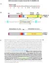
Figure 1. Structural features of the AtWAKL10 protein.
(A) The 14 aa long search motif generated based on conserved and functionally-assigned aa in the catalytic centres of annotated GCs. Amino acid substitutions in the search motif are in square brackets ([ ]); X represents any aa and curly brackets ({ }) define the number of aa. Amino acids in red are functionally assigned residues, hydrogen bonds with the guanine; confers substrate specificity for GTP; binds to the dimer interphase and stabilizes the transition state from GTP to cGMP. (B) Domain organization of AtWAKL10 and AtWAK1 illustrating the location of the predicted signal peptides (SP), extracellular EGF-like domains, TM domains and kinase domains and for AtWAKL10 the GC centre imbedded in the kinase domain. The percentages indicate the determined aa identity and (similarity) between AtWAKL10 and AtWAK1 at the indicated regions. The dashed red vertical lines represent intron locations. The two black triangles demarcate the truncated cytosolic fragment of the AtWAKL10431–700 protein that was expressed as a recombinant. The Arg (R, highlighted in aquamarine) C-terminal of the catalytic centre is a putative metal binding residue and the N-terminal Glu (E, highlighted in aquamarine) is the putative pyrophosphate binding residue. (C) Predicted aa sequence of AtWAKL10. The aa in blue represent the TM domain that separates the extracellular domain from the cytosolic domain. Sequences in bold and demarcated by the two triangles represent the sequence of the recombinant AtWAKL10431–700 protein that was expressed for functional testing. The GC domain is marked in green letters and underlined.
Figure 2
Figure 2. Expression of purified recombinant AtWAKL10 and determination of GC activity.
(A) SDS-PAGE of the purified recombinant AtWAKL10431–700 protein, where M represents the low molecular weight marker while the arrow is marking the recombinant protein band. (B) Cyclic GMP generated following incubation of 10 µg of the purified recombinant AtWAKL10431–700 protein in a reaction system containing 50 mM Tris-HCl; pH 8.0, 1 mM GTP, 2 mM IBMX and either 5 mM Mg2+ or 5 mM Mn2+. The reaction was performed for the indicated time points at room temperature and cGMP levels were determined by enzyme immunoassay. The error bars represent the standard error of the mean (SEM) (n = 3).
Figure 3
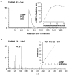
Figure 3. Confirmation of cGMP levels using mass spectrometry.
(A) Extracted mass chromatogram of the m/z 344 [M-1]−1 ion of cGMP generated by recombinant AtWAKL10431–700 after 15 min. Inset: Incubation time course. (B) Mass of the resultant peak in the chromatogram. The inset represents the calibration curve with 1.25, 10 and 40 fmoles of cGMP. The experiment was performed three times and the figure is representative of a typical response.
Figure 4
Figure 4. Testing the recombinant AtWAKL10 for adenylyl cyclase activity.
(A) Extracted mass chromatograms of the m/z 328 [M-1]−1 ion of cAMP and daughter ions generated by 10 µg of recombinant AtWAKL10431–700 with ATP instead of GTP as the substrate, after 15 min at room temperature (24°C). (B) Quantified peak areas of the resultant cAMP chromatograms. The experiment was performed four times and the figure is representative of a typical response.
Figure 5
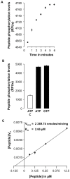
Figure 5. Determination of the kinase activity of the recombinant AtWAKL10.
(A) The calibration curve obtained was produced in the presence 0.1 ng recombinant AtWAKL10431–700 in a reaction system containing 1 mM ATP, 0.2 mM DTT, and 25 µM Ser/Thr-peptide. Phosphorylation was quantified using the Omnia™Ser/Thr-Recombinant system at the indicated time points at 30°C (n = 3). (B) Kinase activity of AtWAKL10431–700 with either ATP or GTP as the substrate. The unfilled bar is a control which contained no recombinant protein (n = 3). (C) Hanes plot for the determination of the reaction kinetics of the recombinant AtWAKL10431–700 indicates the initial velocities for a number of Ser/Thr-peptide concentrations (1.563, 3.125, 6.25, and 12.5 µM) (n = 3). Estimated values for the kinetic constants (Km and Vmax) for the recombinant AtWAKL10 protein were derived from this plot. Km was determined as the negative value of the x-intercept (x = −Km, when y = 0) of the linear fit of the data, while Vmax was calculated from the y-intercept (y = Km/Vmax, when x = 0). Error bars represent the SEM.
Figure 6
Figure 6. Heatmap illustrating fold change in expression of AtWAKL10-ECGG50 in response to select conditions.
A heatmap was constructed to illustrate the fold change (log2) in expression of AtWAKL10 and all genes in the ECGG in response to selected microarray experiments. The experiments presented include; chitooctaose (30 minutes after treatment (mat), GSE8319), elf26 (30 mat, E-MEXP-547), flg22 (1 hour after treatment (hat), NASC-409), NPP1 (1 hat, GSE5615), HrpZ (1 hat, GSE5615), P. infestans (6 hat, NASC-123), B. graminis h (12 hat, GSE12856), E. cichoracearum (3 days after treatment (dat), GSE431), G. orontii (7 dat, Col-0 and eds16/ics1 mutant, GSE13739), B. cinerea (18 hat, NASC-167), OG (1 hat, NASC-409), Pst avrRpm1 (24 hat, NASC-120), BTH treatment (BTH vs. untreated (Col-0) and BTH (npr1) vs. BTH (Col-0), 8 hat, NASC-392), mpk4 (At4g01370, E-MEXP-174), mkk1 (At4g26070) and mkk2 (At4g29810) double mutant (GSE10646), and CHX (3 hat, NASC-189). Details of the microarray experimental conditions are presented in Text S2 (Supporting Information).
Figure 7
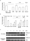
Figure 7. Expression of AtWAKL10 following pathogen and elicitor challenge.
(A) Fold change in AtWAKL10 expression following incubation with the pathogen elicitors, chitooctaose (Col-0 and cerk1 (At3g21630) mutant, 30 mat, GSE8319); elf26 (30 mat, E-MEXP-547), flg22 (1 hat, NASC-409), NPP1 (1 hat, GSE5615), HrpZ (1 hat, GSE5615) and Syringolin A (12 hat, E-MEXP-739) as determined from microarray experiments. (B) Fold change in AtWAKL10 expression following challenge with P. infestans (6 hat, NASC-123), B. graminis h (12 hat, GSE12856), E. cichoracearum (3 dat, GSE431), G. orontii time course (Col-0 and eds16/ics1 mutant, 3, 5 and 7 dat, GSE13739), B. cinerea (18 and 48 hat, NASC-167) and OG treatment (1 hat, NASC-409) as determined from microarray experiments. (C) Semi-quantitative RT-PCR gel image illustrating AtWAKL10 expression over time following inoculation with 5×10⁴ spores/mL of B. cinerea relative to the grape juice control treatment. Expression of AtWAKL10 was induced by B. cinerea biphasically at 1 and notably at 24 and 48 hat. UBQ was used as the “housekeeping” gene to ensure that there was equal amount of template in each sample.
Figure 8
Figure 8. Expression of AtWAKL10 following challenge with different strains of the bacterial P. syringae pathogen.
(A) Fold change in AtWAKL10 expression following challenge with various strains of the bacterial P syringae pathogen including virulent (Pst), avirulent (Pst avrRpm1), and non-host pv. phaseolicola (Psph) as determined from microarray experiments (NASC-120). (B) Semi-quantitative RT-PCR gel image illustrating AtWAKL10 expression over time following inoculation with virulent (Pst) and the avirulent (Pst avrB) strains of P. syringae relative to the control inoculation with 10 mM MgCl2. Expression of AtWAKL10 was induced by Pst and Pst avrB infection. In both cases a biphasic pattern of expression is apparent with the induced expression being faster and stronger in response to the avirulent pathogen as is expected for the compatible interaction. UBQ was used as the “housekeeping” gene to ensure that there was equal amount of template in each sample.
Figure 9
Figure 9. Fold change in expression of AtWAKL10 after chemical treatment and in pathogen related mutants.
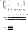
(A) Fold change in AtWAKL10 expression following treatment with the functional synthetic SA analogue, BTH treatment of Col-0 and npr1 genotypes (8 and 24 hat, NASC-392), MeJA (25 hat, NASC-415) and CHX (3 hat, NASC-189) as determined from microarray experiments. Fold change in AtWAKL10 expression was also determined from microarray experiments in a number of pathogen-related mutants including mpk4 (E-MEXP-174), mkk1mkk2 double mutant (GSE10646) and cpr5 (At5g64930, GSE5745). (B) Semi-quantitative RT-PCR confirmed the induction of AtWAKL10 expression in response to 10 µM CHX (3 hat) relative to the DMSO control (C). UBQ was used as the “housekeeping” gene to ensure that there was equal amount of template in each sample.

Similar articles

Cited by

References

    1. Schaap P. Guanylyl cyclases across the tree of life. Front Biosci. 2005;10:1485–1498. - PubMed
    1. Pilz RB, Casteel DE. Regulation of gene expression by cyclic GMP. Circ Res. 2003;93:1034–1046. - PubMed
    1. Newton RP, Smith CJ. Cyclic nucleotides. Phytochemistry. 2004;65:2423–2437. - PubMed
    1. Prado AM, Porterfield DM, Feijo JA. Nitric oxide is involved in growth regulation and re-orientation of pollen tubes. Development. 2004;131:2707–2714. - PubMed
    1. Durner J, Wendehenne D, Klessig DF. Defense gene induction in tobacco by nitric oxide, cyclic GMP, and cyclic ADP-ribose. Proc Natl Acad Sci U S A. 1998;95:10328–10333. - PMC - PubMed

Publication types

MeSH terms