

doi: 10.1039/b919004j.
Epub 2010 Jan 5.
Implementation of a color-capable optofluidic microscope on a RGB CMOS color sensor chip substrate
Affiliations
- PMID: 20126679
- PMCID: PMC10578144
- DOI: 10.1039/b919004j
Item in Clipboard
Implementation of a color-capable optofluidic microscope on a RGB CMOS color sensor chip substrate
Lab Chip.
.
Abstract
We report the implementation of a color-capable on-chip lensless microscope system, termed color optofluidic microscope (color OFM), and demonstrate imaging of double stained Caenorhabditis elegans with lacZ gene expression at a light intensity about 10 mW/cm(2).
Figures
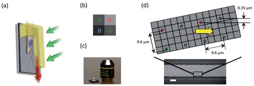
(a) The schematic plot of gravity driven color OFM. (b) The Bayer pattern for the color filter mask of the CMOS sensor. (c) The color OFM device comparing its size with a 10× microscope objective. (d) The schematic plot for the aperture array and the microscope image of an aligned PDMS channel on top of the aperture array. The scale bar is 20 microns.
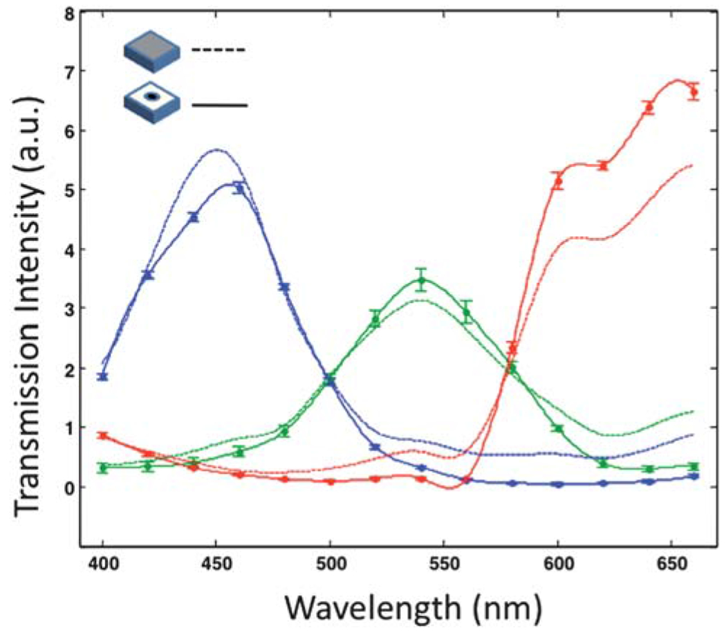
The aperture effect on spectral response of the sensor: the solid lines represent the measured transmission data from a color OFM device; the dash lines are the response from a CMOS sensor without any fabrication process. All the transmission data are normalized at 400 nm.
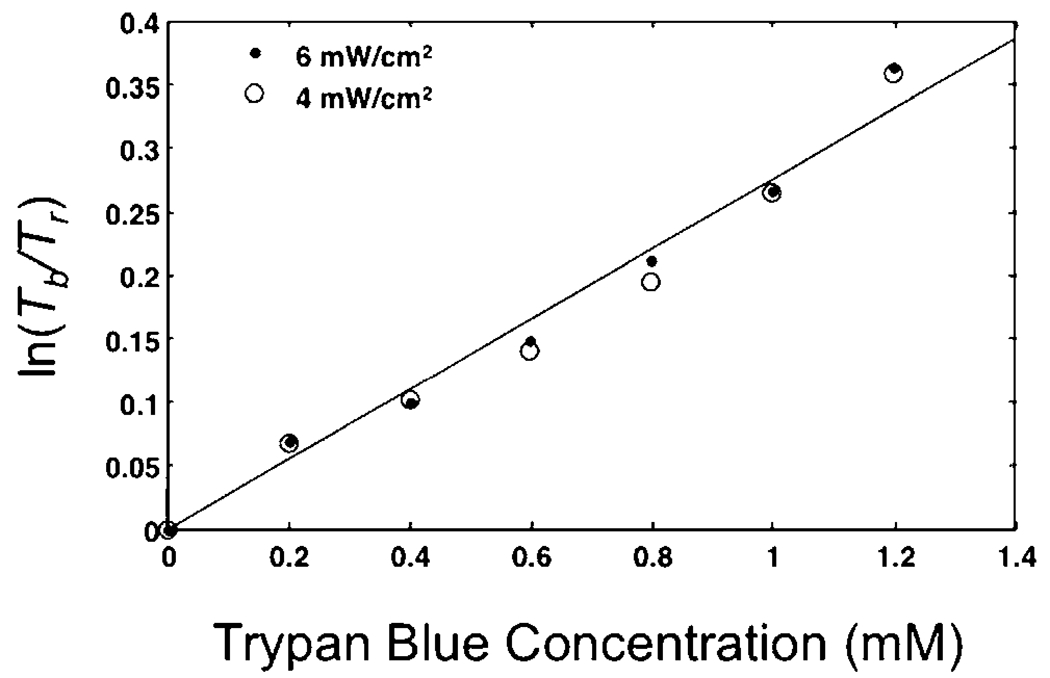
Dye concentration measured by the logarithm of relative intensity of two different colors. The illumination light passes through Trypan Blue solutions with two intensities of 6mW/cm2 and 4mW/cm2.
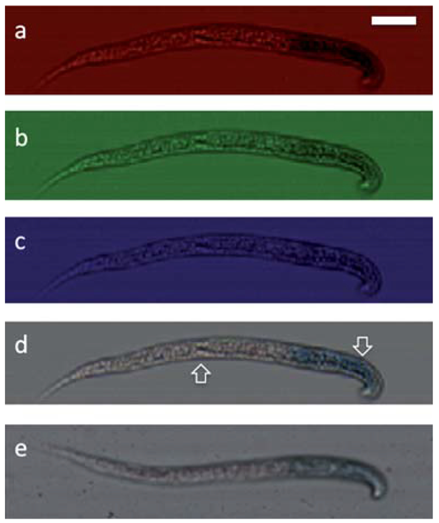
Construction of a color OFM image of C. elegans with LacZ expression. (a)–(c) From top to bottom are the images from red, green, blue pixels of a C. elegans with blue Xgal stain and Ponceau stain image, (d) the constructed color OFM image shows the nonspecific Ponceau stain (left arrow) and the LacZ staining of the pharyngeal muscles (right arrow), (e) the conventional 10× microscope image of the same sample. The scale bar is 20 microns.
Similar articles
-
Lensless high-resolution on-chip optofluidic microscopes for Caenorhabditis elegans and cell imaging.Proc Natl Acad Sci U S A. 2008 Aug 5;105(31):10670-5. doi: 10.1073/pnas.0804612105. Epub 2008 Jul 28. Proc Natl Acad Sci U S A. 2008. PMID: 18663227 Free PMC article.
-
Optofluidic microscopy--a method for implementing a high resolution optical microscope on a chip.Lab Chip. 2006 Oct;6(10):1274-6. doi: 10.1039/b604676b. Epub 2006 Aug 4. Lab Chip. 2006. PMID: 17102839
-
The application of on-chip optofluidic microscopy for imaging Giardia lamblia trophozoites and cysts.Biomed Microdevices. 2009 Oct;11(5):951-8. doi: 10.1007/s10544-009-9312-x. Epub 2009 Apr 14. Biomed Microdevices. 2009. PMID: 19365730 Free PMC article.
-
Lensfree optofluidic microscopy and tomography.Ann Biomed Eng. 2012 Feb;40(2):251-62. doi: 10.1007/s10439-011-0385-3. Epub 2011 Sep 2. Ann Biomed Eng. 2012. PMID: 21887590 Review.
-
Label-free optical imaging of nonfluorescent molecules by stimulated radiation.Curr Opin Chem Biol. 2011 Dec;15(6):831-7. doi: 10.1016/j.cbpa.2011.10.005. Epub 2011 Nov 4. Curr Opin Chem Biol. 2011. PMID: 22055495 Review.
Cited by
-
Automated cell viability assessment using a microfluidics based portable imaging flow analyzer.Biomicrofluidics. 2015 Apr 28;9(2):024123. doi: 10.1063/1.4919402. eCollection 2015 Mar. Biomicrofluidics. 2015. PMID: 26015835 Free PMC article.
-
Long-term imaging of three-dimensional hyphal development using the ePetri dish.Biomed Opt Express. 2024 Jun 17;15(7):4292-4299. doi: 10.1364/BOE.530483. eCollection 2024 Jul 1. Biomed Opt Express. 2024. PMID: 39022548 Free PMC article.
-
On-chip biomedical imaging.IEEE Rev Biomed Eng. 2013;6:29-46. doi: 10.1109/RBME.2012.2215847. IEEE Rev Biomed Eng. 2013. PMID: 23558399 Free PMC article. Review.
-
Imaging without lenses: achievements and remaining challenges of wide-field on-chip microscopy.Nat Methods. 2012 Sep;9(9):889-95. doi: 10.1038/nmeth.2114. Epub 2012 Aug 30. Nat Methods. 2012. PMID: 22936170 Free PMC article.
-
Fluorescence microscopy imaging with a Fresnel zone plate array based optofluidic microscope.Lab Chip. 2011 Nov 7;11(21):3698-702. doi: 10.1039/c1lc20654k. Epub 2011 Sep 21. Lab Chip. 2011. PMID: 21935556 Free PMC article.
References
-
- Heng X, Erickson D, Baugh LR, Yaqoob Z, Sternberg PW, Psaltis D and Yang CH, Lab Chip, 2006, 6, 1274–1276. - PubMed
-
- Cui XQ, Lew M and Yang CH, Appl. Phys. Lett, 2008, 93,091113.
-
- Heng X, Cui XQ, Knapp DW, Wu JG, Yaqoob Z, McDowell EJ, Psaltis D and Yang CH, Opt. Express, 2006, 14, 10410–10425. - PubMed
Publication types
MeSH terms
Substances
Grants and funding
LinkOut - more resources
Full Text Sources
Other Literature Sources