

Hsp40 proteins modulate humoral and cellular immune response in rheumatoid arthritis patients
- PMID: 20127215
- PMCID: PMC3006627
- DOI: 10.1007/s12192-010-0168-z
Hsp40 proteins modulate humoral and cellular immune response in rheumatoid arthritis patients
Abstract
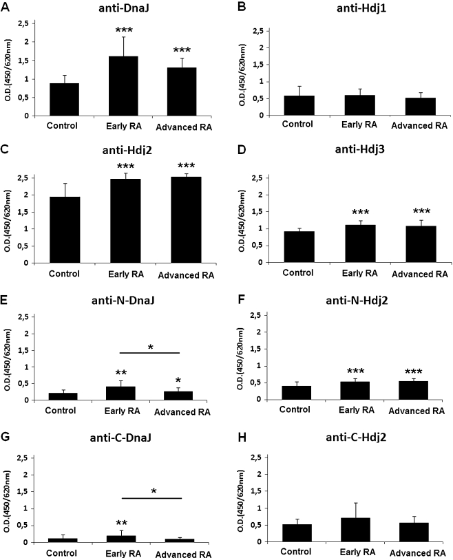
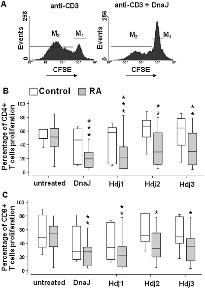
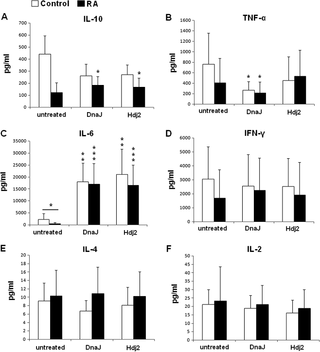
Recent research on the heat shock proteins (Hsps) in chronic inflammatory diseases indicates that Hsps may have disease-suppressive activities. Our aim was to characterize immune response directed to bacterial (DnaJ) and human Hsp40s in patients with rheumatoid arthritis (RA). We found elevated levels of anti-DnaJ, anti-Hdj2, and anti-Hdj3 (but not ant-Hdj1) serum antibodies in the RA patients (P < or = 0.001) compared to healthy controls. In peripheral blood mononuclear cells (PBMCs) culture, all tested Hsp40 proteins significantly inhibited the divisions of CD4+ and CD8+ T cells of the RA patients but not those of the controls. Both DnaJ and Hdj2 stimulated secretion of the main anti-inflammatory cytokine IL-10 by PBMCs of the RA patients (P < 0.05), and of IL-6 by PBMCs of the RA (P < 0.001) and control (P < 0.01) groups. DnaJ reduced TNFalpha secretion (P < 0.05) by both groups of PBMCs. Our results show for the first time that the RA patients have an increased humoral response to human Hsp40 proteins Hdj2 and Hdj3. This is also the first description of immunomodulatory effect of human Hsp40s on T cells and cytokine secretion in RA, suggesting that Hsp40s act as natural anti-inflammatory agents in RA.
Figures
Similar articles
-
Cytokines of the Th1 and Th2 type in sera of rheumatoid arthritis patients; correlations with anti-Hsp40 immune response and diagnostic markers.Acta Biochim Pol. 2010;57(3):327-32. Epub 2010 Sep 9. Acta Biochim Pol. 2010. PMID: 20827447
-
Human Hsp40 proteins, DNAJA1 and DNAJA2, as potential targets of the immune response triggered by bacterial DnaJ in rheumatoid arthritis.Cell Stress Chaperones. 2013 Sep;18(5):653-9. doi: 10.1007/s12192-013-0407-1. Epub 2013 Feb 14. Cell Stress Chaperones. 2013. PMID: 23408083 Free PMC article.
-
Anti-inflammatory effects of atorvastatin on peripheral blood mononuclear cells and synovial fibroblasts in rheumatoid arthritis.Scand J Rheumatol. 2009;38(4):235-9. doi: 10.1080/03009740802572475. Scand J Rheumatol. 2009. PMID: 19247848
-
Interferon-gamma production in response to in vitro stimulation with collagen type II in rheumatoid arthritis is associated with HLA-DRB1(*)0401 and HLA-DQ8.Arthritis Res. 2000;2(1):75-84. doi: 10.1186/ar71. Arthritis Res. 2000. PMID: 11219392 Free PMC article.
-
Effects of sinomenine on the proliferation, cytokine production, and regulatory T-cell frequency in peripheral blood mononuclear cells of rheumatoid arthritis patients.Drug Dev Res. 2021 Apr;82(2):251-258. doi: 10.1002/ddr.21748. Epub 2020 Oct 2. Drug Dev Res. 2021. PMID: 33006164
Cited by
-
Regulation of p53 and Cancer Signaling by Heat Shock Protein 40/J-Domain Protein Family Members.Int J Mol Sci. 2021 Dec 16;22(24):13527. doi: 10.3390/ijms222413527. Int J Mol Sci. 2021. PMID: 34948322 Free PMC article. Review.
-
Autoantibodies to heat shock protein 60, 70, and 90 are not altered in the anti-SARS-CoV-2 IgG-seropositive humans without or with mild symptoms.Cell Stress Chaperones. 2021 Jul;26(4):735-740. doi: 10.1007/s12192-021-01215-3. Epub 2021 Jun 2. Cell Stress Chaperones. 2021. PMID: 34080135 Free PMC article.
-
Do reciprocal interactions between cell stress proteins and cytokines create a new intra-/extra-cellular signalling nexus?Cell Stress Chaperones. 2013 Nov;18(6):685-701. doi: 10.1007/s12192-013-0444-9. Epub 2013 Jul 25. Cell Stress Chaperones. 2013. PMID: 23884786 Free PMC article.
-
General Structural and Functional Features of Molecular Chaperones.Adv Exp Med Biol. 2021;1340:11-73. doi: 10.1007/978-3-030-78397-6_2. Adv Exp Med Biol. 2021. PMID: 34569020
-
Heat Shock Proteins as Immunomodulants.Molecules. 2018 Nov 1;23(11):2846. doi: 10.3390/molecules23112846. Molecules. 2018. PMID: 30388847 Free PMC article. Review.
References
-
- Albani S, Carson DA, Roudier J. Genetic and environmental factors in the immune pathogenesis of rheumatoid arthritis. Rheum Dis Clin North Am. 1992;18:729–740. - PubMed
Publication types
MeSH terms
Substances
LinkOut - more resources
Full Text Sources
Medical
Research Materials
