

Findings from CT, MRI, and PET/CT of a primary malignant melanoma arising in a spinal nerve root
- PMID: 20127497
- PMCID: PMC2899619
- DOI: 10.1007/s00586-010-1285-1
Findings from CT, MRI, and PET/CT of a primary malignant melanoma arising in a spinal nerve root
Abstract
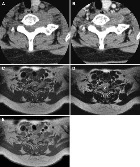
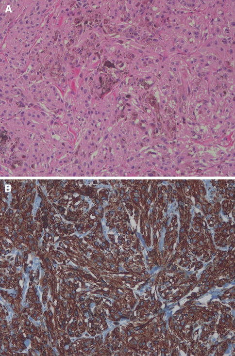
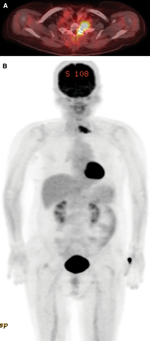
Primary spinal malignant melanoma is an extremely rare condition. We here describe a case of a 71-year-old Asian female presenting with left upper extremity tingling sensation. Computed tomography (CT) showed a homogeneously enhanced mass occupying the left neural foramen at the C6-7 level. Magnetic resonance imaging revealed enhanced mass in intra- and extradural space compressing the spinal cord at this level. It also widened the neural foramen mimicking neurofibroma or schwannoma. Partial resection of the mass was performed. Pathologic diagnosis of the mass was malignant melanoma. Postoperative whole body positron emission tomography/CT scan demonstrated an intense (18)F-FDG uptake at the residual mass site without abnormal uptake at other sites in the body.
Figures
Similar articles
-
Spinal cord herniation into pseudomeningocele after traumatic nerve root avulsion: case report and review of the literature.Eur Spine J. 2008 Sep;17 Suppl 2(Suppl 2):S263-6. doi: 10.1007/s00586-007-0537-1. Epub 2007 Nov 7. Eur Spine J. 2008. PMID: 17987326 Free PMC article. Review.
-
Cervical epidural arteriovenous fistula with radiculopathy mimicking cervical spondylosis.Neurol Med Chir (Tokyo). 2009 Mar;49(3):108-13. doi: 10.2176/nmc.49.108. Neurol Med Chir (Tokyo). 2009. PMID: 19318735
-
Slow progression and benign course of a primary malign melanoma of a lumbar nerve root.Clin Neurol Neurosurg. 2012 Feb;114(2):166-8. doi: 10.1016/j.clineuro.2011.09.012. Epub 2011 Oct 20. Clin Neurol Neurosurg. 2012. PMID: 22018994 No abstract available.
-
Cervical dumbbell intra-extradural hemangioblastoma: total removal through the lateral approach: technical case report.Neurosurgery. 2005 Mar;56(3):E625; discussion E625. Neurosurgery. 2005. PMID: 15730592 Review.
-
Spontaneous CSF leak treated with percutaneous CT-guided fibrin glue.Neurology. 2005 May 24;64(10):1818-9. doi: 10.1212/01.WNL.0000162029.96759.D2. Neurology. 2005. PMID: 15911828 No abstract available.
Cited by
-
Easily misdiagnosed delayed metastatic intraspinal extradural melanoma of the lumbar spine: A case report and review of the literature.Oncol Lett. 2013 Jun;5(6):1799-1802. doi: 10.3892/ol.2013.1299. Epub 2013 Apr 10. Oncol Lett. 2013. PMID: 23833644 Free PMC article.
-
A rare case of primary spinal cord melanoma.Radiol Case Rep. 2018 Feb 20;13(2):424-426. doi: 10.1016/j.radcr.2018.01.009. eCollection 2018 Apr. Radiol Case Rep. 2018. PMID: 29904488 Free PMC article.
-
Rare case of extradural spinal metastasis from primary lung malignant melanoma detected with fluorine-18 fluorodeoxyglucose-positron emission tomography/computed tomography.Indian J Nucl Med. 2014 Jan;29(1):57-8. doi: 10.4103/0972-3919.125782. Indian J Nucl Med. 2014. PMID: 24591789 Free PMC article. No abstract available.
-
Primary Spinal Malignant Melanoma Mimicking a Cervical Nerve Root Schwannoma: Case Report and Literature Review.Asian J Neurosurg. 2024 May 27;19(3):540-550. doi: 10.1055/s-0044-1787081. eCollection 2024 Sep. Asian J Neurosurg. 2024. PMID: 39205889 Free PMC article.
-
Primary cervicothoracic melanoma of spinal cord: a case report and literature review.Front Oncol. 2024 May 28;14:1417268. doi: 10.3389/fonc.2024.1417268. eCollection 2024. Front Oncol. 2024. PMID: 38863638 Free PMC article.
References
-
- Fuster D, Chiang S, Johnson G, Schuchter LM, Zhuang H, Alavi A. Is FDG-PET more accurate than standard diagnostic procedures in the detection of suspected recurrent melanoma? J Nucl Med. 2004;45:1323–1327. - PubMed
Publication types
MeSH terms
LinkOut - more resources
Full Text Sources
Medical
