

Comparative anatomy of the locus coeruleus in humans and nonhuman primates
- PMID: 20127761
- PMCID: PMC2820586
- DOI: 10.1002/cne.22249
Comparative anatomy of the locus coeruleus in humans and nonhuman primates
Abstract
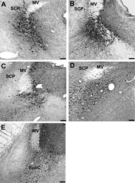
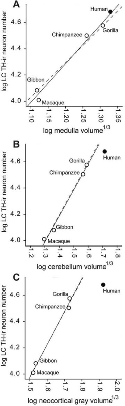
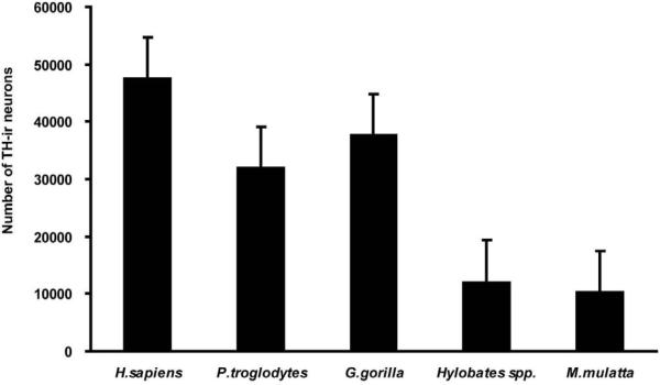
The locus coeruleus (LC) is a dense cluster of neurons that projects axons throughout the neuroaxis and is located in the rostral pontine tegmentum extending from the level of the inferior colliculus to the motor nucleus of the trigeminal nerve. LC neurons are lost in the course of several neurodegenerative disorders, including Alzheimer's and Parkinson's diseases. In this study we used Nissl staining and tyrosine hydroxylase (TH) immunoreactivity to compare the human LC with that of closely related primate species, including great and lesser apes, and macaque monkeys. TH catalyzes the initial and rate-limiting step in catecholamine biosynthesis. The number of TH-immunoreactive (TH-ir) neurons was estimated in each species using stereologic methods. In the LC of humans the mean total number of TH-ir neurons was significantly higher compared to the other primates. Because the total number of TH-ir neurons in the LC was highly correlated with the species mean volume of the medulla oblongata, cerebellum, and neocortical gray matter, we conclude that much of the observed phylogenetic variation can be explained by anatomical scaling. Notably, the total number of LC neurons in humans was most closely predicted by the nonhuman allometric scaling relationship relative to medulla size, whereas the number of LC neurons in humans was considerably lower than predicted according to neocortex and cerebellum volume.
(c) 2009 Wiley-Liss, Inc.
Figures
References
-
- Aston-Jones G, Chiang C, Alexinsky T. Discharge of noradrenergic locus coeruleus neurons in behaving rats and monkeys suggests a role in vigilance. Prog Brain Res. 1991;88:501–520. - PubMed
-
- Aston-Jones G, Rajkowski J, Cohen J. Locus coeruleus and regulation of behavioral flexibility and attention. Prog Brain Res. 2000;126:165–182. - PubMed
-
- Baker KG, Tork I, Hornung JP, Halasz P. The human locus coeruleus complex: an immunohistochemical and three dimensional reconstruction study. Exp Brain Res. 1989;77:257–270. - PubMed
Publication types
MeSH terms
Substances
Grants and funding
LinkOut - more resources
Full Text Sources
