. 2010 Apr;28(4):832-41.
doi: 10.1002/stem.313.

A novel molecule integrating therapeutic and diagnostic activities reveals multiple aspects of stem cell-based therapy

Affiliations

A novel molecule integrating therapeutic and diagnostic activities reveals multiple aspects of stem cell-based therapy

Shawn D Hingtgen et al. Stem Cells. 2010 Apr.

Abstract

Stem cells are promising therapeutic delivery vehicles; however pre-clinical and clinical applications of stem cell-based therapy would benefit significantly from the ability to simultaneously determine therapeutic efficacy and pharmacokinetics of therapies delivered by engineered stem cells. In this study, we engineered and screened numerous fusion variants that contained therapeutic (TRAIL) and diagnostic (luciferase) domains designed to allow simultaneous investigation of multiple events in stem cell-based therapy in vivo. When various stem cell lines were engineered with the optimized molecule, SRL(O)L(2)TR, diagnostic imaging showed marked differences in the levels and duration of secretion between stem cell lines, while the therapeutic activity of the molecule showed the different secretion levels translated to significant variability in tumor cell killing. In vivo, simultaneous diagnostic and therapeutic monitoring revealed that stem cell-based delivery significantly improved pharmacokinetics and anti-tumor effectiveness of the therapy compared to intravenous or intratumoral delivery. As treatment for highly malignant brain tumor xenografts, tracking SRL(O)L(2)TR showed stable stem cell-mediated delivery significantly regressed peripheral and intracranial tumors. Together, the integrated diagnostic and therapeutic properties of SRL(O)L(2)TR answer critical questions necessary for successful utilization of stem cells as novel therapeutic vehicles.


Figures

Figure 1
Figure 1
Engineering and screening of multiple S-TRAIL and luciferase fusions in vitro. (A): Schematic representations of lentiviral transfer vectors bearing IRES-GFP cassettes and encoding various fusions between secreted variant of the pro-apoptotic protein tumor necrosis factor-related apoptosis-inducing ligand (S-TRAIL) and different luciferase proteins. Direct fusion variants: (1) TRAIL-Rluc, (2) TRAIL-Fluc, (3) TRAIL-GpLuc, (4) GpLuc-TRAIL. Variants to test intramolecular spacing: (5) GpLuc-linker 1-TRAIL, (6) GpLuc-linker 2-TRAIL. Variants to test modification of secretion sequence: (7) SGpLuc-Linker 2-TRAIL, (8) SRlucO-linker 2-TRAIL. To screen the various fusion molecules, 293T cells were transduced with lentiviral vectors encoding the designated fusion variant. Bioluminescence imaging and enzyme-linked immunosorbent assay were performed on conditioned medium from the transduced cells to determine diagnostic luciferase activity or concentration of S-TRAIL, respectively. Therapeutic activity of each variant was determined by luciferase-based assay on human Gli36-EGFRvIII cells 24 hours after incubation with equal volumes of conditioned media from lentiviral transduced 293T cells. Abbreviations: GpL1TR, GpLuc-linker 1-TRAIL; GpL2TR, GpLuc-linker 2-TRAIL; GpTR, GpLuc-TRAIL; SGpL2TR, SGpLuc-Linker 2-TRAIL; SRLOL2TR, SRlucO-linker 2-TRAIL; TRFL, TRAIL-Fluc; TRGp, TRAIL-GpLuc; TRRL, TRAIL-Rluc.
Figure 2
Figure 2
Screening S-TRAIL and luciferase fusion variants in vivo. (A): Western blot analysis of lysates from 293T cells transduced with LV demonstrating expression of SGpL2TR and SRLOL2TR. (B): Representative green fluorescent protein (GFP) photomicrograph (large micrograph, 4×; inset, 10×) of human U251 glioma cells co-transduced with equal MOI of lentiviral vectors (LV) encoding SGpL2TR, SRLOL2TR, or control virus and GFP-FLuc. (C–D): U251 glioma cells co-expressing GFP-FLuc and SGpL2TR, SRLOL2TR, or control virus were implanted subcutaneously in mice and imaged on days 1, 3, and 15 to monitor secretion of TRAIL fusion proteins (GpLuc or RLucO intensities, (C)) and on days 2, 7, and 15 to follow changes in tumor volume (FLuc intensities, (D)). (E): Representative images and summary data of similar experiments as those described in (C and D) instead using nontherapeutic SGpLuc or SRLucO. Subcutaneous tumors were imaged on days 1, 5, and 10 for FLuc intensities to determine tumor volume or to monitor secretion of SGpLuc or SRLucO by coelenterazine injection. Representative day 10 images are shown. In all panels, *, p < .05 versus control. Abbreviations: SGpL2TR, SGpLuc-Linker 2-TRAIL; SRLOL2TR, SRlucO-linker 2-TRAIL; S-TRAIL, secreted variant of the pro-apoptotic protein tumor necrosis factor-related apoptosis-inducing ligand.
Figure 3
Figure 3
Imaging of SRLOL2TR reveals differences in stem cell secretion and cancer cell killing. (A): Representative images of mNSC, hNSC, and mMSC transduced with LV encoding SRLOL2TR. (B): Summary data demonstrating differences in transduction efficiency between mNSC, hNSC, and mMSC 24 hours post-transduction with increasing MOI of LV-SRLOL2TR. Green fluorescent protein (GFP)-positive cells were counted and expressed as a ratio of total cell number for each stem cell type. (C): Photon emission from mNSC, hNSC, and mMSC transduced with LV-SRLOL2TR were assayed at days 0, 2, 7, and 14 post-transduction. (D and E): Representative images and summary graphs demonstrating the effects of different stem cell lines secreting SRLOL2TR co-cultured at increasing stem cell to tumor cell ratios with Gli36-EGFRvIII (D) or U251 human cancer cells (E). After 24 hours of co-culture, levels of SRLOL2TR secretion by the stem cells were visualized by RLucO bioluminescence imaging and tumor cell killing was visualized by Fluc bioluminescence imaging and quantified using a luminometer. (F): Western blot analysis of cell lysates from mNSC or Gli36-EGFRvIII tumor cells demonstrating the expression of DR4 in each cell line. (G): Immunocytochemical analysis of undifferentiated mNSC stained with an antibody against NSC marker Nestin (a), or following 10 days of differentiation using antibodies against glial fibrillary acidic protein (GFAP) (b) or Olig-2 (c). (H): Representative photomicrographs demonstrating the migration of transduced mNSC towards gliomas over time. GFP-expressing mNSC were implanted 1 mm lateral to established Gli36-EGFRvIIIFD intracranial gliomas. On days 2 (a), 5 (b), and 10 (c) post-mNSC, implantation mice were sacrificed, brains were removed and sectioned, and both mNSC and glioma volumes were visualized using fluorescence confocal microscopy. Panels a and b: 4x magnification; Panel c: 10x magnification. (I and J): Human Gli36-EGFRvIII glioma cells were incubated with conditioned media from mNSC transduced with control vector, SRLOL2TR, or purified S-TRAIL and caspase-3/7 activity (I), cleaved caspase-8 levels (J), and cleaved PARP levels (J) were determined by luciferase-based caspase 3/7 assay (I) and Western blot analysis (J). In all panels, *, p < .05 versus control. Abbreviations: hNSC, human neural stem cells; mMSC, primary mouse mesenchymal stem cells; mNSC, primary mouse neural stem cells; SRLucOL2TR, SRlucO-linker 2-TRAIL.
Figure 4
Figure 4
Delivery by engineered stem cells improves SRLOL2TR pharmacokinetics in vivo. (A): Representative images and summary graphs showing SRLOL2TR levels when delivered to tumors by engineered stem cells. Gli36-EGFRvIII-FD glioma cells were implanted subcutaneously in mice, and 24 hours later FLuc imaging was performed to demonstrate the localization of the tumor. 24 hours post-imaging, mNSC secreting SRLOL2TR were injected around one of the established tumors, and SRLOL2TR imaging was performed to visualize the secretion of SRLOL2TR. (B): Summary graph showing the effects on tumor volume of control mNSC or mNSC secreting SRLOL2TR 48 hours after implantation around established Gli36-EGFRvIII-FD tumors assessed by FLuc imaging. (C): Ex vivo analysis of biodistribution of mNSC-delivered fusion proteins assessed by RLucO imaging of organs removed 1-hour post injection of coelenterazine. (D–I): In vivo bioluminescence imaging of conditioned medium from LV-SRLOL2TR transduced cells injected into mice bearing established Gli36-EGFRvIII-FD subcutaneous tumors by i.v. infusion (D) or direct intratumoral administration (G) and analyzed at different time points after coelenterazine injection. Ex vivo bioluminescence imaging of organs and tumor tissue from mice 1-hour post-injection of media administered by i.v. infusion (E) or direct injection (H) followed by coelenterazine. Forty-eight hours after media injection, Fluc imaging was performed to determine changes in tumor volumes (F, I). In all panels, *, p < .05 versus control. Abbreviations: SRLOL2TR, SRlucO-linker 2-TRAIL.
Figure 5
Figure 5
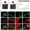
Stem cells efficiently deliver SRLOL2TR to eradicate intracranial glioblastoma. (A): Representative FLuc bioluminescent images and summary data of mice implanted intracranially with mNSC transduced with LV-GFP-FLuc, mixed with Gli36-EGFRvIII, and serially imaged for 15 days. (B–D): mNSC were transduced with control vector or SRLOL2TR, and implanted with Gli36 EGFRvIII-FD intracranially in mice. On days 2, 6, 9, and 12 post-implantation, SRLOL2TR mice were injected with coelenterazine and RLucO imaging was performed to visualize SRLOL2TR secretion (B). Mice were injected with D-Luciferin and FLuc imaging was performed to visualize changes in glioma on days 1, 3, 6, 9, 13, and 21 post-implantation. (C): Representative images and summary data are shown. C = Control; T = SRLOL2TR. (D): Immunohisto-chemistry was performed on sections from brains containing GFP-FLuc-expressing mNSC 4 days post-implantation. Representative merged images are shown of brain sections containing mNSC (GFP) and stained with antibodies (Red) against nestin (a, e, i), glial fibrillary acidic protein (GFAP) (b, f, j), Tuj-1 (c, g, k), or Ki67 (d, h, l). Nb = normal brain; T = tumor. In all panels, *, p < .05 versus control. Abbreviations: SRLOL2TR, SRlucO-linker 2-TRAIL.

Similar articles

Cited by

References

    1. Corsten MF, Shah K. Therapeutic stem-cells for cancer treatment: Hopes and hurdles in tactical warfare. Lancet Oncology. 2008;9:376–384. - PubMed
    1. Daley GQ, Scadden DT. Prospects for stem cell-based therapy. Cell. 2008;132:544–548. - PubMed
    1. Sasportas LS, Kasmieh R, Wakimoto H, et al. Assessment of therapeutic efficacy and fate of engineered human mesenchymal stem cells for cancer therapy. Proc Natl Acad Sci U S A. 2009;106:4822–4827. - PMC - PubMed
    1. Corsten MF, Miranda R, Kasmieh R, et al. MicroRNA-21 knockdown disrupts glioma growth in vivo and displays synergistic cytotoxicity with neural precursor cell delivered S-TRAIL in human gliomas. Cancer Research. 2007;67:8994–9000. - PubMed
    1. Yip S, Sabetrasekh R, Sidman RL, et al. Neural stem cells as novel cancer therapeutic vehicles. Eur J Cancer. 2006;42:1298–1308. - PubMed

Publication types