

Postnatal changes in tryptophan hydroxylase and serotonin transporter immunoreactivity in multiple brainstem nuclei of the rat: implications for a sensitive period
- PMID: 20127812
- PMCID: PMC3694345
- DOI: 10.1002/cne.22265
Postnatal changes in tryptophan hydroxylase and serotonin transporter immunoreactivity in multiple brainstem nuclei of the rat: implications for a sensitive period
Abstract
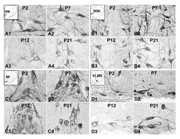
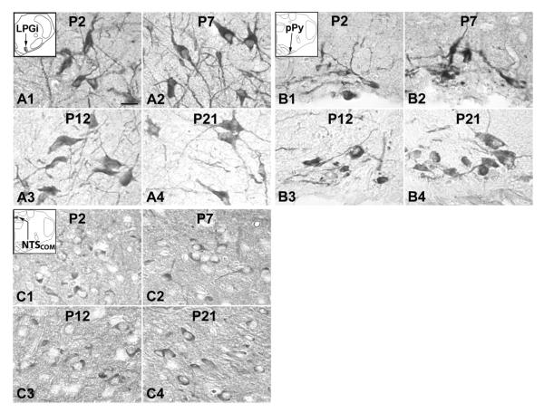
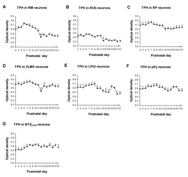
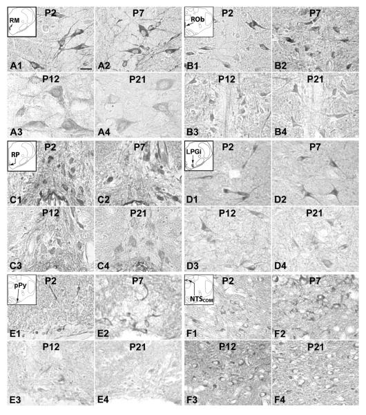
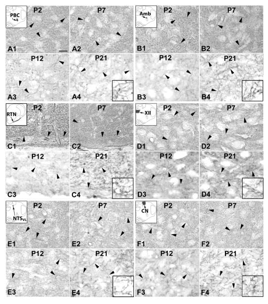
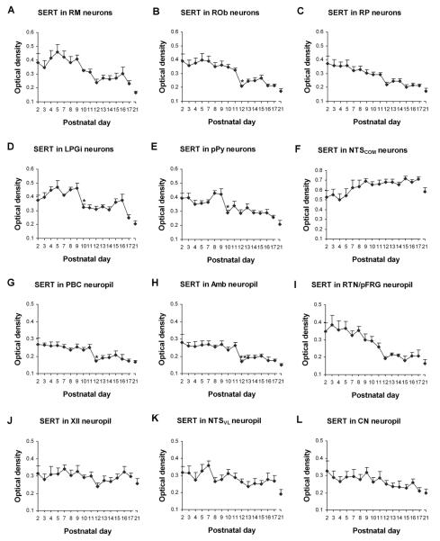
Previously, we found that the brainstem neuronal network in normal rats undergoes abrupt neurochemical, metabolic, and physiological changes around postnatal days (P) 12-13, a critical period when the animal's response to hypoxia is also the weakest. This has special implications for sudden infant death syndrome (SIDS), insofar as seemingly normal infants succumb to SIDS when exposed to respiratory stressors (e.g., hypoxia) during a narrow postnatal window. Because an abnormal serotonergic system has recently been implicated in SIDS, we conducted a large-scale investigation of the 5-HT-synthesizing enzyme tryptophan hydroxylase (TPH) and serotonin transporter (SERT) with semiquantitative immunohistochemistry in multiple brainstem nuclei of normal rats aged P2-21. We found that 1) TPH and SERT immunoreactivity in neurons of raphé magnus, obscurus, and pallidus and SERT in the neuropil of the pre-Bötzinger complex, nucleus ambiguus, and retrotrapezoid nucleus were high at P2-11 but decreased markedly at P12 and plateaued thereafter until P21; 2) SERT labeling in neurons of the lateral paragigantocellular nucleus (LPGi) and parapyramidal region (pPy) was high at P2-9 but fell significantly at P10, followed by a gradual decline until P21; 3) TPH labeling in neurons of the ventrolateral medullary surface was stable except for a significant fall at P12; and 4) TPH and SERT immunoreactivity in a number of other nuclei was relatively stable from P2 to P21. Thus, multiple brainstem nuclei exhibited a significant decline in TPH and SERT immunoreactivity during the critical period, suggesting that such normal development can contribute to a narrow window of vulnerability in postnatal animals.
(c) 2009 Wiley-Liss, Inc.
Figures
Similar articles
-
Effects of neonatal hyperoxia on the critical period of postnatal development of neurochemical expressions in brain stem respiratory-related nuclei in the rat.Physiol Rep. 2018 Mar;6(5):e13627. doi: 10.14814/phy2.13627. Physiol Rep. 2018. PMID: 29516654 Free PMC article.
-
Postnatal changes in the expressions of serotonin 1A, 1B, and 2A receptors in ten brain stem nuclei of the rat: implication for a sensitive period.Neuroscience. 2010 Jan 13;165(1):61-78. doi: 10.1016/j.neuroscience.2009.09.078. Epub 2009 Oct 2. Neuroscience. 2010. PMID: 19800944 Free PMC article.
-
Postnatal development of N-methyl-D-aspartate receptor subunits 2A, 2B, 2C, 2D, and 3B immunoreactivity in brain stem respiratory nuclei of the rat.Neuroscience. 2010 Dec 15;171(3):637-54. doi: 10.1016/j.neuroscience.2010.09.055. Epub 2010 Sep 29. Neuroscience. 2010. PMID: 20887777 Free PMC article.
-
Serotonin innervation of basal ganglia in monkeys and humans.J Chem Neuroanat. 2011 Jul;41(4):256-65. doi: 10.1016/j.jchemneu.2011.04.005. Epub 2011 Jun 1. J Chem Neuroanat. 2011. PMID: 21664455 Review.
-
Neurochemical and physiological correlates of a critical period of respiratory development in the rat.Respir Physiol Neurobiol. 2008 Dec 10;164(1-2):28-37. doi: 10.1016/j.resp.2008.04.014. Respir Physiol Neurobiol. 2008. PMID: 18524695 Free PMC article. Review.
Cited by
-
Peripheral-central chemoreceptor interaction and the significance of a critical period in the development of respiratory control.Respir Physiol Neurobiol. 2013 Jan 1;185(1):156-69. doi: 10.1016/j.resp.2012.05.026. Epub 2012 Jun 8. Respir Physiol Neurobiol. 2013. PMID: 22684042 Free PMC article. Review.
-
Excitatory-inhibitory imbalance in hypoglossal neurons during the critical period of postnatal development in the rat.J Physiol. 2011 Apr 15;589(Pt 8):1991-2006. doi: 10.1113/jphysiol.2010.198945. Epub 2011 Feb 28. J Physiol. 2011. PMID: 21486774 Free PMC article.
-
Microglia modulate brainstem serotonergic expression following neonatal sustained hypoxia exposure: implications for sudden infant death syndrome.J Physiol. 2016 Jun 1;594(11):3079-94. doi: 10.1113/JP271845. Epub 2016 Feb 21. J Physiol. 2016. PMID: 26659585 Free PMC article.
-
Mortality and ventilatory effects of central serotonin deficiency during postnatal development depend on age but not sex.Physiol Rep. 2021 Jul;9(13):e14946. doi: 10.14814/phy2.14946. Physiol Rep. 2021. PMID: 34228894 Free PMC article.
-
Apnea of prematurity and sudden infant death syndrome.Handb Clin Neurol. 2022;189:43-52. doi: 10.1016/B978-0-323-91532-8.00010-0. Handb Clin Neurol. 2022. PMID: 36031315 Free PMC article. Review.
References
-
- Azmitia EC. Modern views on an ancient chemical: serotonin effects on cell proliferation, maturation, and apoptosis. Brain Res Bull. 2001;56:413–424. - PubMed
-
- Ballanyi K. Neuromodulation of the perinatal respiratory network. Curr Neuropharm. 2004;2:221–243. - PubMed
-
- Bieger D, Hopkins DA. Viscerotopic representation of the upper alimentary tract in the medulla oblongata in the rat: the nucleus ambiguus. J Comp Neurol. 1987;262:546–562. - PubMed
-
- Bonham AC. Neurotransmitters in the CNS control of breathing. Respir Physiol. 1995;101:219–230. - PubMed
Publication types
MeSH terms
Substances
Grants and funding
LinkOut - more resources
Full Text Sources
Molecular Biology Databases
