. 2010 Apr 1;518(7):1098-112.
doi: 10.1002/cne.22266.

Ret-PCP2 colocalizes with protein kinase C in a subset of primate ON cone bipolar cells

Affiliations

Ret-PCP2 colocalizes with protein kinase C in a subset of primate ON cone bipolar cells

Pyroja Sulaiman et al. J Comp Neurol. .

Abstract

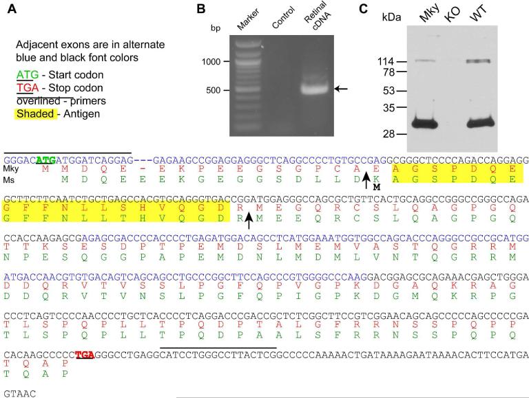
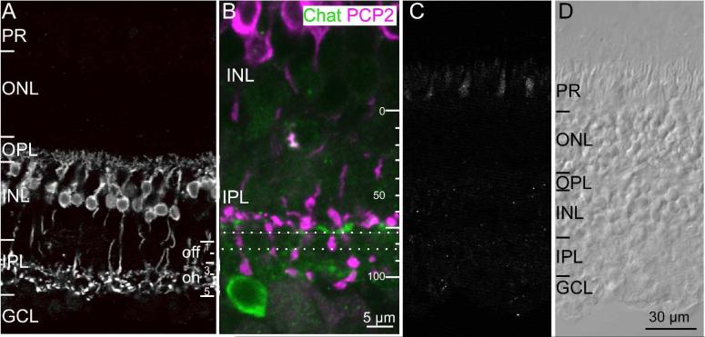
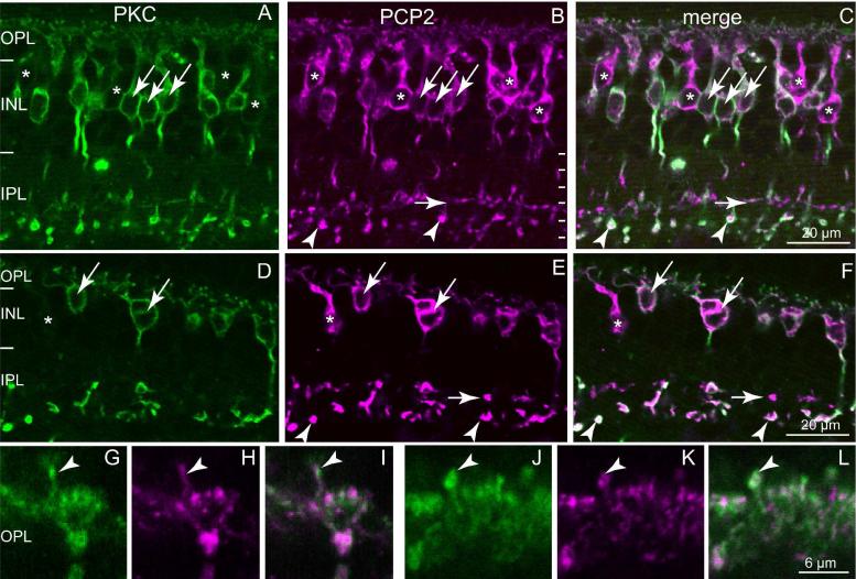
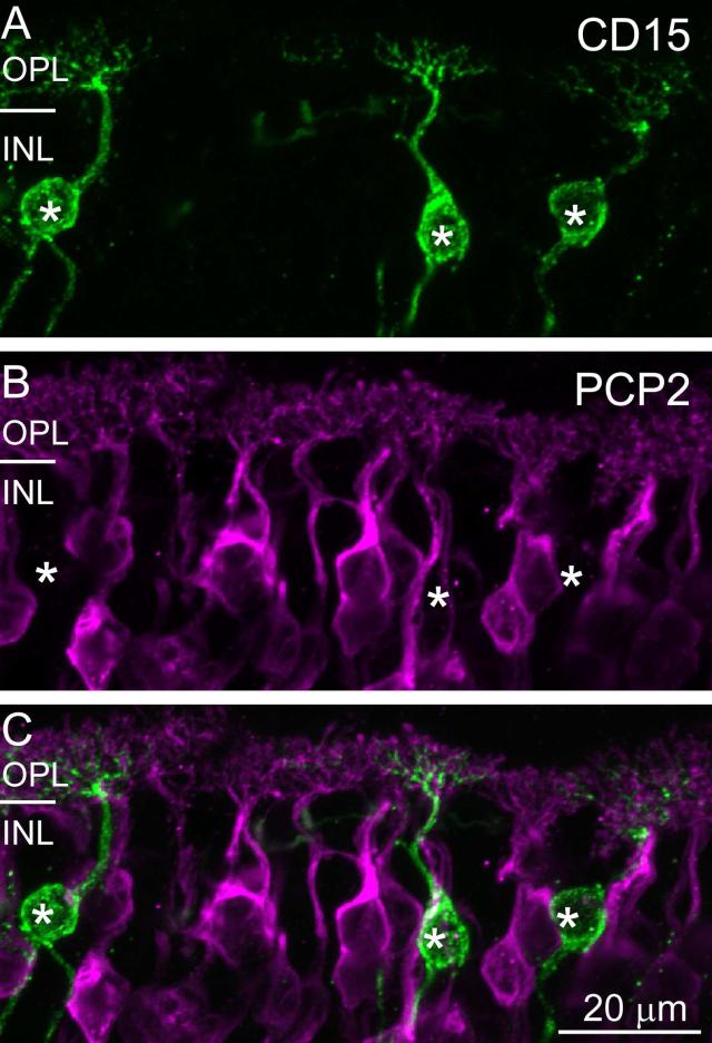
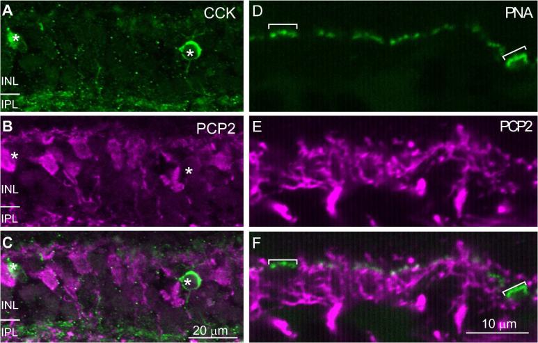
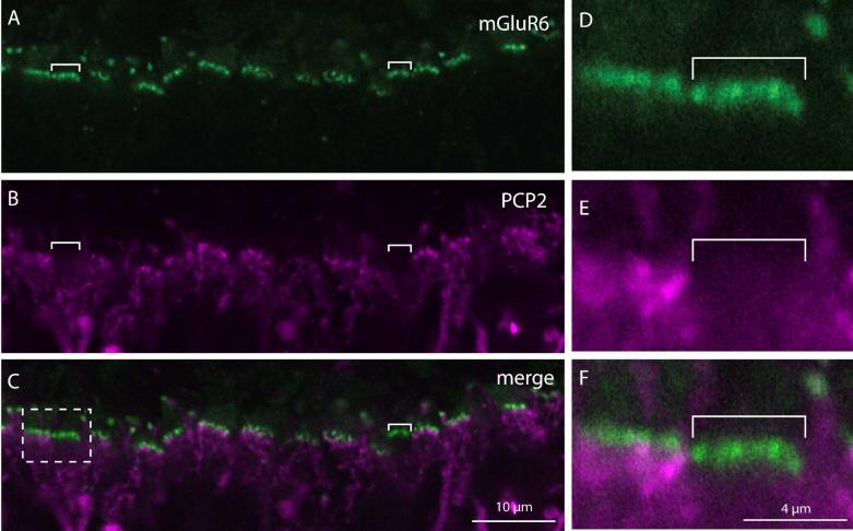
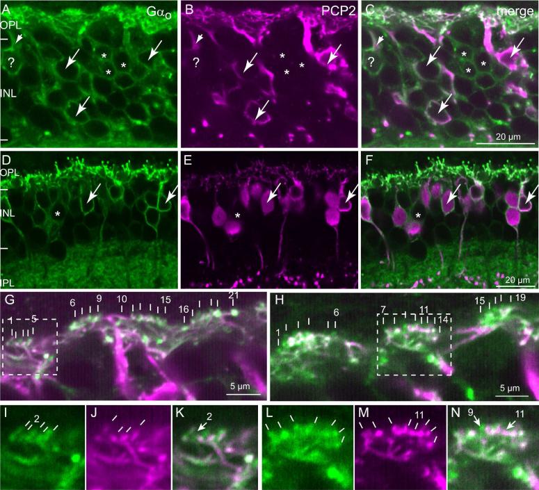
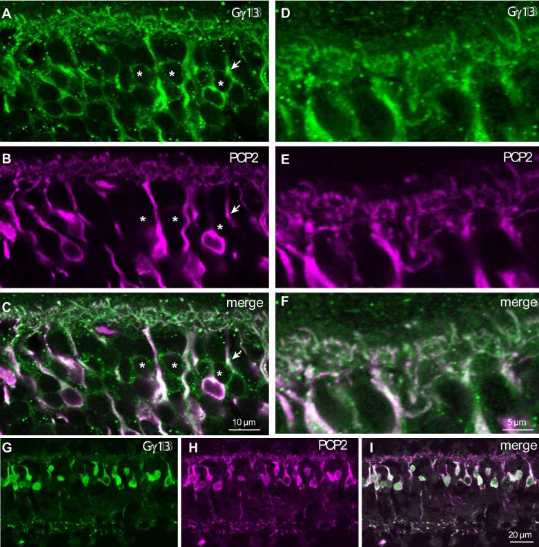
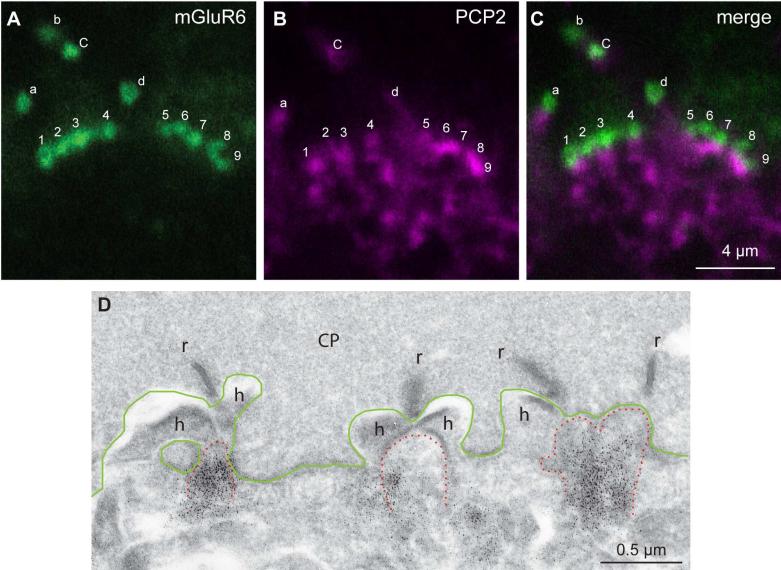
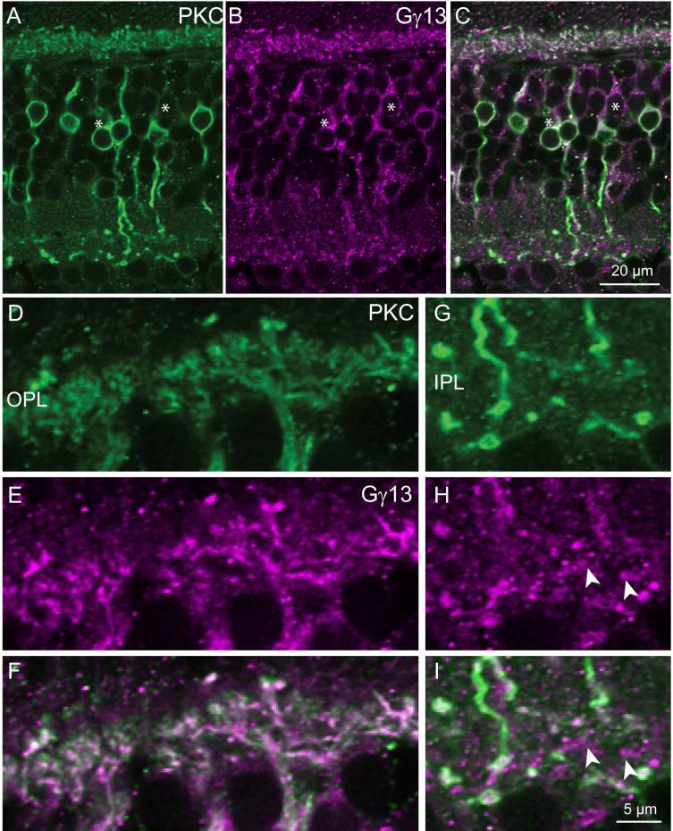
Purkinje cell protein 2 (PCP2), a member of the family of guanine dissociation inhibitors and a strong interactor with the G-protein subunit G alpha(o), localizes to retinal ON bipolar cells. The retina-specific splice variant of PCP2, Ret-PCP2, accelerates the light response of rod bipolar cells by modulating the mGluR6 transduction cascade. All ON cone bipolar cells express mGluR6 and G alpha(o), but only a subset expresses Ret-PCP2. Here we test the hypothesis that Ret-PCP2 contributes to shaping the various temporal bandwidths of ON cone bipolar cells in monkey retina. We found that the retinal splice variants in monkey and mouse are similar and longer than the cerebellar variants. Ret-PCP2 is strongly expressed by diffuse cone bipolar type 4 cells (DB4; marked with anti-PKCalpha) and weakly expressed by midget bipolar dendrites (labeled by antibodies against G alpha(o), G gamma 13, or mGluR6). Ret-PCP2 is absent from diffuse cone bipolar type 6 (DB6; marked with anti-CD15) and blue cone bipolar cells (marked with anti-CCK precursor). Thus, cone bipolar cells that terminate in stratum 3 of the inner plexiform layer (DB4) express more Ret-PCP2 than those that terminate in strata 3 + 4 (midget bipolar cells), and these in turn express more than those that terminate in stratum 5 (DB6 and blue cone bipolar cells). This expression pattern approximates the arborization of ganglion cells (GC) with different temporal bandwidths: parasol GCs stratifying near stratum 3 are faster than midget GCs stratifying in strata 3 + 4, and these are probably faster than the sluggish GCs that arborize in stratum 5.


Figures

Fig. 1
Fig. 1. Retina expresses a new splice variant of PCP2 transcript, Ret-PCP2
A. The sequence of the PCR reaction product (in blue, now called Ret-PCP2) and its predicted translation in monkey (Mky, red). Mouse (Ms) Ret-PCP2 (green) is shown for reference. The sequences are similar. The arrows mark the translation start point of human cerebellar form-B (first arrow) and form-A (second arrow). Lines above the sequence mark the primers and yellow highlights mark the amino acid sequence used to generate the antibody. B. A prominent PCR reaction product of ~450 bp (arrow) results from PCR amplification of retinal cDNA. Control includes water instead of cDNA sample. C. Western blots of monkey (Mky) and wild type mouse (WT) retinal proteins show a single prominent band that migrates with similar mobility (~29 kDa). This band is missing in the PCP2-knockout mouse (KO).
Fig. 2
Fig. 2. Ret-PCP2 is expressed by ON cone bipolar cells
A. Immunostaining for Ret-PCP2 (radial view). Staining is restricted to bipolar cells whose somas are located high in the INL and whose axons terminate in the ON sublamina of the IPL. Note that the arborizations in the IPL appear in two wavy laminas in stratum 3 and 5. For this and all figures: PR, photoreceptors; ONL, outer nuclear layer; OPL, outer plexiform layer; INL, inner nuclear layer; IPL, inner plexiform layer; GCL, ganglion cell layer. B. Immunostaining for Ret-PCP (magenta) and choline acetyl transferase (ChAT;green) shows that the level of cone bipolar arborization lies just above the stratification of the starburst amacrine cells at around 65% depth. PCP2-stained bipolar terminals hardly arborize between the dotted lines, but axons of rod bipolar cells do cross through this sublamina to arborize in sublamina 5. C. Preabsorption control. All staining was eliminated in a retinal section that was incubated in antibody that was pre-absorbed with the antigenic peptide (excess of x10). To show some structure, contrast was enhanced relative to the image in B. D. Image of the same section as in C taken under differential interference contrast.
Fig. 3
Fig. 3. Ret-PCP2 is expressed throughout diffuse bipolar type 4 (DB4) cells
Double-labeling for Ret-PCP2 (magenta) and PKC (green). A-C. Foveal sections. D-F. Peripheral sections. All PKC-labeled somas are Ret-PCP2-positive (arrows); certain somas are Ret-PCP2-positive but PKC-negative (*). Rod bipolar terminals in sublamina 5 are strongly stained by both antibodies (arrowheads) whereas DB4 terminals are strongly stained for PCP2 but faintly stained for PKC (horizontal short arrows). G-L. Two examples at higher magnification of dendrites approaching the cones in the OPL: there is a high degree of correlation between the two stains. Arrowheads point to dendrites of rod bipolar cells recognized by their position above the cone bipolar clusters.
Fig. 4
Fig. 4. Ret-PCP2 is not expressed in DB6 cells
Double staining for Ret-PCP2 (magenta) and CD15 (green). The images represent a projection of 2-3 confocal images. The two stains did not colocalize in either the somas (*) or the dendrites.
Fig. 5
Fig. 5. Ret-PCP2 is not expressed in blue cone bipolar cells
A-C. Double staining for Ret-PCP2 (magenta) and CCK (green). The images represent a projection of 2-3 confocal images. The two stains did not colocalize in the somas (*). D-F. Double staining using peanut agglutinin (PNA; green) and anti-Ret-PCP2 (magenta). Peanut agglutinin highlights the blue cone pedicles more brightly (brackets) than the other pedicles. These pedicles did not receive Ret-PCP2-stained dendrites.
Fig. 6
Fig. 6. A subset of cones (probably blue-sensitive) does not receive Ret-PCP2-expressing dendrites
Double staining for Ret-PCP2 (magenta) and mGluR6 (green). A-C. An example of a section where two clusters of mGluR6 puncta are not approached by Ret-PCP2 stained dendrites (brackets). D-F. Higher magnification of the dashed square in C. The pedicle on the left received multiple Ret-PCP2-expressing cone bipolar dendrites while that on the right received none.
Fig. 7
Fig. 7. Ret-PCP2 is localized to the dendritic tips of midget ON bipolar cells
Double labeling for Ret-PCP2 (magenta) and Gαo (green). A-F. All Ret-PCP2-positive somas are positive for Gαo (arrows), but certain Gαo-positive somas are devoid of Ret-PCP2 staining (*); Gαo-positive PCP2-negative somas in the fovea (A-C) is much greater than that in the periphery (D-F). Note the Gαo-labeled soma that is marked by ?; it is stained for Ret-PCP2 in its primary dendrite (very short arrow) but not in its soma. Note that the staining for Gαo is restricted to the soma's outline (membrane associated) while that for Ret-PCP2 is in the cytosol. G–N. High magnification of the outer plexiform layer. G, H are merged images. Each vertical white bar “points” to an ON bipolar invaginating dendrite. Each cluster of dendrites probably resides under a cone pedicle. The first and last dendrites within a cluster are numbered. Dashed squares denote the areas of higher magnification shown in I-N for each stain separately. In I (Gαo), there are 5 resolvable dendrites; only four can be seen in J (Ret-PCP2) (no. 2 is lacking PCP2; arrow in K). In L (Gαo), 7 dendrites are resolvable, while in M (Ret-PCP2) 8 are resolvable. Dendrite no. 11 is strongly stained for Ret-PCP2, but either unstained or weakly stained for Gαo (arrow in N). The slight white color of twig 11 in the merged image (shown by arrow) is due to diffused green background at this spot; the green appearance of twig 9 results from Gαo in this dendrite that extends beyond Ret-PCP2. The orientation of the lines in I-N intends to show the orientation of the dendrites.
Fig. 8
Fig. 8. Ret-PCP2 is localized to a subset of ON bipolar cell somas, but to most bipolar cell dendrites
Double staining for Ret-PCP2 (magenta) and Gγ13 (green). A-C. Central retina, certain somas that stain for Gγ13 are not stained (or are very weakly stained) for Ret-PCP2 (*). The arrow points to a primary dendrite that is stained for both Gγ13 and Ret-PCP2, but its soma is stained only for Gγ13. D-F. Central section shows the OPL at higher magnification. All dendrites appear stained for both proteins. G-I. In a different region where soma density is lower (mid periphery), all somas stain for both proteins.
Fig. 9
Fig. 9. Ret-PCP2 is expressed in most ON bipolar cell dendrites
Double staining for Ret-PCP2 (magenta) and mGluR6 (green). A-C. Two clusters of mGluR6-stained puncta are shown; the lettered puncta are rod bipolar dendritic tips (identified as such because these are large puncta located above the level of the cone pedicles) and the numbered puncta are cone bipolar dendritic tips. mGluR6 concentrates slightly above Ret-PCP2. Rod bipolar magenta punctum b is not visible in this focal plane and d is weak. All cone bipolar puncta can be correlated with Ret-PCP2 staining just below mGluR6. D. Electron micrograph of monkey retina stained for Ret-PCP2 shows a region of a cone pedicle (CP, its base is outlined in green) with 4 synaptic ribbons (r) and 3 postsynaptic triads. The triads consists of stained (black dots) central elements (ON bipolar dendrites; tip is outlined in dotted red lines) and two lateral elements (horizontal cell process, h).
Fig. 10
Fig. 10. PKC colocalizes with Gγ13 in the ON bipolar cell dendrites
Double staining for PKC (green) and Gγ13 (magenta). A-C. Low magnification shows that some somas are stained only for Gγ13 (*). D-F. High magnification of OPL shows close to 100% colocalization. G-I. High magnification of IPL shows that certain bipolar terminals (arrowheads) are stained only for Gγ13. Scale bar in I applies to D-I

Similar articles

Cited by

References

    1. Awatramani GB, Slaughter MM. Origin of transient and sustained responses in ganglion cells of the retina. J Neurosci. 2000;20:7087–7095. - PMC - PubMed
    1. Berrebi AS, Mugnaini E. Characteristics of labeling of the cerebellar Purkinje neuron by L7 antiserum. J Chem Neuroanat. 1992;5:235–243. - PubMed
    1. Berrebi AS, Oberdick J, Sangameswaran L, Christakos S, Morgan JI, Mugnaini E. Cerebellar Purkinje cell markers are expressed in retinal bipolar neurons. J Comp Neurol. 1991;308:630–649. - PubMed
    1. Calvert PD, Strissel KJ, Schiesser WE, Pugh EN, Jr., Arshavsky VY. Light-driven translocation of signaling proteins in vertebrate photoreceptors. Trends Cell Biol. 2006;16:560–568. - PubMed
    1. Chan TL, Martin PR, Grünert U. Immunocytochemical identification and analysis of the diffuse bipolar cell type DB6 in macaque monkey retina. Eur J Neurosci. 2001;13:829–832. - PubMed

Publication types

MeSH terms