. 2010 Nov 15;127(10):2334-50.
doi: 10.1002/ijc.25222.

Oral benzo[a]pyrene-induced cancer: two distinct types in different target organs depend on the mouse Cyp1 genotype

Affiliations

Oral benzo[a]pyrene-induced cancer: two distinct types in different target organs depend on the mouse Cyp1 genotype

Zhanquan Shi et al. Int J Cancer. .

Abstract

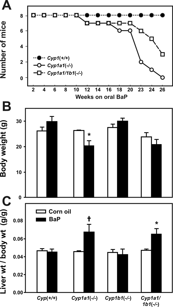
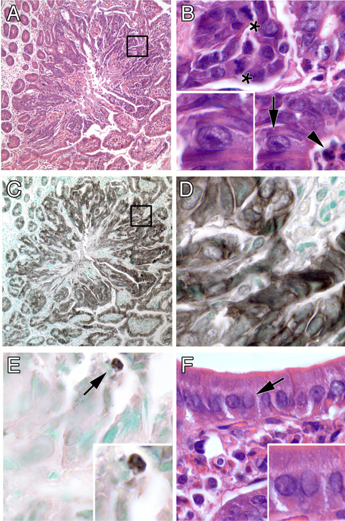
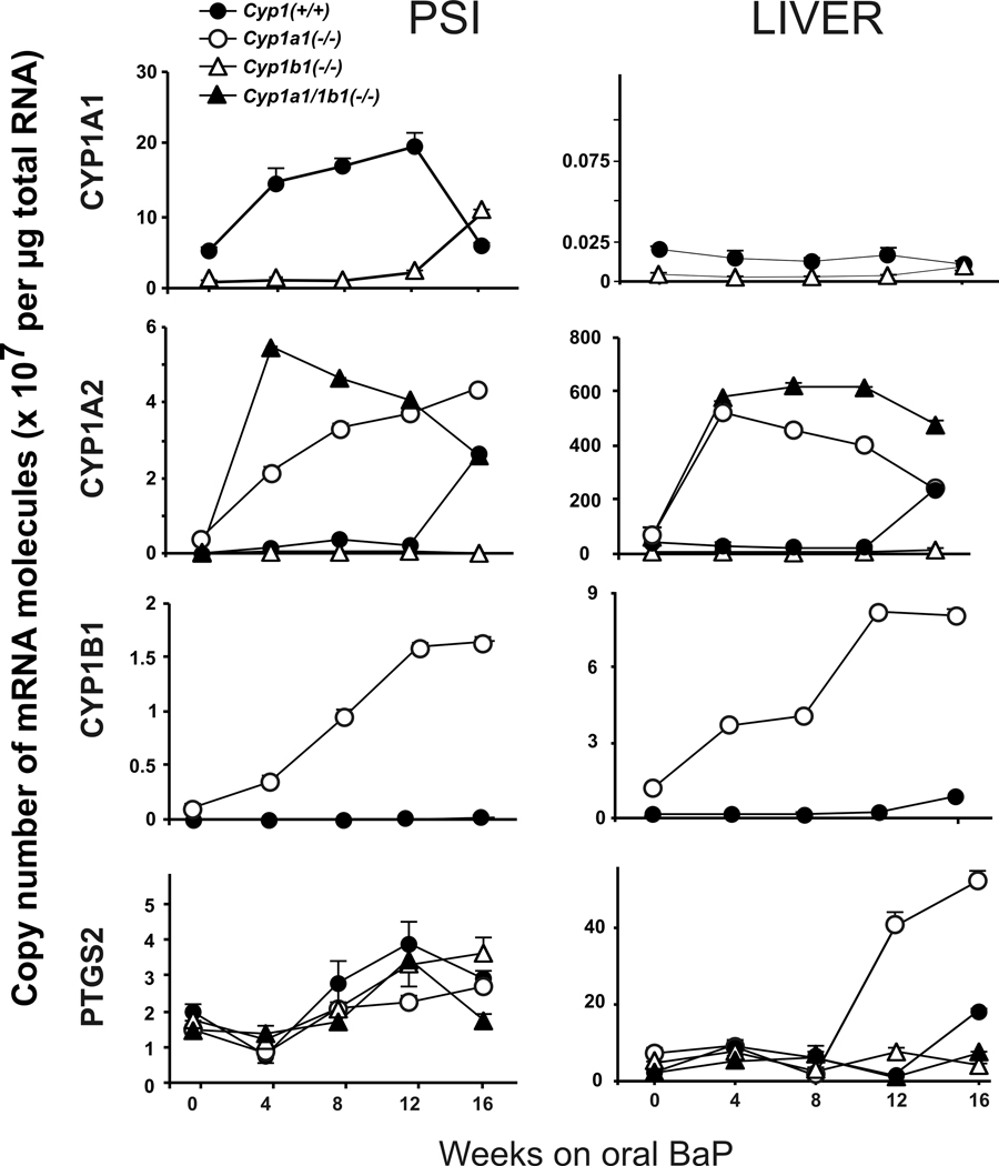
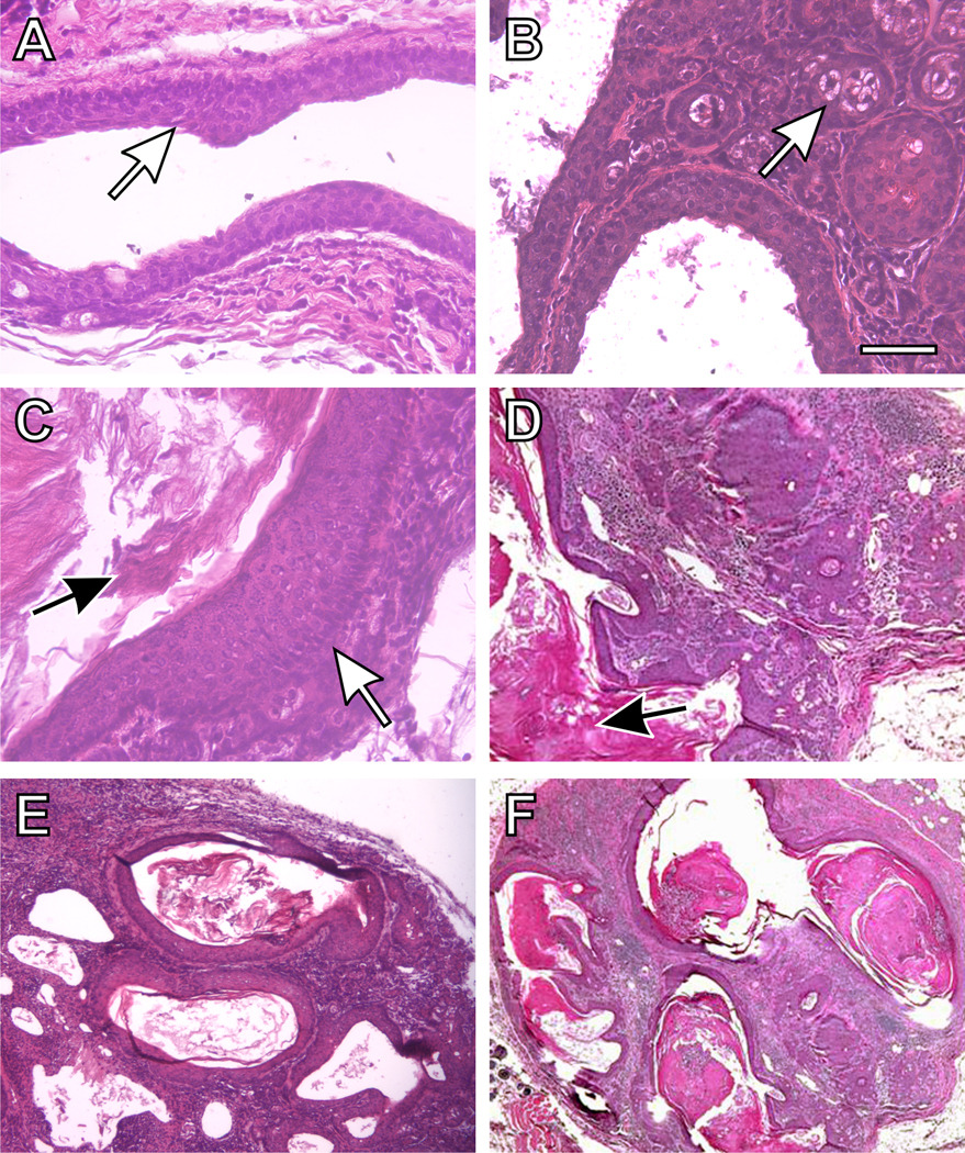
Benzo[a]pyrene (BaP) is a prototypical polycyclic aromatic hydrocarbon (PAH) found in combustion processes. Cytochrome P450 1A1 and 1B1 enzymes (CYP1A1 and CYP1B1) can both detoxify PAHs and activate them to cancer-causing reactive intermediates. Following high dosage of oral BaP (125 mg/kg/day), ablation of the mouse Cyp1a1 gene causes immunosuppression and death within ∼28 days, whereas Cyp1(+/+) wild-type mice remain healthy for >12 months on this regimen. In this study, male Cyp1(+/+) wild-type, Cyp1a1(-/-) and Cyp1b1(-/-) single-knockout and Cyp1a1/1b1(-/-) double-knockout mice received a lower dose (12.5 mg/kg/day) of oral BaP. Tissues from 16 different organs-including proximal small intestine (PSI), liver and preputial gland duct (PGD)-were evaluated; microarray cDNA expression and >30 mRNA levels were measured. Cyp1a1(-/-) mice revealed markedly increased CYP1B1 mRNA levels in the PSI, and between 8 and 12 weeks developed unique PSI adenomas and adenocarcinomas. Cyp1a1/1b1(-/-) mice showed no PSI tumors but instead developed squamous cell carcinoma of the PGD. Cyp1(+/+) and Cyp1b1(-/-) mice remained healthy with no remarkable abnormalities in any tissue examined. PSI adenocarcinomas exhibited striking upregulation of the Xist gene, suggesting epigenetic silencing of specific genes on the Y-chromosome; the Rab30 oncogene was upregulated; the Nr0b2 tumor suppressor gene was downregulated; paradoxical overexpression of numerous immunoglobulin kappa- and heavy-chain variable genes was found-although the adenocarcinoma showed no immunohistochemical evidence of being lymphatic in origin. This oral BaP mouse paradigm represents an example of "gene-environment interactions" in which the same exposure of carcinogen results in altered target organ and tumor type, as a function of just 1 or 2 globally absent genes.


Figures

Figure 1
Figure 1
Survival rates, body weights, and liver-weight-per-total-body-weight ratio. Survival rates (A) during exposure to oral BaP at 12.5 mg/kg/day (N = 8 per genotype). Cyp1(+/+) wild-type (closed circles) are compared with Cyp1a1(−/−) knockout (open circles) and Cyp1a1/1b1(−/−) double-knockout (open squares) mice. At every oral BaP dose tested, Cyp1a1(−/−) mice always show significantly (P<0.001) more toxicity than Cyp1a1/1b1(−/−) mice. This is because CYP1A1 is the major inducible CYP in the GI tract and, with oral BaP in Cyp1(+/+) wild-type mice, CYP1A1 becomes so highly induced that there is major detoxication and elimination of BaP metabolites in the feces. Without CYP1A1 in the GI tract in Cyp1a1(−/−) mice, at least 25-fold higher amounts of oral BaP reach distal tissues, including the spleen-thymus-bone marrow where BaP causes CYP1B1-dependent immunosuppression. Without CYP1A1 and CYP1B1 in the GI tract of Cyp1a1/1b1(−/−) mice, at least 75-fold higher amounts of oral BaP reach distal tissues; however, BaP is ineffective at causing immunosuppression because there is no CYP1B1 in the spleen-thymus-bone marrow to metabolize BaP to reactive oxygenated intermediates. Similar to wild-type mice, no Cyp1b1(−/−) deaths were seen during the 26-week experiment (not shown). Body weights (B) and liver-weight-per-total-body-weight ratios (C) in the four genotypes, following corn oil alone (open bars) versus oral BaP at 12.5 mg/kg/day (closed bars) for 12 weeks. Cyp1a1(−/−) and Cyp1b1(−/−) single-knockout and Cyp1a1/1b1(−/−) double-knockout were compared with Cyp1(+/+) wild-type. *P<0.05. P =.025.
Figure 2
Figure 2
Progressive atypical histology of the PSI in Cyp1a1(−/−) knockout mice (following 12 weeks of oral BaP, 12.5 mg/kg/day), showing normal intestinal villi together with nests of neoplastic cells near the basement membrane. A, area of normal proximal small intestine. (B, C, & D) areas involved by a progression from epithelial dysplasia to carcinoma in situ (white arrows). (E & F) unequivocal areas of invasive carcinoma into the stroma and the muscle layer (white arrows). Inset: a small region of metastasis (white arrow) found in a nearby lymph node. Few (if any) lymphocytes or other immune cells implicating possible inflammation are present. Bar, upper right = 100 microns for all images.
Figure 3
Figure 3
Features of a typical PSI tumor. (A) low magnification showing expanding boundary of tumor (inset enlarged in B). (B) high magnification of disarrayed tumor cells showing mitotic figures (asterisks), enlarged nuclei with prominent nucleoli (arrow; enlarged in inset), and an apoptotic body (arrowhead). (C & D) immunohistochemical staining for a keratin epithelial marker, low and high magnification, respectively. (E) immunohistochemical staining for the apoptosis marker cleaved caspase-3. (F) normal-appearing intestinal epithelium away from the tumor, showing orderly cell arrangement and small nuclei without enlarged nucleoli (compare insets of panelsF & B). Original optical magnifications 100x and 1000x.
Figure 4
Figure 4
Time-course of CYP1A1, CYP1A2, CYP1B1, and PTGS2 mRNA levels in the PSI, liver and PGD from Cyp1(+/+), Cyp1a1(−/−) and Cyp1a1/1b1(−/−) mice over the course of 16 weeks of oral BaP at 12.5 mg/kg/day. Brackets denote S.E.M.; when bracket cannot be seen, the S.E.M. is within the size of the symbol.
Figure 5
Figure 5
Histology of a typical preputial gland duct of Cyp1a1/1b1(−/−) double-knockout mice (following 12 weeks of oral BaP, 12.5 mg/kg/day), showing squamous cell carcinoma of the duct with invasion into the surrounding tissues. (A) normal preputial duct morphology with a stratified squamous cell epithelial lining of the larger ducts (white arrow). (B) cuboidal holocrine secretory acinar cells (white arrow). (C) a site of thickening of the squamous epithelium (white arrow), and increased keratin production can be seen (C & D black arrows). {D, E & F) excessive accumulation of keratin within ducts and resulting dilatation, and an epithelial morphology characteristic of squamous cell carcinoma. There is thickening of the lining of the tubular glands, with an abscess and excessive inflammation. The overall architecture is disorganized, and the amount of stroma between glands is increased. Bar = 100 microns for all images.

Similar articles

Cited by

References

    1. Pelkonen O, Nebert DW. Metabolism of polycyclic aromatic hydrocarbons: etiologic role in carcinogenesis. Pharmacol Rev. 1982;34:189–222. - PubMed
    1. Conney AH, Chang RL, Jerina DM, Wei SJ. Studies on the metabolism of benzo[a]pyrene and dose-dependent differences in the mutagenic profile of its ultimate carcinogenic metabolite. Drug Metab Rev. 1994;26:125–163. - PubMed
    1. Nebert DW, Dalton TP, Okey AB, Gonzalez FJ. Role of aryl hydrocarbon receptor-mediated induction of the CYP1 enzymes in environmental toxicity and cancer. J Biol Chem. 2004;279:23847–23850. - PubMed
    1. Nebert DW. The Ah locus: genetic differences in toxicity, cancer, mutation, and birth defects. Crit Rev Toxicol. 1989;20:153–174. - PubMed
    1. Miller KP, Ramos KS. Impact of cellular metabolism on the biological effects of benzo[a]pyrene and related hydrocarbons. Drug Metab Rev. 2001;33:1–35. - PubMed

Publication types

MeSH terms