. 2010 Mar;159(5):1174-86.
doi: 10.1111/j.1476-5381.2009.00595.x. Epub 2010 Jan 29.

Microsomal prostaglandin E synthase-1 and cyclooxygenase-2 are both required for ischaemic excitotoxicity

Affiliations

Microsomal prostaglandin E synthase-1 and cyclooxygenase-2 are both required for ischaemic excitotoxicity

Y Ikeda-Matsuo et al. Br J Pharmacol. 2010 Mar.

Abstract

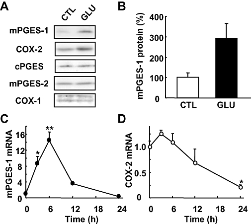
Background and purpose: Although both microsomal prostaglandin E synthase (mPGES)-1 and cyclooxygenase (COX)-2 are critical factors in stroke injury, but the interactions between these enzymes in the ischaemic brain is still obscure. This study examines the hypothesis that mPGES-1 activity is required for COX-2 to cause neuronal damage in ischaemic injury.

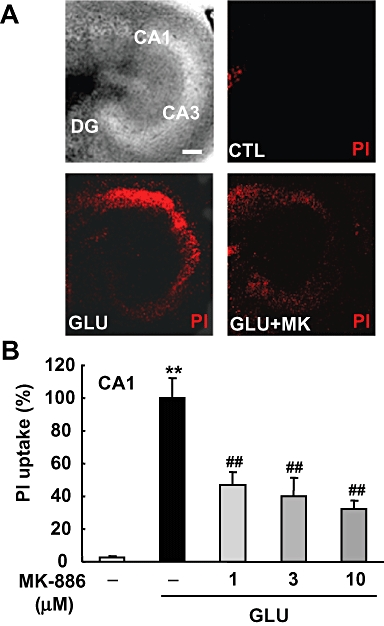
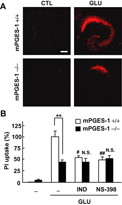
Experimental approach: We used a glutamate-induced excitotoxicity model in cultures of rat or mouse hippocampal slices and a mouse middle cerebral artery occlusion-reperfusion model in vivo. The effect of a COX-2 inhibitor on neuronal damage in mPGES-1 knockout (KO) mice was compared with that in wild-type (WT) mice.

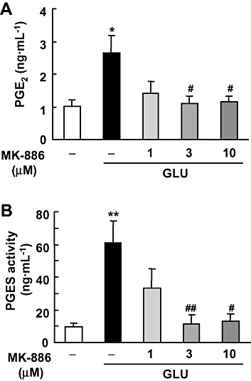
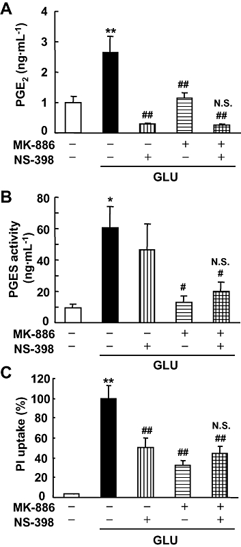
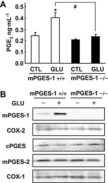
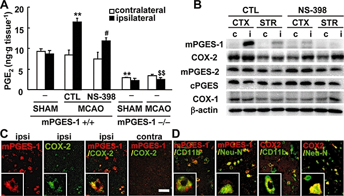
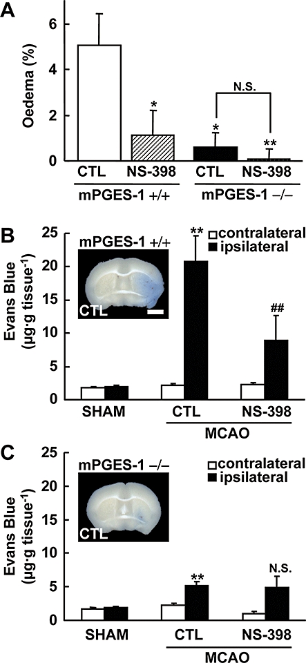
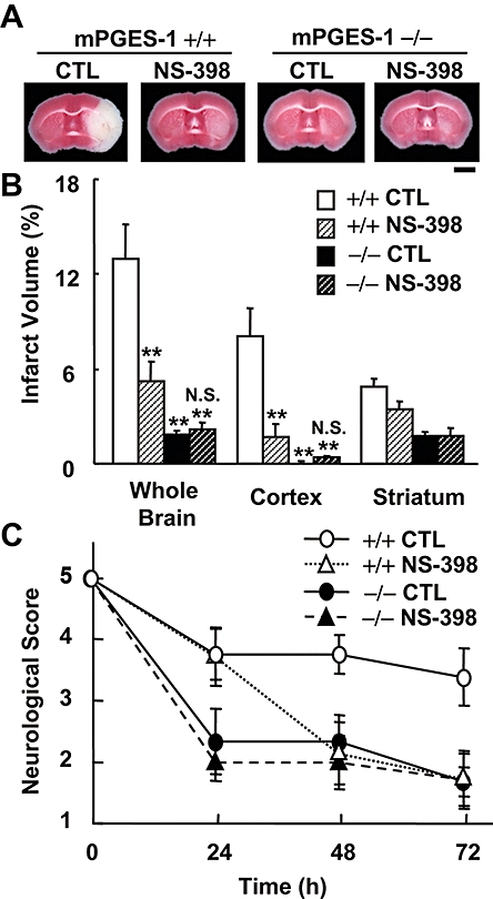
Key results: In rat hippocampal slices, glutamate-induced excitotoxicity, as well as prostaglandin (PG) E(2) production and PGES activation, was significantly attenuated by either MK-886 or NS-398, inhibitors of mPGES-1 and COX-2 respectively; however, co-application of these inhibitors had neither an additive nor a synergistic effect. The protective effect of NS-398 on the excitotoxicity observed in WT slices was completely abolished in mPGES-1 KO slices, which showed less excitotoxicity than WT slices. In the transient focal ischaemia model, mPGES-1 and COX-2 were co-localized in the infarct region of the cortex. Injection of NS-398 reduced not only ischaemic PGE(2) production, but also ischaemic injuries in WT mice, but not in mPGES-1 KO mice, which showed less dysfunction than WT mice.

Conclusion and implications: Microsomal prostaglandin E synthase-1 and COX-2 are co-induced by excess glutamate in ischaemic brain. These enzymes are co-localized and act together to exacerbate stroke injury, by excessive PGE(2) production.


Figures

Figure 1
Figure 1
mPGES-1 and COX-2 induction after glutamate exposure in cultures of rat hippocampal slices. (A) Western blot analysis for mPGES-1, COX-2, cPGES, mPGES-2 and COX-1 in the cultures of hippocampal slices 24 h after exposure to 1 mM glutamate (GLU) or vehicle (CTL) for 15 min. Representative data from three separate experiments are presented. (B) Summary data from immunoblotting with mPGES-1 antibody were scaled to a percentage of the control response (n = 3). (C and D) Time course of the expression of mPGES-1 and COX-2 mRNA after glutamate exposure. Quantitated data from real-time PCR analysis with the mPGES-1 (C) and COX-2 (D) primers were normalized to GAPDH (n = 3). **P < 0.01, *P < 0.05 versus the basal level (0 h). COX, cyclooxygenase; cPGES, mPGES, cytosolic/microsomal prostaglandin E synthase.
Figure 2
Figure 2
Effect of MK-886 on glutamate-induced PGE2 synthesis and PGES activity in cultures of rat hippocampal slices. MK-886 (1, 3 and 10 µM) was applied during and after the 1 mM glutamate (GLU) exposure for 15 min. (A) The amount of PGE2 in the culture medium 24 h after glutamate exposure was measured using an EIA kit (n = 4). (B) PGES activity in membrane fraction was measured as the conversion of exogenous PGH2 (0.5 µg) to PGE2 for 30 s by the lysate of these cells (30 µg protein). The amount of PGE2 was measured by the same protocol as in (A) (n = 4–5). **P < 0.01, *P < 0.05 versus the control; ##P < 0.01, #P < 0.05 versus glutamate alone. EIA, enzyme immunoassay; PGES, prostaglandin E synthase.
Figure 3
Figure 3
Effect of MK-886 on glutamate-induced excitotoxicity in cultures of rat hippocampal slices. MK-886 (1, 3 and 10 µM) was applied during and after the 1 mM glutamate exposure for 15 min. Twenty-four h later, PI uptake was analysed. (A) A representative differential interference microscopic image of a cultured hippocampal slice and confocal images of PI fluorescence of a control slice (CTL), a slice that received glutamate exposure (GLU) and a slice that received glutamate exposure with 10 µM of MK-886 (GLU+MK) are shown (scale bar: 200 µm). (B) Summary data from PI uptake analysis in the CA1 region with or without glutamate and/or MK-886 were scaled to a percentage of the response of glutamate alone (n = 11–13 slices per group). **P < 0.01 versus the control; ##P < 0.01 versus glutamate alone. PI, propidium iodide.
Figure 4
Figure 4
Effect of co-application of MK-886 and NS-398 on glutamate-induced PGE2 synthesis, PGES activity and excitotoxicity in cultures of rat hippocampal slices. MK-886 (10 µM) and NS-398 (1 µM) were applied during and after the 1 mM glutamate (GLU) exposure for 15 min. (A) The amount of PGE2 in the culture medium 24 h after glutamate exposure was measured using an EIA kit (n = 4). (B) PGES activity in the membrane fraction was measured as the conversion of exogenous PGH2 (0.5 µg) to PGE2 for 30 s by the lysate of these cells (30 µg protein). The amount of PGE2 was measured by the same protocol as in (A) (n = 4–5). (C) PI uptake was analysed 24 h after glutamate exposure. Summary data from PI uptake analysis in the CA1 region with or without glutamate and/or MK-886 and/or NS-398 were scaled to a percentage of the response of glutamate alone (n = 11–13 slices per group). **P < 0.01, *P < 0.05 versus the control; ##P < 0.01, #P < 0.05, N.S. (not significant) versus glutamate alone. EIA, enzyme immunoassay; PGES, prostaglandin E synthase; PI, propidium iodide.
Figure 6
Figure 6
Effects of COX inhibitors on the glutamate-induced excitotoxicity in an mPGES-1 KO and a WT mouse hippocampal slice culture. PI uptake was analysed 24 h after 1 mM glutamate exposure for 30 min. (A) Representative confocal images of PI fluorescence of a control slice (CTL) and a slice that received glutamate (GLU) exposure in WT (+/+) mice and mPGES-1 KO (−/−) mice are shown (scale bar: 200 µm). (B) Summary data from PI uptake analysis in the CA1 region with or without glutamate and indomethacin (IND, 1 µM) or NS-398 (1 µM) were scaled to a percentage of the glutamate response in slices from WT mice. Indomethacin and NS-398 were applied during and after the glutamate exposure (n = 6–11 slices per group). **P < 0.01, ##P < 0.01, #P < 0.05, versus glutamate alone in a slice from a WT mouse; N.S. (not significant) versus glutamate alone in a slice from a mPGES-1 KO mouse. COX, cyclooxygenase; mPGES, microsomal prostaglandin E synthase; KO, knockout; PI, propidium iodide; WT, wild-type.
Figure 5
Figure 5
mPGES-1 is an essential component for PGE2 production after glutamate exposure in cultures of mice hippocampal slices. (A) The production of PGE2 in the culture medium of slices from mPGES-1 KO (−/−) and WT (+/+) mice 24 h after 1 mM glutamate (GLU) exposure for 30 min and in control hippocampal slices (CTL) (n = 3). (B) Western blot analysis for mPGES-1, COX-2, cPGES, mPGES-2 and COX-1 in cultures of hippocampal slices 24 h after glutamate exposure. Representative data from three separate experiments are presented. *P < 0.05 versus the control; #P < 0.05 versus glutamate alone in slices from WT mice. COX, cyclooxygenase; cPGES, mPGES, cytosolic/microsomal prostaglandin E synthase; KO, knockout; WT, wild-type.
Figure 7
Figure 7
Co-induction of mPGES-1 and COX-2 contributes to post-ischaemic PGE2 production. (A) The production of PGE2 in the ipsilateral or contralateral cortex of mPGES-1 KO (−/−) or WT (+/+) mice injected with NS-398 or vehicle (CTL), 24 h after MCA occlusion (MCAO) and sham operation (SHAM) (n = 4–6 mice per group). (B) Western blot analysis for mPGES-1, COX-2, cPGES, mPGES-2, COX-1 and β-actin in the ipsilateral (i) or contralateral (c) cerebral cortex (CTX) and striatum (STR) of WT mice injected with NS-398 or vehicle (CTL). Representative data from three separate experiments are presented. (C) The double-immunostaining of mPGES-1 (red) and COX-2 (green) in the ischaemic regions of the ipsilateral (ipsi) and contralateral (contra) cortex. Insets show high magnification of staining. (D) The double-immunostaining of mPGES-1 (red) or COX-2 (red) and CD11b (green) in the ischaemic core region or Neu-N (green) in the peri-infarct region of the ipsilateral cortex. Insets show high magnification of staining. The photos shown here are representative examples from three separate experiments (scale bar, 20 µm; insets, 5 µm). **P < 0.01 versus the ipsilateral cortex of sham-operated WT mice or contralateral cortex of WT mice after MCA occlusion; #P < 0.05 versus the ipsilateral cortex of CTL-injected WT mice after MCA occlusion; $$P < 0.01 versus the ipsilateral cortex of WT mice after MCA occlusion. COX, cyclooxygenase; cPGES, mPGES, cytosolic/microsomal prostaglandin E synthase; KO, knockout; MCA, middle cerebral artery; Neu-N, neuron-specific nuclear protein; WT, wild-type.
Figure 9
Figure 9
Protective effect of NS-398 on oedema and Evans Blue extravasation after ischaemia in WT mice but not in mPGES-1 KO mice. NS-398 (10 mg·kg−1; i.p.) or vehicle was administered twice daily starting 10 min after MCA occlusion. (A) The corrected oedema percentage in the NS-398 or vehicle (CTL)-injected WT (+/+) and mPGES-1 KO (−/−) mice (n = 7–8 mice per group). **P < 0.01, *P < 0.05 versus the vehicle-injected WT mice. (B and C) The Evans Blue contents of the ipsilateral and contralateral cortex after MCA occlusion (MCAO) or sham-operation (SHAM) in WT (B) and mPGES-1 KO (C) mice, injected with NS-398 or vehicle were measured 48 h after transient ischaemia (n = 6–7 mice per group). **P < 0.01 versus the contralateral cortex of mice after MCA occlusion or the ipsilateral cortex of sham-operated mice; ##P < 0.01, N.S. (not significant) versus the ipsilateral cortex of vehicle-treated mice after MCA occlusion The insets in (B) and (C) show representative results of Evans Blue-stained brain slices of a vehicle-injected WT (+/+) and mPGES-1 KO (−/−) mouse respectively (scale bar: 2 mm). KO, knockout; MCA, middle cerebral artery; mPGES, microsomal prostaglandin E synthase; WT, wild-type.
Figure 8
Figure 8
Protective effect of NS-398 on post-ischaemic symptoms in WT mice but not in mPGES-1 KO mice. NS-398 (10 mg·kg−1; i.p.) or vehicle was administered twice daily starting 10 min after MCA occlusion. (A) Representative TTC-stained coronal sections 72 h after MCA occlusion of the WT (+/+) mice and mPGES-1 KO (−/−) mice injected with vehicle (CTL) or NS-398 (scale bar: 2 mm). (B) The volume of infarcted cortex 72 h after ischaemia was estimated and expressed as a percentage of the corrected tissue volume (n = 7–9 mice per group). (C) Neurological dysfunction in the NS-398-injected WT and mPGES-1 KO mice 24, 48 and 72 h after ischaemia (n = 7–9 mice per group); **P < 0.01 versus WT control mice; N.S. (not significant) versus mPGSE-1 KO control mice. KO, knockout; MCA, middle cerebral artery; mPGES, microsomal prostaglandin E synthase; WT, wild-type.

Similar articles

Cited by

References

    1. Ahmad AS, Ahmad M, de Brum-Fernandes AJ, Doré S. Prostaglandin EP4 receptor agonist protects against acute neurotoxicity. Brain Res. 2005;1066:71–77. - PubMed
    1. Ahmad AS, Saleem S, Ahmad M, Doré S. Prostaglandin EP1 receptor contributes to excitotoxicity and focal ischemic brain damage. Toxicol Sci. 2006;89:265–270. - PubMed
    1. Ahmad M, Zhang Y, Liu H, Rose ME, Graham SH. Prolonged opportunity for neuroprotection in experimental stroke with selective blockade of cyclooxygenase-2 activity. Brain Res. 2009;1279:168–173. - PMC - PubMed
    1. Alexander SPH, Mathie A, Peters JA. Guide to Receptors and Channels (GRAC), 3rd edn. Br J Pharmacol. 2008;153(Suppl 2):S1–S209. - PMC - PubMed
    1. Armstead WM, Mirro R, Busija DW, Leffler CW. post-ischaemic generation of superoxide anion by newborn pig brain. Am J Physiol. 1988;255:H401–H403. - PubMed

Publication types

MeSH terms