. 2010 Mar;159(6):1326-38.
doi: 10.1111/j.1476-5381.2009.00607.x. Epub 2010 Jan 28.

Oxaliplatin responses in colorectal cancer cells are modulated by CHK2 kinase inhibitors

Affiliations

Oxaliplatin responses in colorectal cancer cells are modulated by CHK2 kinase inhibitors

I M Pires et al. Br J Pharmacol. 2010 Mar.

Abstract

Background and purpose: Checkpoint kinase 2 (CHK2) is activated by DNA damage and can contribute to p53 stabilization, modulating growth arrest and/or apoptosis. We investigated the contribution of CHK2 to oxaliplatin-mediated toxicity in a colorectal cancer model.

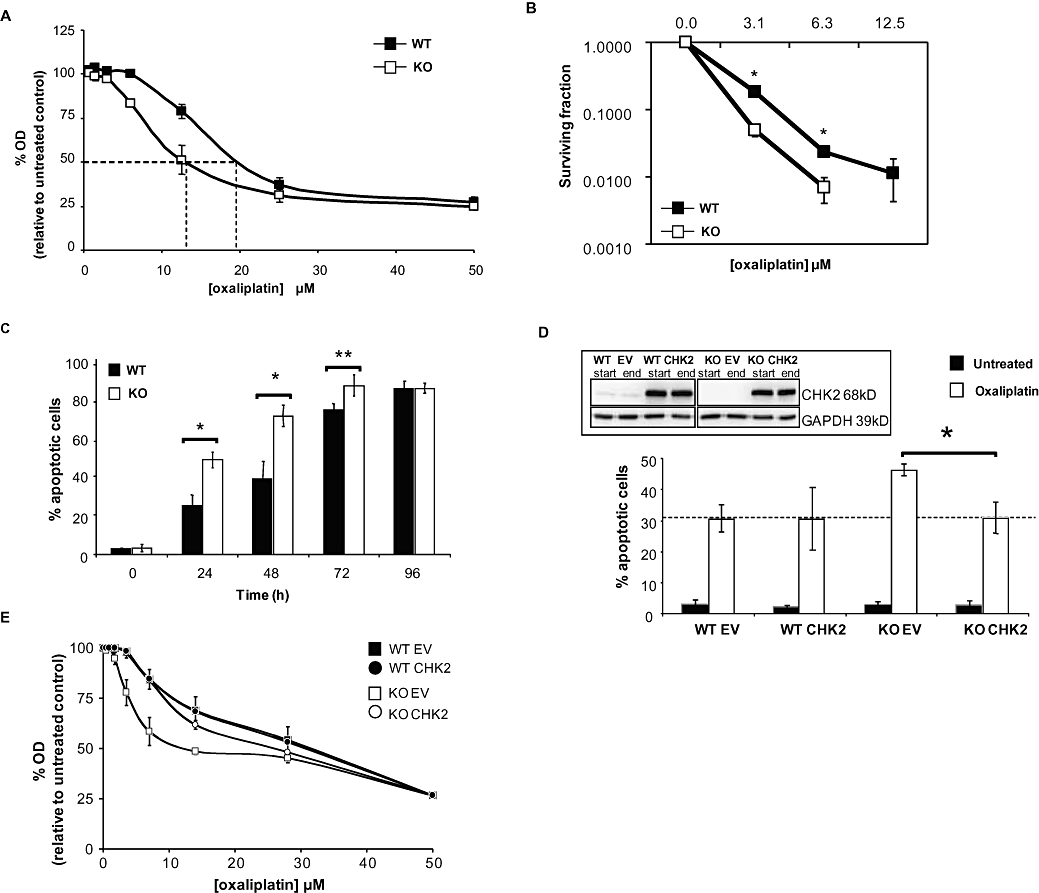
Experimental approach: We evaluated the ability of CHK2 small molecule inhibitors to potentiate oxaliplatin-induced toxicity. The role of CHK2 in oxaliplatin-induced apoptosis was investigated in HCT116 cells that were wild-type (WT) or KO for CHK2. Small molecule inhibitors of CHK2 were used in combination studies with oxaliplatin in this cell model.

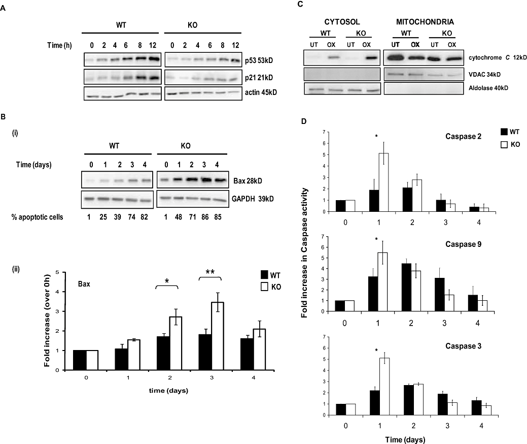
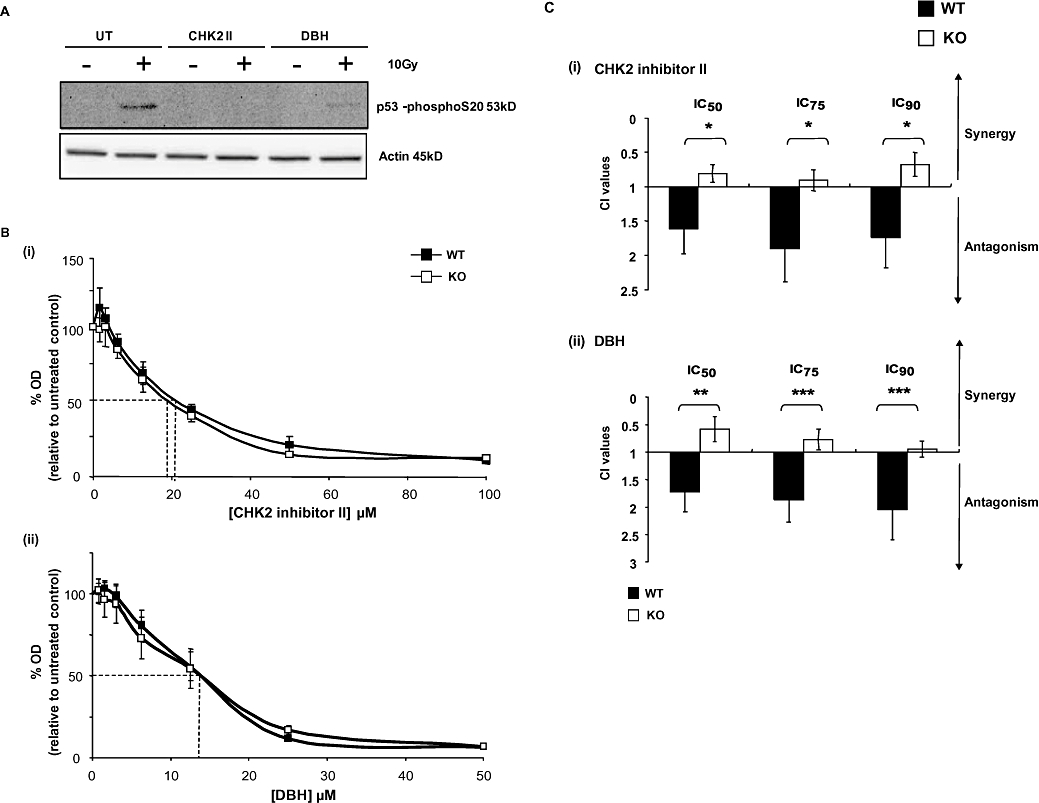
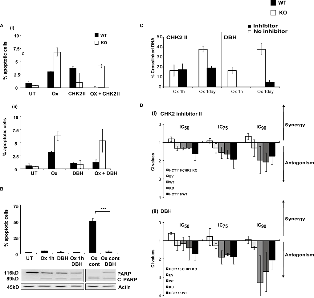
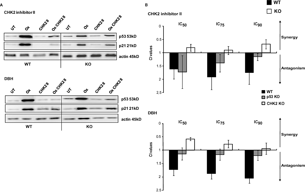
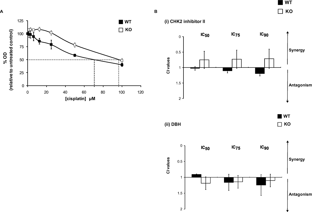
Key results: In oxaliplatin-treated CHK2 KO cells, accelerated apoptosis was accompanied by attenuated p53 stabilization and p21(WAF-1) up-regulation correlating with increased Bax expression, cytochrome c release and elevated caspase activity. The higher levels of apoptosis in CHK2 KO cells were restored to control (WT) levels when CHK2 was re-introduced. This 'uncoupling' of p53 stabilization and Bax up-regulation in CHK2 KO cells suggested oxaliplatin-induced apoptosis was due to a p53-independent response. Combination studies revealed that CHK2 inhibitor II or debromohymenialdisine antagonized the responses to oxaliplatin. This inhibitory effect correlated with decreases in apoptosis, p53 stabilization and DNA inter-strand cross-link formation, and was dependent on the presence (but not activity) of CHK2.

Conclusions and implications: Combinations of CHK2 inhibitors with oxaliplatin should further sensitize cells to oxaliplatin treatment. However, these inhibitors produced an antagonistic effect on the response to oxaliplatin, which was reversed on the re-introduction of CHK2. These observations may have implications for the use of oxaliplatin in colorectal cancer therapy in combination with therapies targeting CHK2.


Figures

Figure 1
Figure 1
Characterization of the effect of oxaliplatin on HCT116 checkpoint kinase 2 (CHK2) wild-type (WT) and KO cell lines. Responses of HCT116 CHK2 WT and CHK2 KO to treatment with oxaliplatin for 1 h. (A) Sulforhodamine-B (SRB) concentration–response curves, dashed lines indicate the IC50 doses. (B) Clonogenic survival curves. (C) Oxaliplatin-induced apoptosis kinetics for the CHK2 WT and CHK2 KO following 40 µM continuous treatment with oxaliplatin. Data represent the percentage of apoptotic cells based on DAPI (4′-6′-diamino-2-phenylindole di-hydrochloride) stained nuclear morphology (condensation and fragmentation). (D) CHK2 WT or CHK KO cells were transfected with either empty vector (EV) or CHK2-expressing vector (CHK2) then exposed continuously to 40 µM oxaliplatin or to vehicle control for 24 h. The percentages of apoptotic cells were determined as in (C). The data represented in (A–D) are the average of three independent experiments, ±SE. *P < 0.05 and **P < 0.01, Student's t-test. Inset: western blot, representative of three independent experiments, showing expression of CHK2 in cell lines following transfection, just prior to oxaliplatin treatment (start) and 24 h following (end) of treatment. GAPDH was used as a loading control. (E) SRB concentration–response curves of HCT116 CHK2 WT and CHK2 KO transfected with either an EV or CHK2-expressing vector (CHK2) following treatment with oxaliplatin for 1 h (dashed lines indicate the IC50 doses).
Figure 2
Figure 2
Kinetics of apoptosis of HCT116 wild-type (WT) and checkpoint kinase 2 (CHK2) KO cells induced by oxaliplatin. (A) Cells were continuously exposed to 40 µM oxaliplatin for up to 12 h. Western blots (50 µg protein per lane) show the kinetics of p53 stabilization and up-regulation of p21WAF-1. (B) (i) Western blots of HCT116 WT and CHK2 KO cells following continuous exposure to oxaliplatin for up to 4 days. (ii) Densitometry analysis of blots depicted in B (i). (C) Western blots showing cytochrome c levels in cytosolic and mitochondrial fractions from WT or CHK2 KO cells treated with 40 µM oxaliplatin (OX) for 2 days. GAPDH was used as a loading control. Validation of purity and loading were confirmed using VDAC for the mitochondrial fraction and aldolase for the cytosolic fraction. (D) Fluorogenic measurement of active caspase-2, 3 and 9 levels in HCT116 WT and CHK2 KO cells during a 4 day continuous challenge with 40 µM oxaliplatin. Blots are representative of three independent experiments. Actin was used as a loading control; error bars represent ±SE. *P < 0.05 and **P < 0.025, Student's t-test.
Figure 3
Figure 3
Effect of the checkpoint kinase inhibitors checkpoint kinase 2 (CHK2) inhibitor II and debromohymenialdisine (DBH) on the responses of the different cell lines to oxaliplatin. (A) Validation of CHK2 inhibition by CHK2 inhibitor II and DBH. CHK2 wild-type (WT) and CHK2 KO cells, either untreated (UT) or treated (CHK2II, DBH) were subsequently exposed to 10 Gy of ionizing radiation. Cells were harvested 30 min later and subjected to western blotting to determine the phosphorylation status of serine 20 on p53 Western blots are representative of two independent experiments and actin was used as a protein loading control. (B) Effect of a 72 h continuous exposure to either CHK2 inhibitor II (i) or DBH (ii) on CHK2 WT and KO cells. Cell population growth was determined using the sulforhodamine-B (SRB) assay as described and data plotted as a percentage of untreated controls. Data points are the average of three independent experiments error bars represent ±SE. Dashed lines indicate the IC50 doses. (C) The Chou and Talalay combination index (CI) values following fixed drug concentration ratios of oxaliplatin and either (i) CHK2 inhibitor II (CHK2 II) or (ii) DBH. CI values are representative of the IC50, IC75 and IC90 effective doses. WT and CHK2 KO were treated for 1 h with oxaliplatin; inhibitors were added 24 h before and continuously following oxaliplatin treatment. Data shown are for the average of three independent experiments. *P < 0.05, **P < 0.01 and ***P < 0.005, Student's t-test.
Figure 4
Figure 4
(A) Apoptotic responses of HCT116 checkpoint kinase 2 (CHK2) wild-type (WT) and CHK2 KO cells following treatment with a combination of CHK inhibitors and oxaliplatin. HCT116 WT or CHK2 KO were either left untreated (UT) or treated with (i) oxaliplatin alone (20 µM, Ox), CHK2 inhibitor II alone (19–21 µM) or with the combination (Ox + CHK2 II), (ii) debromohymenialdisine (DBH) alone (13 µM) or the combination of oxaliplatin and DBH (Ox + DBH). Oxaliplatin treatment was for 1 h and CHK2 II or DBH was added 24 h before oxaliplatin and continuously thereafter. (B) Effect of duration of exposure to oxaliplatin in combination with CHK2 inhibitor DBH in HCT116 WT cells on apoptotic response. HCT116 WT cells were either left untreated (UT), treated with DBH alone, oxaliplatin (20 µM) alone [1 h or continuously (cont)] or in combination with DBH (13 µM). DBH was added 24 h prior to oxaliplatin and maintained throughout the experiment. The percentage of apoptotic cells was determined by characteristic changes in nuclear morphology. Data are the average of three independent experiments; error bars represent ±SE and ***P < 0.005, Student's t-test. A western blot (representative of the 3 experiments) of PARP and cleaved PARP (C PARP, inset) levels are also shown. (C) Kinetics of DNA cross-linking after treatment of HCT116 WT and KO cells with oxaliplatin alone or in combination with CHK inhibitors and effects of the CHK2 status of the HCT116 cells. HCT116 WT cells were treated with oxaliplatin alone or in combination with CHK2 II or DBH. Cells were treated with the inhibitor 24 h prior to a 1 h pulse or 24 h treatment with oxaliplatin. Samples were harvested immediately following the treatment with oxaliplatin into fresh medium + inhibitor. The levels (%) of DNA cross-links were measured using the comet-X assay, as described in the Methods section. Results are representative of the average of three independent experiments each consisting of 25 cells counted on each of two independent slides. (D) Effect of re-introduction and overexpression of CHK2 on the response of WT and CHK2 KO cells to oxaliplatin and the combination of oxaliplatin and the checkpoint inhibitors. Both HCT116 WT and CHK2 KO cells were transfected with empty vector (EV), CHK2 wild-type (CHK2 WT) or CHK2 kinase dead (CHK2 KD) expressing vectors. Chou and Talalay combination index (CI) determinations using fixed drug concentrations ratios (IC50) were then performed following a 1 h oxaliplatin treatment in combination with either CHK2 inhibitor II or DBH. Inhibitors were added 24 h prior to oxaliplatin treatment and maintained throughout the experiment. Data plotted are the average percentage of apoptotic cells determined from three independent experiments; error bars represent ±SE.
Figure 5
Figure 5
The effect of the checkpoint inhibitors on oxaliplatin-mediated p53 stabilization and p21 up-regulation. (A) HCT116 wild-type (WT) and checkpoint kinase 2 (CHK2) KO cells were left either untreated (UT) or treated with either oxaliplatin (Ox), CHK2 II, debromohymenialdisine (DBH) or with a combination of the inhibitor and a 1 h pulse treatment of oxaliplatin. Inhibitors were added 24 h prior to, and continuously after, oxaliplatin treatment. The levels of p53 or p21 were assessed by western blotting. Actin was used as a protein-loading control. The western blot depicted is representative of three independent experiments. (B) Chou and Talalay combination index (CI) values following fixed drug concentration-ratios of oxaliplatin and either CHK2 II or DBH. CI values are representative of the IC50, IC75 and IC90 effective doses. WT, CHK2 KO and p53 KO cells were treated with oxaliplatin for 1 h. Inhibitors were added 24 h before and continuously following platinum treatment. Data shown are for the average of three independent experiments.
Figure 6
Figure 6
Effects of the checkpoint kinase 2 (CHK2) status of HCT116 cells on the response to cisplatin. (A) Response of HCT116 CHK2 WT and CHK2 KO following treatment with cisplatin for 1 h. Sulforhodamine-B (SRB) concentration–response curves, dashed lines indicate the IC50 doses. (B) Chou and Talalay combination index (CI) values following fixed drug concentration-ratios of cisplatin and either CHK2 II or debromohymenialdisine (DBH), as described in the legend of Figure 4.

Similar articles

Cited by

References

    1. Ahn J, Urist M, Prives C. Questioning the role of checkpoint kinase 2 in the p53 DNA damage response. J Biol Chem. 2003;278(23):20480–20489. - PubMed
    1. Almeida GM, Duarte TL, Steward WP, Jones GD. Detection of oxaliplatin-induced DNA crosslinks in vitro and in cancer patients using the alkaline comet assay. DNA Repair (Amst) 2006;5(2):219–225. - PubMed
    1. Antoni L, Sodha N, Collins I, Garrett MD. CHK2 kinase: cancer susceptibility and cancer therapy – two sides of the same coin? Nat Rev Cancer. 2007;7(12):925–936. - PubMed
    1. Arango D, Wilson AJ, Shi Q, Corner GA, Aranes MJ, Nicholas C, et al. Molecular mechanisms of action and prediction of response to oxaliplatin in colorectal cancer cells. Br J Cancer. 2004;91(11):1931–1946. - PMC - PubMed
    1. Arienti KL, Brunmark A, Axe FU, McClure K, Lee A, Blevitt J, et al. Checkpoint kinase inhibitors: SAR and radioprotective properties of a series of 2-arylbenzimidazoles. J Med Chem. 2005;48(6):1873–1885. - PubMed

Publication types

MeSH terms