. 2009 Dec 3;30(11):2064-76.
doi: 10.1111/j.1460-9568.2009.07016.x. Epub 2009 Nov 25.

Engineering neuronal nicotinic acetylcholine receptors with functional sensitivity to alpha-bungarotoxin: a novel alpha3-knock-in mouse

Affiliations

Engineering neuronal nicotinic acetylcholine receptors with functional sensitivity to alpha-bungarotoxin: a novel alpha3-knock-in mouse

Philip M Caffery et al. Eur J Neurosci. .

Abstract

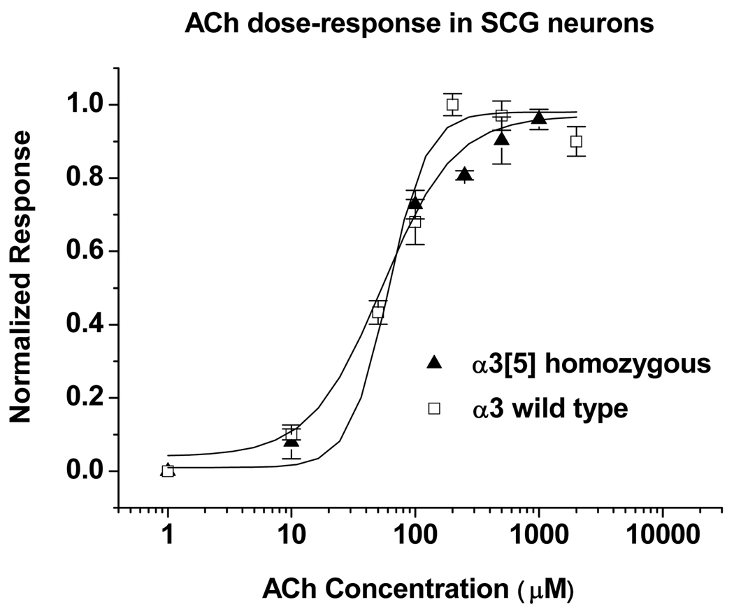
We report here the construction of a novel knock-in mouse expressing chimeric alpha3 nicotinic acetylcholine receptor (nAChR) subunits with pharmacological sensitivity to alpha-bungarotoxin (alphaBTX). Sensitivity was generated by substituting five amino acids in the loop C (beta9-beta10) region of the mouse alpha3 subunit with the corresponding residues from the alpha1 subunit of the muscle type receptor from Torpedo californica. To demonstrate the utility of the underlying concept, expressed alpha3[5] subunits were characterized in the superior cervical ganglia (SCG) of homozygous knock-in mice, where the synaptic architecture of postsynaptic alpha3-containing nAChR clusters could now, for the first time, be directly visualized and interrogated by live-staining with rhodamine-conjugated alphaBTX. Consistent with the postsynaptic localization of ganglionic nAChRs, the alphaBTX-labeled puncta colocalized with a marker for synaptic varicosities. Following in vivo deafferentation, these puncta persisted but with significant changes in intensity and distribution that varied with the length of the recovery period. Compound action potentials and excitatory postsynaptic potentials recorded from SCG of mice homozygous for alpha3[5] were abolished by 100 nmalphaBTX, even in an alpha7 null background, demonstrating that synaptic throughput in the SCG is completely dependent on the alpha3-subunit. In addition, we observed that the genetic background of various inbred and outbred mouse lines greatly affects the functional expression of alpha3[5]-nAChRs, suggesting a powerful new approach for exploring the molecular mechanisms underlying receptor assembly and trafficking. As alphaBTX-sensitive sequences can be readily introduced into other nicotinic receptor subunits normally insensitive to alphaBTX, the findings described here should be applicable to many other receptors.


Figures

Figure 1
Figure 1. Overview of the α3[5] knock-in targeting strategy and supporting genetic analysis
(A) A genetic map depicts significant landmarks in the WT Chrna3 allele, α3[5] targeting DNA, and recombinant allele before (Chrna3tm1.0Hwrt) and after (Chrna3tm1.1Hwrt) deletion of the neomycin cassette, along with anticipated regions of homologous recombination and restriction cleavage sites. WT exons are represented as solid rectangles. The mutated ExonV is shown as a hatched rectangle. (B) Southern analysis of targeted ES cell clone. A single band at ~17.5 kb is detected by the 3' probe in the WT control (left, lane 1). In targeted ES cell cone A6, the ~17.5 kb WT band is detected along with a second band at ~10.5 kb, the anticipated size of the 3’ KpnI fragment in the recombinant allele (left, lane 2). A single band is detected at ~17.5 kb in the WT sample with the 5' probe (right, lane 1). In targeted ES cell clone A6, the ~17.5 kb band appeared along with a second band at ~8.9 kb, the anticipated size of the 5' KpnI fragment in the mutant allele (right, lane 2). (C) Southern analysis of α3[5] knock-in mice. Following deletion of the neomycin cassette, the mutant allele is reduced by ~2 kb from 8.9 kb in an α3[5] heterozygote (+neo, lane 1) to 6.9 kb in an α3[5] homozygote (-neo, lane 2). (D) RT-PCR analysis of SCG tissue extract. cDNAs were prepared from WT (+/+), α3[5]-heterozygous mice (+/tm), and α3[5]-homozygous mice (tm/tm). The WT-specific primer amplified 480 bp PCR products from the α3[5] (+/tm) and WT samples, but not with cDNAs from the α3[5] (tm/tm). The α3[5]-specific primer amplified 480 bp PCR-products with cDNAs prepared from heterozygote and homozygote mice, and no bands are observable with WT cDNA. Chrna3tm1.1Hwrt mice (i.e., neo-deleted) on a mixed C57BL/6-129S background at age <P20 were used in the RT-PCR experiments.
Figure 2
Figure 2. Labeling of nicotinic α3[5]-containing receptors using rhodamine-conjugated αBTX in α3[5]-homozygous mice
(A) Rhodamine-labeled αBTX (rhoBTX, red) clusters colocalize with a majority of the staining for vesicular acetylcholine transporter (VAChT, green; see panel D below) in SCG from α3[5]-homozygous mice (left, α3tm/tm) but not in SCG from WT mice (right). Immunostaining for neurofilament (NFM, blue) shows the preganglionic axons. These results were seen in 5/5 mice and indicate that α3[5]-containing receptors are targeted to synapses. Inset shows regions of NFM-labeled axons that have VAChT-positive varicosities that colocalize with receptor clusters. (B) RhoBTX-labeled clusters (red) colocalize with postsynaptic density 93 (PSD93, green; see panel D below). All rhoBTX-clusters colocalize with PSD93, but many PSD93 puncta do not colocalize with detectable rhoBTX clusters.(C) RhoBTX-labeling is abolished by pre-incubating SCG from α3[5]-homozygous mice with unlabeled αBTX (100 nM, left), but is normal when SCG are pre-incubated with MLA (50 nM, middle). RhoBTX-labeled clusters present on SCG neurons from double homozygous (α3[5]-homozygous and α7 (−/−)) mice (right). Chrna3tm1.1Hwrt mice on a mixed C57BL/6-CD-1 background aged 2–3 months were used in these experiments. Scale bar is 2 µm for A, B, and C, and 0.5 µm for the insets in A. (D) Quantitation of puncta from images obtained in the studies described in panels A, B and C indicate that all rhoBTX labeled clusters are colocalized with VAChT and PSD93 labeled puncta. (E) Quantitative analysis of puncta from images obtained in the studies described in panels B and C indicate that ~60% of VAChT stained puncta have αBTX clusters associated with them and ~30% of the PSD93 stained puncta have αBTX clusters associated with them. (F) Quantitative analysis of αBTX labeled puncta obtained in the studies described in panels A, B, and C indicate no detectable puncta in WT α3 mice, but comparable densities of αBTX labeled puncta in α3[5]-homozygous mice in either the null α7 (−/−) genetic background or with wild-type α7 background.
Figure 3
Figure 3. Fast synaptic transmission in SCG from α3[5]-homozygous mice is blocked by αBTX
(A) Extracellular compound action potentials (CAP) recorded from the sympathetic trunk in 2-month old WT (top row) and a 2-month old α3[5]-homozygous mouse (bottom row); the traces on the right show CAPs recorded from the same ganglia in the presence of 100 nM αBTX. CAPs recorded from α3[5]-homozygous SCG are approximately 10–20% the amplitude of control and are completely blocked by αBTX. Similar results were seen in 12 WT mice and in 15 C57BL/6/CD-1 Chrna3tm1.1Hwrt homozygous mice greater than 2 months of age. (B) Nerve-evoked EPSPs recorded intracellularly from sympathetic neurons in 2-month old WT (top row), 2-month old C57BL/6/CD-1 Chrna3tm1.1Hwrt homozygous (middle row) and 5-month old homozygous double mutant (α3[5]-homozygous α7(−/−)) SCG (bottom row). Suprathreshold stimulation of the preganglionic nerve evokes small ~3mV EPSPs on sympathetic neurons from 5-month old α3[5]-homozygous SCG that are completely blocked by αBTX. Similar results were seen for 20 EPSPs from WT neurons and 30 EPSPs from α3[5]-homozygous SCG neurons dissected from mice greater than 2 months of age.
Figure 4
Figure 4. The dose-response characteristics of α3/α1[5]-containing receptors compared to WT α3 nicotinic receptors measured in cultured SCG neurons
Chrna3tm1.1Hwrt mice congenic in the C57BL/6 strain were crossed with BALB/cJ mice and resulting heterozygotes were mated in an F2 cross to prepare Chrna3tm1.1Hwrt homozygotes in the mixed C57BL/6/BALB/cJ background. WT α3 F2 littermates were used as the source of WT α3 for comparison with α3[5]-containing receptors. Dissociated SCG primary cultures were prepared and maintained at 37°C. Two-three days before the ACh dose-response study, the cultures were shifted to 30°C. The fitted curves yielded an EC50 of 48±2 µM for α3[5]-containing receptors and a value of 64±3 µM for WT α3 receptors. Data points shown are the mean ± SEM (n=3–4 per data point).
Figure 5
Figure 5. Time course of αBTX block and washout in cultured SCG neurons from Chrna3tm1.1Hwrt homozygous mice
Chrna3tm1.1Hwrt mice congenic in the C57BL/6 strain were crossed with BALB/cJ mice and resulting heterozygotes were mated in an F2 cross to prepare Chrna3tm1.1Hwrt homozygotes in the mixed C57BL/6/BALB/cJ background. WT α3 F2 littermates were used as the source of WT α3 for comparison with α3[5]-containing receptors. Dissociated SCG primary cultures were prepared and maintained at 37°C. Two-three days before recording, the cultures were shifted to 30°C. The data points show normalized peak currents evoked by brief (2 sec) application of 200 µM ACh. The current density of the ACh evoked responses in the SCG neurons prior to αBTX application was 11.8±2.1 pA/pF (n=3). Immediately after the first control data point was obtained, the cells were perfused with external solution containing 100 nM αBTX for 5 minutes. Current responses were recorded during this 5 minute time interval with a co-application of 200 µM ACh and 100 nM αBTX. At time point 0, the washout of the αBTX was begun, and test pulses of 200 µM ACh were applied to monitor recovery from αBTX block. Error bars indicate SEM, n=3.
Figure 6
Figure 6. ACh-evoked whole-cell currents in cultured SCG neurons from neonatal α3[5]-homozygous C57BL/6 mice are sensitive to αBTX block and their detection is facilitated by short term culture at 30°C
(A) Bar graph showing whole-cell current density measurements recorded from cultured SCG neurons from WT C57BL/6 mice (<P20). Reducing the incubation temperature to 30°C for 1 day has no significant effect on the mean current density in WT SCG neurons (middle, P=0.10). Under these conditions, the mean current density is unaffected following a 1 h incubation with 100 nM αBTX (right, P=0.74). Values represent the mean (n=15) and SEM from 3 mice (5 cells/mouse). (B) SCG neurons from α3[5]-homozygous mice congenic on C57BL/6 (<P20) are unresponsive to ACh application when cultured at 37°C (left). Following 1 day at 30°C, SCG neurons from α3[5]-homozygous mice generate ACh-evoked currents (note change in scale of y-axis) which are unaffected by the presence or absence of the α7 subunit (middle, P values >0.70). Under these conditions, the ACh responses are greatly decreased following 1 h incubation with 100 nM αBTX (right). No significant differences in the levels of αBTX block were observed among the three α7 genotypes (P values >0.1). Values represent the mean and SEM (n=5) from 1 mouse. (C) Data points representing ACh-evoked current densities in cultured SCG neurons either from α3[5]-homozygous mice (<P20) congenic on C57BL/6, from F2 mixed-background inbred (C3H, DBA, and BALB/cJ), or from outbred (ICR) strains. P values were calculated using an unpaired t test.
Figure 7
Figure 7. Time course of αBTX-labeled nAChR cluster size, average cluster intensity, and surface density in the SCG following preganglionic denervation
(A) Shows the size distribution of rhoBTX labeled receptor clusters in innervated α3[5]-homozygous SCG (n=5 ganglia). (B) Size distribution of rhoBTX labeled receptor clusters in α3[5]-homozygous SCG two days after denervation (n=5 ganglia). There is an increase in the number and the size of puncta. (C) Size distribution of rhoBTX labeled receptor puncta in α3[5]-homozygous SCG one week after denervation (n=5 ganglia). The number and the size of clusters one week following denervation are decreased slightly compared with control. (D) Size distribution of rhoBTX labeled receptor cluster in α3[5]-homozygous SCG two weeks after denervation (n=5 ganglia). Cluster number and size are markedly decreased two weeks following denervation. For A–D, The red lines are gaussian fits of the data, and the insets show representative images following live staining with rhoBTX and post-fix immunostaining for VAChT. Scale bars are 1 µm. (E) Mean cluster density and intensity (a.u.) at different times post denervation. (number of clusters counted for each time point is >1000). Chrna3tm1.1Hwrt mice on a mixed C57BL/6-CD-1 background were used in these experiments.

Similar articles

Cited by

References

    1. Bogdanov Y, Michels G, Armstrong-Gold C, Haydon PG, Lindstrom J, Pangalos M, Moss SJ. Synaptic GABAA receptors are directly recruited from their extrasynaptic counterparts. EMBO J. 2006;25:4381–4389. - PMC - PubMed
    1. Cangiano A. Denervation supersensitivity as a model for the neural control of muscle. Neuroscience. 1985;14:963–971. - PubMed
    1. Colquhoun D, Unwin N, Shelley C, Hatton C, Sivilotti L. Nicotinic acetylcholine receptors. In: Abraham D, editor. Burger's Medicinal Chemistry Vol II Drug Discovery and Drug Development. New York: Wiley; 2003. pp. 357–406.
    1. Cooper ST, Harkness PC, Baker ER, Millar NS. Up-regulation of cell-surface alpha4beta2 neuronal nicotinic receptors by lower temperature and expression of chimeric subunits. J Biol Chem. 1999;274:27145–27152. - PubMed
    1. Cuevas J, Roth AL, Berg DK. Two distinct classes of functional 7-containing nicotinic receptor on rat superior cervical ganglion neurons. J Physiol. 2000;525(Pt 3):735–746. - PMC - PubMed

Publication types

MeSH terms