. 2009 Dec 3;30(11):2112-20.
doi: 10.1111/j.1460-9568.2009.07001.x. Epub 2009 Nov 25.

Long-term synaptic plasticity is impaired in rats with lesions of the ventrolateral preoptic nucleus

Affiliations

Long-term synaptic plasticity is impaired in rats with lesions of the ventrolateral preoptic nucleus

Elda Arrigoni et al. Eur J Neurosci. .

Abstract

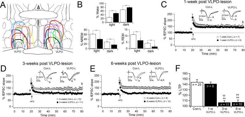
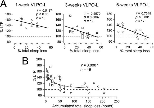
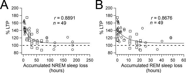
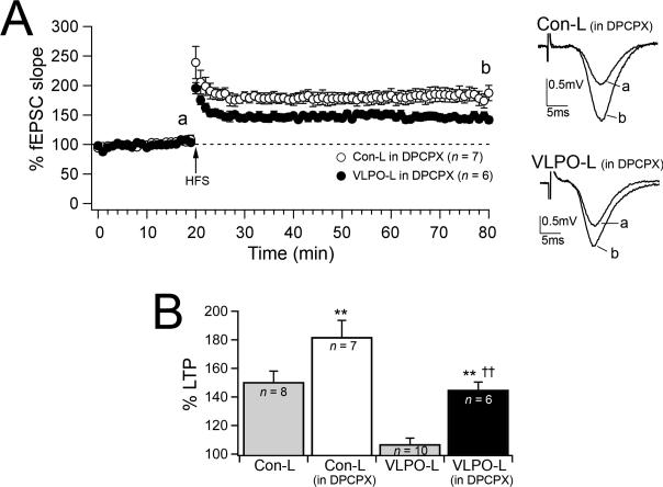
Impairment of memory functions has been frequently reported in models of sleep deprivation. Similarly, hippocampal long-term synaptic plasticity has been shown to be sensitive to sleep loss caused by acute sleep restriction. However, such approaches are limited by the stressful nature of sleep deprivation, and because it is difficult to study long-term sleep restriction in animals. Here, we report the effects of chronic sleep loss on hippocampal long-term potentiation (LTP) in a rodent model of chronic partial sleep deprivation. We studied LTP of the Schaffer collateral-CA1 synapses in hippocampal slices prepared from rats with lesions of the ventrolateral preoptic nucleus (VLPO), which suffered reductions in total sleep time for several weeks after lesions. In slices prepared from VLPO-lesioned rats, LTP was impaired proportionally to the amount of sleep loss, and the decline in LTP followed a single exponential function over the amount of accumulated sleep debt. As compared with sham-lesioned controls, hippocampal slices from VLPO-lesioned rats showed a greater response to adenosine antagonists and greater paired-pulse facilitation (PPF). However, exogenous adenosine depressed evoked synaptic transmission and increased PPF in VLPO-lesioned and sham-lesioned rats by equal amounts, suggesting that the greater endogenous adenosine inhibitory tone in the VLPO-lesioned rats is associated with greater ligand accumulation rather than a change in adenosine receptor sensitivity or adenosine-mediated neurotransmitter release probability. LTP in VLPO-lesioned animals was partially restored by adenosine antagonists, suggesting that adenosine accumulation in VLPO-lesioned animals could account for some of the observed synaptic plasticity deficits.


Figures

Figure 1
Figure 1
Hippocampal LTP is impaired in VLPO-lesioned animals. A, Camera lucida drawings illustrating 10 examples of lesions in the VLPO region. B, Graphs represent the % of wake, NREM sleep and REM sleep during the light (7:00 – 19:00) and dark (19:00 7:00) cycles in the Con-L (white bars) and the VLPO-L (black bars) rats used for the LTP experiments in panels C-F. Con-L (n = 13 rats) and VLPO-L (n = 16 rats), ** p < 0.01 unpaired t test. C, D, E, Graphs compare CA1-schaffer collateral LTP in slices prepared from VLPO-L (total sleep loss > 20%) and Con-L at three time points: 1-week, 3-weeks and 6-weeks post VLPO-lesions and post sham-lesions. Data are represented as mean ± S.E.M. percent changes in fEPSP slope before and after high frequency stimulation (HFS, 100 pulses at 100 Hz) and n = number of recordings. Representative fEPSCs (average of 6 consecutive traces) were recorded over 2 minute periods before HSF (a) and 60 minutes after HSF (b). F, Summarized results of mean percent changes in fEPSP slope (60 min after HFS) in slices from control-lesioned animals (Con-L = grouped data from 1-week, 3-week and 6-week Con-L animals; one-way ANOVA between Con-L groups, F = 0.43, p = 0.654) and 1-week, 3-week and 6-week VLPO lesioned animals (one-way ANOVA test between animal groups: F = 17.5, p < 0.001; Fisher's PLSD: ** Con-L > 3-W VLPO-L, p < 0.01 and Con-L > 6-week VLPO-L, p < 0.01; †† 1-W VLPO-L > 3-W VLPO-L, p < 0.01; and 1-W VLPO-L > 6-week VLPO-L, p < 0.01).
Figure 2
Figure 2
LTP is impaired in proportion to the amount of sleep loss and LTP progressively decays with the accumulated sleep loss. A, Correlation of the percent LTP (% change of fEPSP slope 60 min after HFS) vs % daily total sleep loss (NREM + REM sleep) across animals of the three animal groups (1-week, 3-week, and 6-week VLPO-L rats). Regression analysis shows a strong negative correlation between the amount of sleep loss and percentage of hippocampal LTP in the 3-week VLPO-L rats and the 6-week VLPO-L rats, but no statistically significant correlation in the 1-week VLPO-L rats. B, The graph represents percent LTP (% change of fEPSP slope 60 min after HFS) vs accumulated total sleep loss (see Materials and Methods) from all VLPO-lesioned animals (triangles = 1-week VLPO-L; square = 3-week VLPO-L and circles = 6-week VLPO-L animals). Data was fit with a single exponential function (fitting parameters ± standard deviation: A = 45.7 ± 5.8%; τ = 33.4 ± 11.4 h; C = 104.8 ± 4.7%).
Figure 3
Figure 3
LTP declines as a single exponential function over both accumulated NREM and REM sleep losses. A, Percent LTP (% change of fEPSP slope 60 min after HFS) vs accumulated NREM sleep loss (see Materials and Methods) from all VLPO-lesioned animals (triangles = 1-week VLPO-L; square = 3-week VLPO-L and circles = 6-week VLPO-L animals). Data was fit with a single exponential function (fitting parameters ± standard deviation: A = 45.7 ± 5.7%; τ = 22.3 ± 7.7 h; C = 106.1 ± 4.3%). B, Percent LTP vs accumulated REM sleep loss (triangles = 1-week VLPO-L; square = 3-week VLPO-L and circles = 6-week VLPO-L animals). Data was fit with a single exponential function (fitting parameters ± standard deviation: A = 34.9 ± 7.6%; τ = 9.2 ± 5.9 h; C = 105.8 ± 7.4%).
Figure 4
Figure 4
Hippocampal slices from VLPO-lesioned animals are under greater inhibitory tone by endogenous adenosine. A1-2, The adenosine A1 antagonist DPCPX (400 nM) increases fEPSC slope and this effect is greater in slices prepared from VLPO-L rats (6-weeks VLPOL) compared to Con-L rats (6-weeks Con-L). A1, Representative responses to DPCPX in Con-L and VLPO-L animals (average of 6 consecutive fEPSCs recorded at the end of: 10 min in control ACSF; “Con” and 20 min in DPCPX). A2, Shows the time course of the response to DPCPX (100% is the averaged fEPSP slopes during 15 min preceding DPCPX). B1-2, Paired-pulse facilitation mediated by endogenous adenosine is greater in VLPO-lesioned animals compared to control lesioned animals. B1, Representative fEPSCs (paired-pulses test: 40 msec interstimulus interval) recorded before and during DPCPX. Scaled traces to match fEPSC (1st pulses) are shown on the right. B2, Shows the averaged results of PPR in Con-L and VLPO-L animals recorded in control ACSF and in DPCPX (one-way ANOVA test between animal groups and treatments: F = 30.6, p < 0.001; Fisher's PLSD: ** VLPO-L > Con-L, p < 0.001; †† Con-L > Con-L in DPCPX, p < 0.001; ‡‡ VLPO-L > VLPO-L in DPCPX, p < 0.001). C1-3, Effects of exogenous adenosine on fEPSC slope and PPF in slices from Con-L and VLPO-L rats. C1 shows fEPSCs (paired-pulse test: 50 msec interstimulus interval) recorded before (Con) and during adenosine 20 μM (AD). C2-3, Two graphs showing the effects of exogenous adenosine on fEPSC slope and PPF in slices from Con-L and VLPO-L rats. There is no statistically significant difference in the response to adenosine between Con-L and VLPO-L rats (AD-inhibition: p = 0.59, unpaired t test; AD-PPR: p = 0.75, unpaired t test). All data are represented as mean ± S.E.M. (n = number of recordings).
Figure 5
Figure 5
Effects of blocking adenosine A1 receptors. A, LTP in the presence of DPCPX (400 nM) in slices from Con-L and VLPO-L rats (6-week Con-L and 6-week VLPO-L rats; mean ± S.E.M. percent changes in fEPSP slope before and after HFS; n = number of recordings). Representative fEPSCs (average of 6 consecutive traces) in slices from Con-L and VLPO-L rats recorded in the presence of DPCPX (400 nM) before HSF (a) and 60 minutes after HSF (b). B, Summarized results of mean percent change in fEPSP slope (60 min after HFS) in slices from Con-L and VLPO-L rats recorded in control ACSF and in the presence of DPCPX (one-way ANOVA test between animal groups and treatments: F = 19.2, p < 0.001; Fisher's PLSD: ** Con-L (in DPCPX) > Con-L, p = 0.006 and VLPO-L (in DPCPX) > VLPO-L, p =0.001; †† Con-L (in DPCPX) > VLPO-L (in DPCPX), p = 0.003).

References

    1. Alanko L, Heiskanen S, Stenberg D, Porkka-Heiskanen T. Adenosine kinase and 5'-nucleotidase activity after prolonged wakefulness in the cortex and the basal forebrain of rat. Neurochem Int. 2003a;42:449–454. - PubMed
    1. Alanko L, Stenberg D, Porkka-Heiskanen T. Nitrobenzylthioinosine (NBMPR) binding and nucleoside transporter ENT1 mRNA expression after prolonged wakefulness and recovery sleep in the cortex and basal forebrain of rat. J Sleep Res. 2003b;12:299–304. - PubMed
    1. Arai A, Kessler M, Lynch G. The effects of adenosine on the development of long-term potentiation. Neurosci Lett. 1990;119:41–44. - PubMed
    1. Arai A, Lynch G. Factors regulating the magnitude of long-term potentiation induced by theta pattern stimulation. Brain Res. 1992;598:173–184. - PubMed
    1. Arrigoni E, Rosenberg PA. Nitric oxide-induced adenosine inhibition of hippocampal synaptic transmission depends on adenosine kinase inhibition and is cyclic GMP independent. Eur J Neurosci. 2006;24:2471–2480. - PubMed

Publication types

MeSH terms