Review
. 2010 Feb;9(1):1-20.

Unconventional nuclides for radiopharmaceuticals

Affiliations
Review

Unconventional nuclides for radiopharmaceuticals

Jason P Holland et al. Mol Imaging. 2010 Feb.

Abstract

Rapid and widespread growth in the use of nuclear medicine for both diagnosis and therapy of disease has been the driving force behind burgeoning research interests in the design of novel radiopharmaceuticals. Until recently, the majority of clinical and basic science research has focused on the development of 11C-, 13N-, 15O-, and 18F-radiopharmaceuticals for use with positron emission tomography (PET) and 99mTc-labeled agents for use with single-photon emission computed tomography (SPECT). With the increased availability of small, low-energy cyclotrons and improvements in both cyclotron targetry and purification chemistries, the use of "nonstandard" radionuclides is becoming more prevalent. This brief review describes the physical characteristics of 60 radionuclides, including beta+, beta-, gamma-ray, and alpha-particle emitters, which have the potential for use in the design and synthesis of the next generation of diagnostic and/or radiotherapeutic drugs. As the decay processes of many of the radionuclides described herein involve emission of high-energy gamma-rays, relevant shielding and radiation safety issues are also considered. In particular, the properties and safety considerations associated with the increasingly prevalent PET nuclides 64Cu, 68Ga, 86Y, 89Zr, and 124I are discussed.


Figures

Figure 1
Figure 1
Pictures of (A) a 68Ge/68Ga generator; (B) the focusing magnets; and (C) the multihead target assembly of the Memorial Sloan-Kettering Cancer Center EBCO TR19/9 cyclotron and (D) the core of the University of Missouri Research Reactor (MURR). Courtesy of David Nickolaus, MURR Center.
Figure 2
Figure 2
Global supply of 99Mo in 2009. Source: http://www.iaea.org/worldatom/rrdb/.
Figure 3
Figure 3
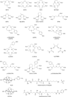
Structures of common direct and conjugate-based (prosthetic groups based on Bolton-Hunter compounds) reagents used in the radiohalogenation of small molecules, peptides, and antibodies with Br, I, and At radionuclides. In particular, the reactions with tyrosine and histidine amino acid side chains are shown.
Figure 4
Figure 4
Selected bifunctional chelates often employed in conjugate labeling strategies.,–
Figure 5
Figure 5
Structure of the DOTA-TOC ligand used in the preparation of the somatostatin analogue [68Ga]-DOTA-TOC.
Figure 6
Figure 6
Images of a 56-year-old woman with multiple liver and lymph node metastases. The patient was referred for restaging after surgery and chemotherapy. The CT image shows the presence of liver and lymph node metastases but was negative for bone lesions. A, [68Ga]-DOTA-TOC showed all the visceral metastases, as well as additional osteoblastic and osteolytic bone metastases. Conventional scintigraphy, (B) anterior and (C) posterior views, failed to delineate the majority of bone metastases. Osteoblastic bone lesions were confirmed by [18F]-NaF PET (D). Retrospective CT analysis after image fusion revealed some but not all of these bone metastases. Reprinted by permission of the Society of Nuclear Medicine from: Gabriel M, Decristoforo C, Kendler D, et al. 68Ga-DOTA-Tyr3-octreotide PET in neuroendocrine tumors: comparison with somatostatin receptor scintigraphy and CT. J Nucl Med 2007;48:508–18. Figure 2.
Figure 7
Figure 7
Stack plot showing the γ-ray emission spectra of the positron-emitting radionuclides 18F, 86Y, 124I, and 89Zr. Details of the emission characteristics are presented in Table 1 and Table 3.
Figure 8
Figure 8
Immuno-PET images with [89Zr]-DFO-U36 of a head and neck cancer patient with a tumor in the left tonsil (large arrow) and lymph node metastases (small arrows) at the left (levels II and III) and right (level 11) sides of the neck. Images were obtained 72 hours postinjection. A, Sagittal; B, transaxial; and C, coronal image. Reprinted with permission from the American Association for Cancer Research: Borjesson PKE, Jauw YWS, Boellaard R, et al. Performance of immunopositron emission tomography with zirconium-89-labeled chimeric monoclonal antibody U36 in the detection of lymph node metastases in head and neck cancer patients. Clin Cancer Res 2006;12:2133–40. Figure 2.

References

    1. Pagani M, Stone-Elander S, Larsson SA. Alternative positron emission tomography with non-conventional positron emitters: effects of their physical properties on image quality and potential clinical applications. Eur J Nucl Med. 1997;24:1301–1327. - PubMed
    1. Welch MJ, Kilbourn MR, Green MA. Radiopharmaceuticals labeled with short-lived positron-emitting radionuclides. Radioisotopes. 1985;34:170–179. - PubMed
    1. McQuade R, Rowland DJ, Lewis JS, Welch MJ. Positron-emitting isotopes produced on biomedical cyclotrons. Curr Med Chem. 2005;12:807–818. - PubMed
    1. Blower P. Towards molecular imaging and treatment of disease with radionuclides: the role of inorganic chemistry. Dalton Trans. 2006:1705–1711. - PubMed
    1. Lewis JS, Singh RK, Welch MJ. Long lived and unconventional PET radionuclides. In: Pomper MG, Gelovani JG, editors. Molecular imaging in oncology. New York: Informa Healthcare USA Inc; 2009. pp. 283–292.

Publication types

MeSH terms