

Different strategies for reducing intestinal background radioactivity associated with imaging HSV1-tk expression using established radionucleoside probes
- PMID: 20128998
- PMCID: PMC3068838
Different strategies for reducing intestinal background radioactivity associated with imaging HSV1-tk expression using established radionucleoside probes
Abstract
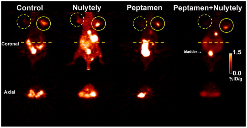
One limitation of HSV1-tk reporter positron emission tomography (PET) with nucleoside analogues is the high background radioactivity in the intestine. We hypothesized that endogenous expression of thymidine kinase in bacterial flora could phosphorylate and trap such radiotracers, contributing to the high radioactivity levels in the bowel, and therefore explored different strategies to increase fecal elimination of radiotracer. Intestinal radioactivity was assessed by in vivo microPET imaging and ex vivo tissue sampling following intravenous injection of 18F-FEAU, 124I-FIAU, or 18F-FHBG in a germ-free mouse strain. We also explored the use of an osmotic laxative agent and/or a 100% enzymatically hydrolyzed liquid diet. No significant differences in intestinal radioactivity were observed between germ-free and normal mice. 18F-FHBG-derived intestinal radioactivity levels were higher than those of 18F-FEAU and 124I-FIAU; the intestine to blood ratio was more than 20-fold higher for 18F-FHBG than for 18F-FEAU and 124I-FIAU. The combination of Peptamen and Nulytely lowered intestinal radioactivity levels and increased (2.2-fold) the HSV1-tk transduced xenograft to intestine ratio for 18F-FEAU. Intestinal bacteria in germ-free mice do not contribute to the high intestinal levels of radioactivity following injection of radionucleoside analogues. The combination of Peptamen and Nulytely increased radiotracer elimination by increasing bowel motility without inducing dehydration.
Figures


Similar articles
-
Comparison of [18F]FHPG and [124/125I]FIAU for imaging herpes simplex virus type 1 thymidine kinase gene expression.Eur J Nucl Med. 2001 Jun;28(6):721-9. doi: 10.1007/s002590100526. Eur J Nucl Med. 2001. PMID: 11440032
-
Molecular-genetic PET imaging using an HSV1-tk mutant reporter gene with enhanced specificity to acycloguanosine nucleoside analogs.J Nucl Med. 2009 Mar;50(3):409-16. doi: 10.2967/jnumed.108.058735. Epub 2009 Feb 17. J Nucl Med. 2009. PMID: 19223410
-
Semiautomated radiosynthesis and biological evaluation of [18F]FEAU: a novel PET imaging agent for HSV1-tk/sr39tk reporter gene expression.Mol Imaging Biol. 2008 Mar-Apr;10(2):82-91. doi: 10.1007/s11307-007-0122-3. Epub 2007 Dec 22. Mol Imaging Biol. 2008. PMID: 18157580 Free PMC article.
-
1-(2'-Deoxy-2'-[18F]fluoro-β-d-arabinofuranosyl)-5-iodocytosine.2012 Mar 19 [updated 2012 Apr 11]. In: Molecular Imaging and Contrast Agent Database (MICAD) [Internet]. Bethesda (MD): National Center for Biotechnology Information (US); 2004–2013. 2012 Mar 19 [updated 2012 Apr 11]. In: Molecular Imaging and Contrast Agent Database (MICAD) [Internet]. Bethesda (MD): National Center for Biotechnology Information (US); 2004–2013. PMID: 22514809 Free Books & Documents. Review.
-
2'-Fluoro-2'-deoxy-5'-[123/124/125/131I]iodo-1β-d-arabinofuranosyluracil.2005 Feb 1 [updated 2005 Feb 9]. In: Molecular Imaging and Contrast Agent Database (MICAD) [Internet]. Bethesda (MD): National Center for Biotechnology Information (US); 2004–2013. 2005 Feb 1 [updated 2005 Feb 9]. In: Molecular Imaging and Contrast Agent Database (MICAD) [Internet]. Bethesda (MD): National Center for Biotechnology Information (US); 2004–2013. PMID: 20641413 Free Books & Documents. Review.
Cited by
-
The sodium iodide symporter (NIS) as an imaging reporter for gene, viral, and cell-based therapies.Curr Gene Ther. 2012 Feb 1;12(1):33-47. doi: 10.2174/156652312799789235. Curr Gene Ther. 2012. PMID: 22263922 Free PMC article. Review.
-
Cre/lox-assisted non-invasive in vivo tracking of specific cell populations by positron emission tomography.Nat Commun. 2017 Sep 5;8(1):444. doi: 10.1038/s41467-017-00482-y. Nat Commun. 2017. PMID: 28874662 Free PMC article.
-
Repurposing 18F-FMISO as a PET tracer for translational imaging of nitroreductase-based gene directed enzyme prodrug therapy.Theranostics. 2021 Apr 7;11(12):6044-6057. doi: 10.7150/thno.55092. eCollection 2021. Theranostics. 2021. PMID: 33897898 Free PMC article.
-
Cell tracking in cardiac repair: what to image and how to image.Eur Radiol. 2012 Jan;22(1):189-204. doi: 10.1007/s00330-011-2190-7. Epub 2011 Jul 7. Eur Radiol. 2012. PMID: 21735069 Free PMC article. Review.
-
Bacteroides ovatus ATCC 8483 monotherapy is superior to traditional fecal transplant and multi-strain bacteriotherapy in a murine colitis model.Gut Microbes. 2019;10(4):504-520. doi: 10.1080/19490976.2018.1560753. Epub 2019 Jan 21. Gut Microbes. 2019. PMID: 30663928 Free PMC article.
References
-
- Alauddin MM, Shahinian A, Park R, et al. In vivo evaluation of 2′-deoxy-2′-[(18)F]fluoro-5-iodo-1-beta-D-arabinofuranosyluracil ([18F]FIAU) and 2′-deoxy-2′-[18F]fluoro-5-ethyl-1-beta-D-arabinofuranosyluracil ([18F]FEAU) as markers for suicide gene expression. Eur J Nucl Med Mol Imaging. 2007;34:822–9. doi: 10.1007/s00259–006–0305–1. - DOI - PubMed
-
- Tjuvajev JG, Doubrovin M, Akhurst T, et al. Comparison of radiolabeled nucleoside probes (FIAU, FHBG, and FHPG) for PET imaging of HSV1-tk gene expression. J Nucl Med. 2002;43:1072–83. - PubMed
Publication types
MeSH terms
Substances
Grants and funding
LinkOut - more resources
Full Text Sources