. 2010 Jan 19;17(1):53-64.
doi: 10.1016/j.ccr.2009.11.021.

IAP regulation of metastasis

Affiliations

IAP regulation of metastasis

Swarna Mehrotra et al. Cancer Cell. .

Abstract

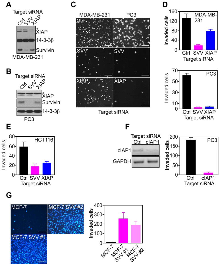
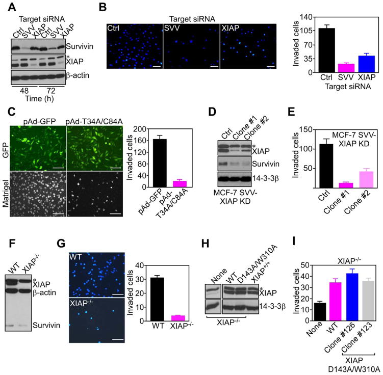
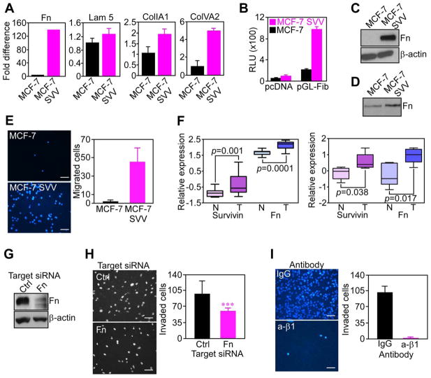
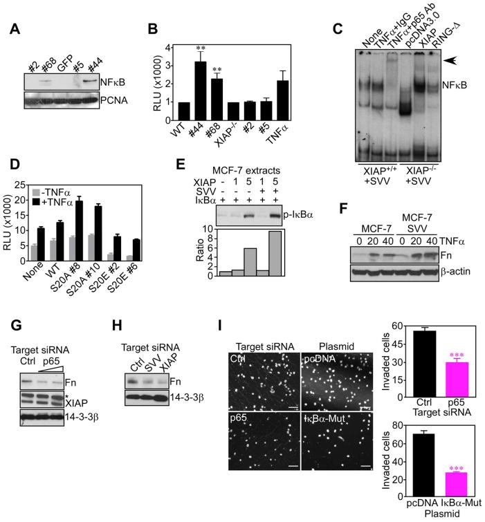
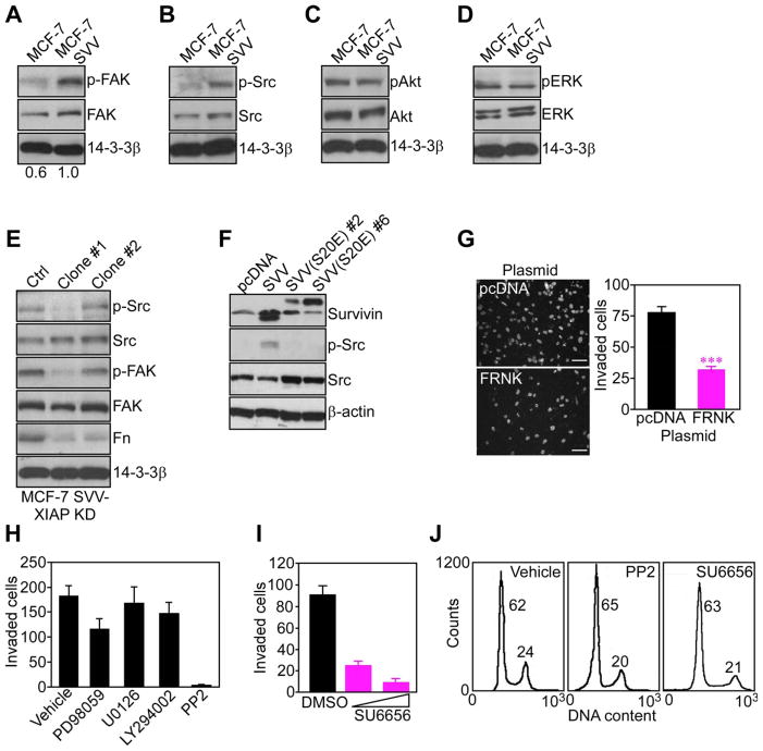
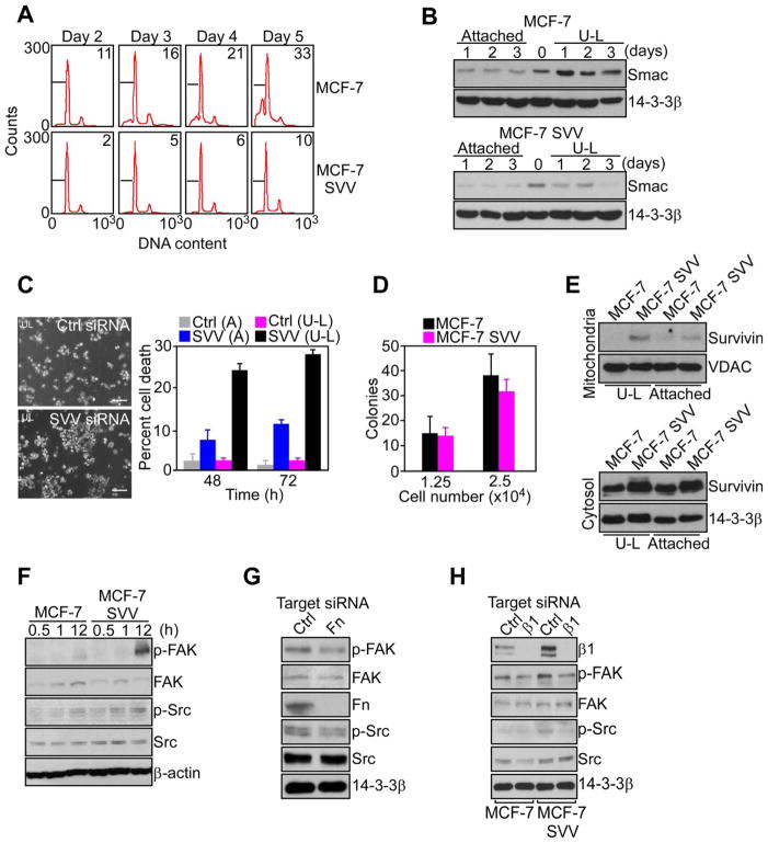
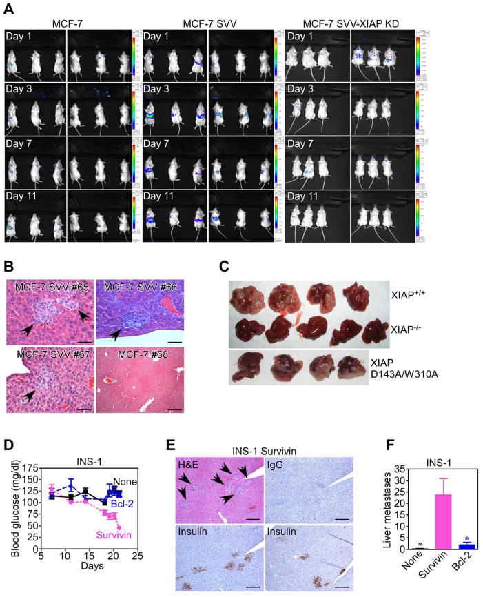
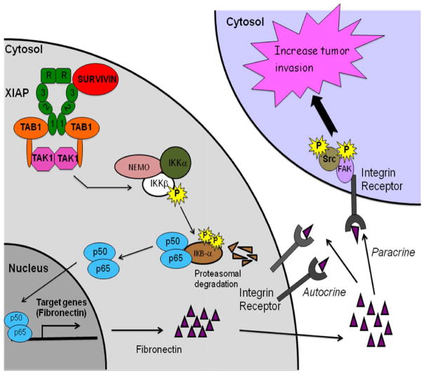
Inhibitor-of-Apoptosis (IAP) proteins contribute to tumor progression, but the requirements of this pathway are not understood. Here, we show that intermolecular cooperation between XIAP and survivin stimulates tumor cell invasion and promotes metastasis. This pathway is independent of IAP inhibition of cell death. Instead, a survivin-XIAP complex activates NF-kappaB, which in turn leads to increased fibronectin gene expression, signaling by beta1 integrins, and activation of cell motility kinases FAK and Src. Therefore, IAPs are direct metastasis genes, and their antagonists could provide antimetastatic therapies in patients with cancer.


Conflict of interest statement

CONFLICT OF INTEREST

The authors declare that they have no competing interest.

Figures

Figure 1
Figure 1. IAP-mediated tumor cell invasion
(A, B) Breast adenocarcinoma MDA-MB-231 (A) or prostate adenocarcinoma PC3 (B) cells transfected with control (Ctrl) or survivin (SVV)- or XIAP-directed siRNA, were analyzed by Western blotting after 48 h. *, non-specific. (C–E) The indicated siRNA transfected cell types were analyzed for invasion through Matrigel-coated Transwell inserts after 6 h by DAPI staining (C), and quantified (D). Scale bars, 200 μm (MDA-MB-231), 100 μm (PC3). (F) PC3 cells were transfected with control (Ctrl) or cIAP1-directed siRNA, and analyzed by PCR (left) and Matrigel invasion after 6 h (right). (G) MCF-7 or MCF-7 SVV cells (clones #1 and #2) were analyzed for Matrigel invasion after 6 h by DAPI staining (left), and quantified (right). Scale bars, 200 μm. For panels D–G data are the mean±SD of duplicates of a representative experiment out of at least two independent determinations. See also Figure S1.
Figure 2
Figure 2. IAP targeting inhibits tumor cell invasion
(A) MCF-7 SVV cells transfected with control (Ctrl) or survivin (SVV)- or XIAP-directed siRNA were analyzed by Western blotting. (B) MCF-7 SVV cells (clone #1) transfected with the indicated siRNA were analyzed for Matrigel invasion after 6 h by DAPI staining (left), and quantified (right). Scale bars, 200 μm. (C) MCF-7 SVV cells were transduced with pAd-GFP or pAd-T34A/C84A survivin mutant, and imaged after 24 h for GFP expression (top), or DAPI staining of Matrigel invasion (bottom). Invaded cells were quantified after 6 h (right). Scale bars, 200 μm. (D, E) MCF-7 SVV cells with stable shRNA knockdown of XIAP (MCF-7 SVV-XIAP KD; clones #1 and #2) were analyzed by Western blotting (D), and quantified for Matrigel invasion after 6 h (E). (F, G) Wild type (WT) or XIAP−/− HCT116 cells were analyzed by Western blotting (F), and characterized for Matrigel invasion after 6 h by DAPI staining (G, left), and quantified (G, right). Scale bars, 200 μm. (H) XIAP−/− HCT116 cells were stably transfected with WT XIAP or D143A/W310A XIAP mutant, and analyzed by Western blotting. Wild type (XIAP+/+) HCT116 cells were used as a control. (I) XIAP−/− HCT116 cells stably expressing WT XIAP or XIAP D143A/W310A mutant (clones #123 and #126) were quantified for Matrigel invasion after 6 h by DAPI staining. For panels B, C, E, G, I, data are the mean±SD of duplicates of a representative experiment out of at least two independent determinations. See also Figure S2.
Figure 3
Figure 3. IAP induction of fibronectin
(A) RNA was amplified for the indicated gene products, and normalized to GAPDH expression. Fn, fibronectin; Lam 5, laminin 5; CollA1, collagen A1; Col IVA2, collagen IV A2. (B) Cells transfected with pcDNA or a 1.9 kb fibronectin promoter-luciferase construct (pGL-Fib) were analyzed after 24 h for β-galactosidase-normalized luciferase activity. RLU, relative luciferase units. (C) Cell extracts were analyzed by Western blotting. (D) Conditioned media collected after 48 h was normalized per cell number and analyzed by Western blotting. (E) Cell migration on uncoated Transwell inserts was analyzed by DAPI staining (left), and quantified (right). Scale bars, 200 μm. (F) Microarray datasets of two breast cancer patient series were analyzed for expression of survivin and fibronectin (Fn) in tumor (T) versus normal (N) tissues. (G) MCF-7 SVV cells transfected with control (Ctrl) or fibronectin (Fn)-directed siRNA were analyzed after 48 h by Western blotting. Scale bars, 200 μm. (H) siRNA-transfected cells as in G were analyzed for Matrigel invasion after 6 h by DAPI staining (left), and quantified (right). Scale bars, 200 μm. ***, p=0.0006. (I) MCF-7 SVV cells were incubated with non-binding IgG or a function-blocking antibody to β1 integrin (β1), and characterized for Matrigel invasion after 6 h by DAPI staining (left), and quantified (right). Scale bars, 200 μm. For panels A, B, E, H, I, data are the mean±SD of duplicates of a representative experiment out of at least two independent determinations. For panel F, data are shown as box plots, where whiskers are the minimum and maximum, and the box is the upper and lower quartile. See also Figure S3.
Figure 4
Figure 4. NFκB induction of fibronectin mediates IAP tumor cell invasion
(A) Nuclear extracts from WT (clones #44 and #68) or XIAP−/− (clones #2 and #5) MEF stably transfected with survivin were analyzed by Western blotting. (B) Survivin-expressing WT or XIAP−/− clones as in (A) were analyzed for β-galactosidase-normalized NFκB-luciferase reporter activity. TNFα-stimulated cells were used as a control. **, p=0.006. (C) Nuclear extracts from WT or XIAP−/− clones expressing survivin as in (A) were incubated with a 32P-labeled NFκB probe in the presence of IgG or an antibody to p65 NFκB, followed by autoradiography. In reconstitution experiments, XIAP−/− cells were transfected with wild type XIAP or RING-less XIAP (RING-Δ). (D) INS-1 clones stably transfected with the indicated survivin mutant were analyzed for β-galactosidase-normalized NFκB luciferase promoter activity, with or without TNFα. For panels B and E, RLU, relative luciferase units. (E) Extracts from MCF-7 cells incubated with recombinant survivin (SVV), XIAP, or IκBα were analyzed by Western blotting. Bottom, densitometric quantification of protein bands. (F) Unstimulated or TNFα (ng/ml)-stimulated cells were analyzed by Western blotting. (G) MCF-7 SVV cells transfected with control (Ctrl) or p65 NFκB-directed siRNA were analyzed after 48 h by Western blotting. *, non-specific. (H) MCF-7 SVV cells transfected with control (Ctrl), survivin (SVV)- or XIAP-directed siRNA were analyzed by Western blotting. (I) MCF-7 SVV cells transfected with control (Ctrl) or p65 NFκB-directed siRNA, or, alternatively, pcDNA or IκBα mutant (IκBα-Mut), were analyzed for Matrigel invasion after 6 h by DAPI staining (left), and quantified (right). Scale bars, 200 μm. ***, p<0.0001. Mean±SD of duplicates of a representative experiment out of at least two independent determinations. For panels B, D, data are the mean±SEM of triplicates of two independent determinations. See also Figure S4.
Figure 5
Figure 5. IAP activation of cell motility kinases
(A-D) Adherent cells were analyzed after 48 h by Western blotting for phosphorylation/expression of FAK (A), Src (B), Akt (C), or ERK1,2 (D). (E) MCF-7 SVV cells with stable shRNA knockdown of XIAP (MCF-7 SVV-XIAP KD; clones #1 and #2) were analyzed by Western blotting. (F) INS-1 cells stably transfected with wild type survivin (SVV) or survivin S20E mutant (clones #2 and #6) were analyzed by Western blotting. (G) MCF-7 SVV cells transfected with pcDNA or FAK dominant negative FRNK mutant were analyzed for Matrigel invasion after 6 h by DAPI staining (left), and quantified (right). Scale bars, 200 μm. ***, p<0.0001. (H) MCF-7 SVV cells treated with the indicated pharmacologic inhibitors were quantified for Matrigel invasion after 6 h. (I) MCF-7 SVV cells treated with the Src inhibitor SU6656 (25–50 μM) were quantified for Matrigel invasion after 6 h. (J) MCF-7 SVV cells were incubated with vehicle (DMSO) or the indicated Src inhibitors, and analyzed for DNA content after 24 h by propidium iodide staining and flow cytometry. The percentage of cells in the G1 or G2/M phase of the cell cycle is indicated. For panels G, H, I, data are the mean±SD of duplicates of a representative experiment out of at least two independent determinations. See also Figure S5.
Figure 6
Figure 6. IAP suppression of anoikis
(A) Cells were maintained in suspension using ultra-low (U-L) attachment plates, and analyzed at the indicated time intervals by propidium iodide staining and flow cytometry. The percentage of cells with hypodiploid (apoptotic) DNA content is indicated. (B) Cytosolic extracts of cultures maintained attached or in suspension (U-L) were analyzed by Western blotting. (C) Cultures maintained attached (A) or in suspension (U-L) were transfected with control (Ctrl) or survivin (SVV)-directed siRNA, and analyzed by phase contrast microscopy (left) and quantified for nuclear morphology of apoptosis after 48 h (right). Scale bars, 200 μm. (D) Cells were plated at the indicated numbers in semi-solid medium, and colonies were scored after 2 weeks by light microscopy. (E) Mitochondria (top) or cytosolic (bottom) extracts of cultures attached or in suspension (U-L) were analyzed by Western blotting. (F) Cultures in suspension were analyzed by Western blotting. (G) MCF-7 SVV cells transfected with control (Ctrl) or fibronectin (Fn)-directed siRNA in suspension were analyzed by Western blotting. (H) Cells transfected with control (Ctrl) or β1 integrin (β1)-directed siRNA were analyzed by Western blotting. For panels C, D, data are the mean±SD of duplicates of a representative experiment out of at least two independent determinations. See also Figure S6.
Figure 7
Figure 7. IAP-mediated metastasis, in vivo
(A) MCF-7, MCF-7 SVV or MCF-7 SVV cells carrying stable knockdown of XIAP (MCF-7 SVV-XIAP KD) stably transfected with luciferase were injected in the spleen of SCID/beige mice, and analyzed for metastatic tumor growth at the indicated time intervals, by bioluminescence imaging. (B) Livers from representative animals (#) reconstituted as indicated were harvested at d. 11, and analyzed by hematoxylin-eosin (H&E) staining and light microscopy. Arrows, metastatic foci. Scale bars, 100 μm, 500 μm. (C) XIAP+/+, XIAP−/− or XIAP D143A/W310A mutant HCT116 cells were injected in the spleen of SCID/beige mice, and resected livers were examined macroscopically after 2 weeks. (D) SCID/beige mice reconstituted with INS-1 cells stably transfected with survivin or Bcl-2 were analyzed for blood glucose levels. (E) Livers of animals reconstituted with INS-1 cells transfected with survivin were stained by H&E or insulin. IgG, non binding antibody. Arrows, metastatic foci. Scale bars, 500 μm. (F) Metastatic foci of INS-1 cells were counted blindly in four independent microscopy fields of serial liver sections harvested from the indicated animal groups. *, p=0.017–0.025. For panels D, F, data are the mean±SEM. See also Figure S7.
Figure 8
Figure 8. Schematic mechanistic model of IAP-induced metastasis
See text for details.

References

    1. Altieri DC. Survivin, cancer networks and pathway-directed drug discovery. Nat Rev Cancer. 2008;8:61–70. - PubMed
    1. Birkey Reffey S, Wurthner JU, Parks WT, Roberts AB, Duckett CS. X-linked inhibitor of apoptosis protein functions as a cofactor in transforming growth factor-beta signaling. J Biol Chem. 2001;276:26542–26549. - PubMed
    1. Bittner M, Meltzer P, Chen Y, Jiang Y, Seftor E, Hendrix M, Radmacher M, Simon R, Yakhini Z, Ben-Dor A, et al. Molecular classification of cutaneous malignant melanoma by gene expression profiling. Nature. 2000;406:536–540. - PubMed
    1. Bouchard V, Harnois C, Demers MJ, Thibodeau S, Laquerre V, Gauthier R, Vezina A, Noel D, Fujita N, Tsuruo T, et al. B1 integrin/Fak/Src signaling in intestinal epithelial crypt cell survival: integration of complex regulatory mechanisms. Apoptosis. 2008;13:531–542. - PubMed
    1. Bredesen DE, Mehlen P, Rabizadeh S. Receptors that mediate cellular dependence. Cell Death Differ. 2005;12:1031–1043. - PubMed

Publication types