. 2010 Jan 19;17(1):89-97.
doi: 10.1016/j.ccr.2009.12.008.

Tobacco smoke promotes lung tumorigenesis by triggering IKKbeta- and JNK1-dependent inflammation

Affiliations

Tobacco smoke promotes lung tumorigenesis by triggering IKKbeta- and JNK1-dependent inflammation

Hiroyuki Takahashi et al. Cancer Cell. .

Abstract

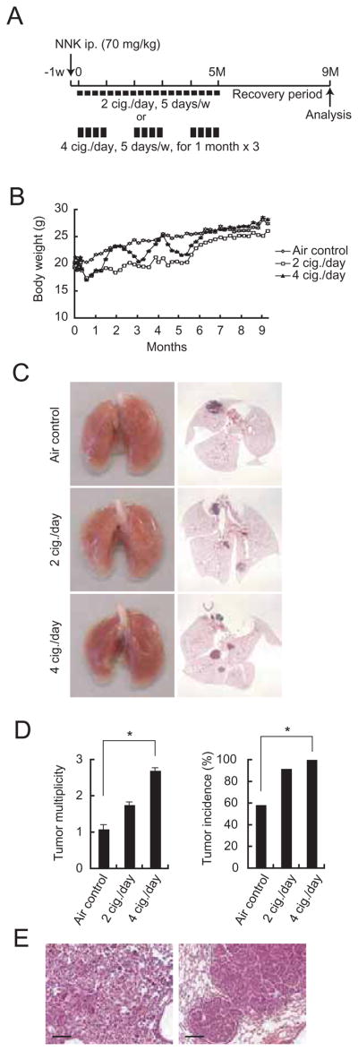
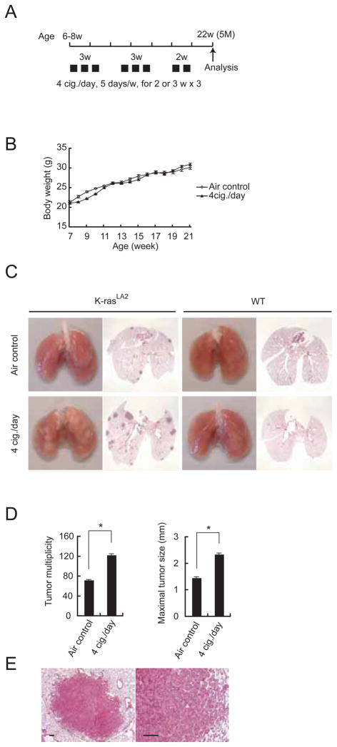
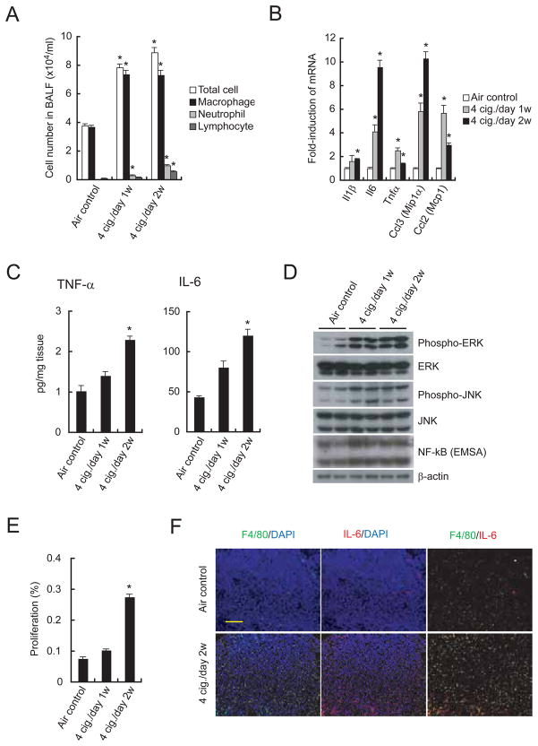
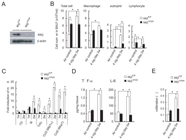
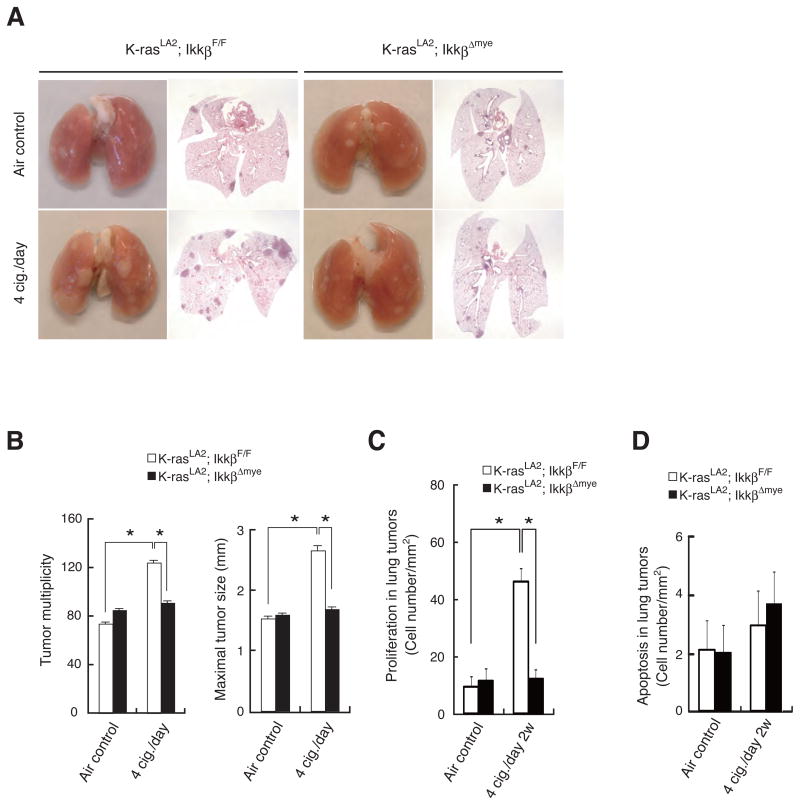
Chronic exposure to tobacco smoke, which contains over 60 tumor-initiating carcinogens, is the major risk factor for development of lung cancer, accounting for a large portion of cancer-related deaths worldwide. It is well established that tobacco smoke is a tumor initiator, but we asked whether it also acts as a tumor promoter once malignant initiation, such as caused by K-ras activation, has taken place. Here we demonstrate that repetitive exposure to tobacco smoke promotes tumor development both in carcinogen-treated mice and in transgenic mice undergoing sporadic K-ras activation in lung epithelial cells. Tumor promotion is due to induction of inflammation that results in enhanced pneumocyte proliferation and is abrogated by IKKbeta ablation in myeloid cells or inactivation of JNK1.


Figures

Figure 1
Figure 1. Tobacco smoke exposure promotes development of chemically-induced lung cancer
(A) Experimental protocols for promotion of NNK-induced lung cancer. 7-week-old male A/J mice were MTS-exposed using two different regimens starting 1 week after NNK injection. Tumor development was analyzed 9 months later. (B) Effect of MTS exposure on weight gain in A/J mice. Results are means ± S.E. (air control: n = 12; 2 cig./day: n = 12; 4 cig./day: n = 13). (C) Lung appearance (left panels) and histology (H&E stain; right panels) in A/J mice 9 months after initiation of the NNK + MTS protocol. (D) Lung tumor multiplicity and incidence were determined by serial sectioning at 350 μm intervals. Incidence = number of tumor-bearing mice divided by the number of mice in each group. Results are means ± S.E. (n: as described in panel B). Significant difference, *P < 0.03. (E) Histological appearance of adenoma (left panel) and adenocarcinoma with bronchial invasion (right panel) in A/J mice given NNK and 4 cig./day MTS. Scale bar = 100 μm. See also Figure S1.
Figure 2
Figure 2. Tobacco smoke exposure promotes development of genetically-induced lung cancer
(A) Outline of the experimental protocol in which K-rasLA2 mice were exposed to MTS and analyzed at 5 months of age. (B) Effect of MTS exposure on weight gain in K-rasLA2 mice. Results are means ± S.E. (air control: n = 4; 4 cig./day: n = 8). (C) Lung appearance (left side) and histology (H&E stain; right side) in 5-month-old K-rasLA2 or WT mice with or without MTS exposure. (D) Lung tumor multiplicity and maximal tumor sizes were determined as above. Results are means ± S.E. (air control: n = 16; 4 cig./day: n = 18). Significant difference, *P < 0.002. (E) Histological appearance of adenoma in MTS-exposed K-rasLA2 mice. Scale bar = 100 μm. See also Figure S2.
Figure 3
Figure 3. Tobacco smoke induces pulmonary inflammation and cell proliferation
(A) Total cell number and leukocyte populations in BALF collected from C57BL6 males 24 hrs after last MTS or air exposure. Cellular composition was analyzed using cytospin preparations. Results are means ± S.E. (n = 8 for each group). Significant difference, *P < 0.02 vs. air control. (B) Induction of inflammatory cytokine and chemokine mRNAs in lungs of MTS-exposed C57BL6 males. Lung RNA was isolated 24 hrs after last MTS exposure and analyzed by real-time PCR. Results are means ± S.E. (n = 5 for each group). Significant difference, *P < 0.03 vs. air control. (C) Elevated cytokine secretion by lungs of MTS-exposed male mice. Fresh lungs were cut into small pieces and incubated in medium at 37°C for 48 hrs. Cytokines in culture supernatants were measured by ELISA. Results are means ± S.E. (air control: n = 7; 4 cig./day 1w: n = 9; 4 cig./day 2w: n = 9). Significant difference, *P < 0.02 vs. air control. (D) ERK and JNK activation and NF-κB DNA binding activity in lungs of MTS-exposed mice. Lung lysates and nuclear extracts prepared 4 hrs after last MTS exposure were analyzed by immunoblotting and EMSA. Nuclear protein content was determined by immunoblotting with β-actin antibody. Shown are results from 2 representative mice per group. (E) Cell proliferation in lungs of air- or MTS-exposed mice was determined by BrdU labeling. Results are means ± S.E. (air control: n = 7; 4 cig./day 1w: n = 5; 4 cig./day 2w: n = 8). Significant difference, *P < 4.0 × 10⁻⁵ vs. air control. (F) Infiltration of IL-6 positive macrophages into K-rasLA2 lung tumors 2 weeks after initiation of MTS exposure. Lung sections prepared 24 hrs after last MTS exposure were analyzed by immunostaining for F4/80 (green) and IL-6 (red). Nuclei were counterstained by DAPI (blue). Scale bar = 100 μm. Results shown are for one representative mouse. For quantitation of the entire experiment see Figure S3F. See also Figure S3.
Figure 4
Figure 4. Myeloid cell IKKβ deletion decreases MTS-induced inflammation and cell proliferation
(A) Expression of IKKβ in alveolar macrophages of 7-week-old IkkβF/F and IkkβΔmye mice. (B) BALF cellular composition in air- or MTS-exposed IkkβF/F and IkkβΔmye mice 24 hrs after last MTS exposure. Results are means ± S.E. (IkkβF/F air control: n = 9, IkkβF/F 4 cig./day 2w: n = 9, IkkβΔmye air control: n = 9, IkkβΔmye 4 cig./day 2w: n = 13). Significant difference, *P < 0.03. (C) Induction of inflammatory cytokine and chemokine mRNAs in lungs of MTS-exposed IkkβF/F and IkkβΔmye mice 24 hrs after last 1 or 2 weeks MTS exposure. Results are means ± S.E. (n = 5 for each group). Significant difference, *P < 0.05. (D) Secretion of cytokines by lungs of MTS-exposed mice was analyzed as in Fig. 3C. Results are means ± S.E. (IkkβF/F air control: n = 14; IkkβF/F 4 cig./day 2w: n = 8; IkkβΔmye air control: n = 12; IkkβΔmye 4 cig./day 2w: n = 13). Significant difference, *P < 0.04. (E) Cell proliferation in lungs of air- or MTS-exposed mice was analyzed as in Fig. 3E. Results are means ± S.E. (n = 7 for each group). Significant difference, *P < 0.03. See also Figure S4.
Figure 5
Figure 5. IKKβ deletion in myeloid cells inhibits MTS-induced lung tumor promotion and malignant cell proliferation in K-rasLA2 mice
(A) Lung appearance (left) and histology (H&E stain; right) in 5-months-old K-rasLA2; IkkβF/F and K-rasLA2; IkkβΔmye mice with or without MTS exposure. (B) Lung tumor multiplicity and maximal tumor sizes were determined as in Figure 2D in K-rasLA2; IkkβF/F and K-rasLA2; IkkβΔmye mice that were either air- or MTS-exposed. Results are means ± S.E. (K-rasLA2; IkkβF/F air control: n = 18; K-rasLA2; IkkβF/F 4 cig./day: n = 17; K-rasLA2; IkkβΔmye air control: n = 20; K-rasLA2; IkkβΔmye 4 cig./day: n = 18). Significant difference, *P < 0.02. (C) Cell proliferation in lung adenomas from air- or MTS-exposed mice of indicated genotypes was determined by BrdU labeling. Results are means ± S.E. (n = 5–6 for each group). Significant difference, *P < 0.001. (D) Apoptosis in lung adenomas from air- or MTS-exposed mice of indicated genotypes was determined by TUNEL staining. Results are means ± S.E. (n = 5–6 for each group). See also Figure S5.
Figure 6
Figure 6. Deletion of JNK1 decreases MTS-induced inflammation, cell proliferation, and lung tumor promotion
(A) Induction of inflammatory cytokine and chemokine mRNAs in lungs of MTS-exposed Jnk1−/− mice was analyzed as in Fig. 3B. Results are means ± S.E. (n = 5 for each group). Significant difference, *P < 0.05. (B) Secretion of cytokines in lungs of MTS-exposed Jnk1−/− mice was analyzed as in Fig. 3C. Results are means ± S.E. (WT air control: n = 12; WT 4 cig./day: n = 9; Jnk1−/− air control: n = 9; Jnk1−/− 4 cig./day: n = 9). Significant difference, *P < 0.05. (C) JNK1 deletion decreases MTS-induced lung cell proliferation. Results are means ± S.E. (WT air control: n = 7; WT 4 cig./day: n = 8; Jnk1−/− air control: n = 10; Jnk1−/− 4 cig./day: n = 9). Significant difference, *P < 0.0003. (D) Lung appearance (left side) and histology (H&E stain; right side) in 5-month-old K-rasLA2 or K-rasLA2; Jnk1−/− mice with or without MTS exposure. (E) Lung tumor multiplicity and maximal tumor sizes were determined as in Fig. 2D. Results are means ± S.E. (WT air control: n = 16; WT 4 cig./day: n = 18; Jnk1−/− air control: n = 13; Jnk1−/− 4 cig./day: n = 14). Significant difference, *P < 0.002. See also Figure S6.

Similar articles

Cited by

References

    1. Barnes PJ. The cytokine network in asthma and chronic obstructive pulmonary disease. J Clin Invest. 2008;118:3546–3556. - PMC - PubMed
    1. Berns A. Cancer. Improved mouse models. Nature. 2001;410:1043–1044. - PubMed
    1. Borm PJ, Driscoll K. Particles, inflammation and respiratory tract carcinogenesis. Toxicol Lett. 1996;88:109–113. - PubMed
    1. Broide DH, Lawrence T, Doherty T, Cho JY, Miller M, McElwain K, McElwain S, Karin M. Allergen-induced peribronchial fibrosis and mucus production mediated by IkappaB kinase beta-dependent genes in airway epithelium. Proc Natl Acad Sci U S A. 2005;102:17723–17728. - PMC - PubMed
    1. Brown BG, Chang CJ, Ayres PH, Lee CK, Doolittle DJ. The effect of cotinine or cigarette smoke co-administration on the formation of O6-methylguanine adducts in the lung and liver of A/J mice treated with 4-(methylnitrosamino)-1-(3-pyridyl)-1-butanone (NNK) Toxicol Sci. 1999;47:33–39. - PubMed

Publication types

MeSH terms