. 2010 Apr 10;399(2):257-69.
doi: 10.1016/j.virol.2009.12.020. Epub 2010 Feb 2.

Identification of N-linked carbohydrates from severe acute respiratory syndrome (SARS) spike glycoprotein

Affiliations

Identification of N-linked carbohydrates from severe acute respiratory syndrome (SARS) spike glycoprotein

Gayle Ritchie et al. Virology. .

Abstract

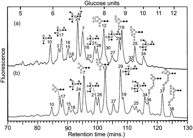
N-glycans were released from the SARS coronavirus (SARS-CoV) spike glycoprotein produced in Vero E6 cells and their structures were determined by a combination of matrix-assisted laser desorption/ionization (MALDI) mass spectrometry, negative ion electrospray collision-induced dissociation time-of-flight mass spectrometry and normal-phase high-performance liquid chromatography with exoglycosidase digestion. Major glycans were high-mannose (Man(5-9)GlcNAc(2)), hybrid and bi-, tri- and tetra-antennary complex with and without bisecting GlcNAc and core fucose. Complex glycans with fewer than the full complement of galactose residues were present and sialylation was negligible. Treatment with the glucosidase inhibitor N-butyl-deoxynojirimycin (NB-DNJ) inhibited N-glycan processing as evidenced by the appearance of glycans of composition Glc(3)Man(7-9)GlcNAc(2). However, some complex glycans remained suggesting the presence of an alpha-endomannosidase. Our data in tissue culture indicate that inhibition of N-glycan processing may be considered as a therapeutic strategy against SARS CoV infections.


Figures

Fig. 1
Fig. 1
SDS-PAGE and western blotting analysis of purified SARS-CoV (strain Tor2) preparation. Left panel: 9% SDS-PAGE analysis of SARS-CoV virion preparation. S = SARS-CoV spike glycoprotein, N = SARS-CoV nucleoprotein, M = SARS-CoV matrix protein. Molecular weight markers are shown on the left. Right panel: Western blotting analysis of SARS-Co V virion preparation using an anti-SARS-CoV S antibody (Imgenex). U, untreated preparation; D, drug(NB-DNJ) treated preparation; M, mock infected preparation.
Fig. 2
Fig. 2
(a) NP-HPLC analysis of SARS-CoV S N-glycans. (b) HPLC analysis of SARS-CoV N-glycans following digestion with Arthrobacter ureafaciens sialidase (ABS, EC 3.2.1.18), (c) HPLC analysis following digestion with a mixture of ABS and bovine testis β-galactosidase (BTG, EC 3.2.1.23) and (d) HPLC analysis following incubation with a mixture of ABS, BTG and Streptococcus pneumoniae hexosaminidase (GUH, EC 3.2.1.30). The broken lines connecting the peaks in the various traces show movement of the peaks following digestion. Symbols used for the structural formulae: ■ = GlcNAc, ○ = mannose, ◊ = galactose, formula image 
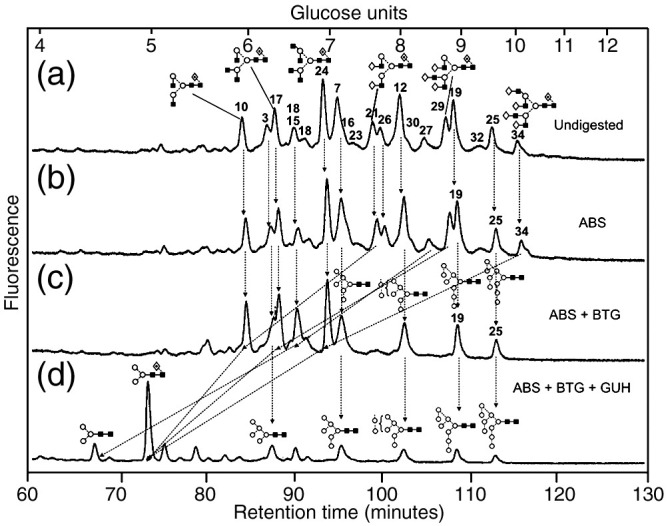
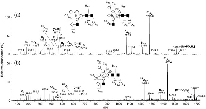
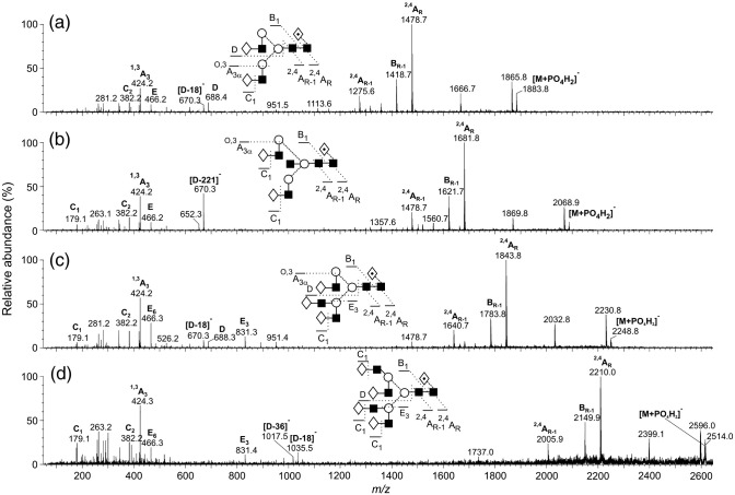
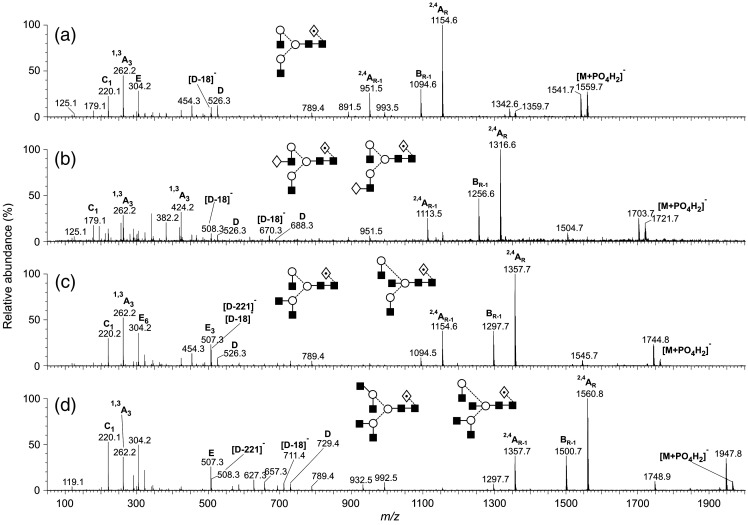
= fucose. The angle of the lines connecting the symbols shows the linkage with full and broken lines specifying β- and α-linkages, respectively. Further details are given in the paper by Harvey et al. (2009).
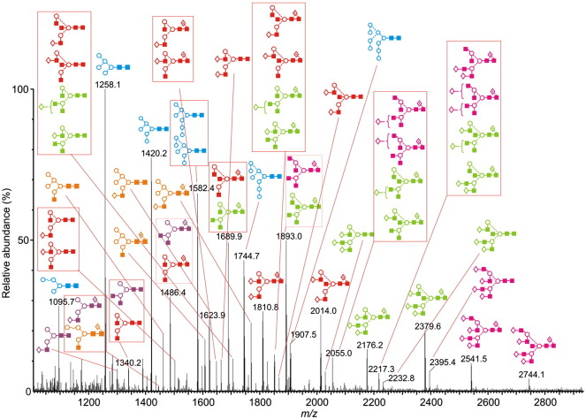
Fig. 3
Fig. 3
Positive ion MALDI-TOF mass spectrum ([M+Na]+ions) of the N-linked glycans obtained from the SARS-CoV spike protein. Peaks are identified in Table 1 and the key to the structural symbols is in the legend to Fig. 2 with the addition of □ = glucose. Glycan types are coloured: blue = high mannose, orange = hybrid, purple = monoantennary, red = biantennary, green = triantennary, pink = tetra-antennary.
Fig. 4
Fig. 4
Negative ion MS/MS spectra of the [M+H2PO4] ions from the high-mannose glycans Man5–9GlcNAc2. Symbols for the structural diagrams are defined in the legend to Fig. 2. Ions are labelled according to the scheme introduced by Domon and Costello (1988).
Fig. 5
Fig. 5
Negative ion MS/MS spectra of the [M+H2PO4] ions from the hybrid glycans 9 and 14. Symbols for the structural diagrams are defined in the legend to Fig. 2.
Fig. 6
Fig. 6
Negative ion MS/MS spectra of the [M+H2PO4] ions from (a) the fucosylated biantennary glycan 21, (b) the bisected, fucosylated biantennary glycan 26, (c) the fucosylated triantennary glycan 29 and (d) the fucosylated tetra-antennary glycan 34. Symbols for the structural diagrams are defined in the legend to Fig. 2.
Fig. 7
Fig. 7
Negative ion MS/MS spectra of the [M+H2PO4] ions from (a) the fucosylated biantennary glycan lacking galactose (10), (b) a mixture of the fucosylated biantennary glycan with one galactose residue on either antenna (15), (c) a mixture of the fucosylated triantennary glycan and the bisected biantennary glycan lacking galactose (17) and (d) a mixture of the fucosylated tetra-antennary glycan and the bisected triantennary glycan lacking galactose (24). Symbols for the structural diagrams are defined in the legend to Fig. 2.
Fig. 8
Fig. 8
(a) NP-HPLC analysis of SARS-CoV N-glycans obtained without (a) and with (b) 100 μg/mL NB-DNJ. Peaks are identified in Table 1. The three glycans with residual glucose residues are highlighted.

Similar articles

Cited by

References

    1. Bergeron E., Vincent M.J., Wickham L., Hamelin J., Basak A., Nichol S.T., Chrétien M., Seidah N.G. Implication of proprotein convertases in the processing and spread of severe acute respiratory syndrome coronavirus. Biochem. Biophys. Res. Commun. 2005;326:554–563. - PMC - PubMed
    1. Bigge J.C., Patel T.P., Bruce J.A., Goulding P.N., Charles S.M., Parekh R.B. Nonselective and efficient fluorescent labeling of glycans using 2-aminobenzamide and anthranilic acid. Anal. Biochem. 1995;230:229–238. - PubMed
    1. Börnsen K.O., Mohr M.D., Widmer H.M. Ion exchange and purification of carbohydrates on a Nafion(R) membrane as a new sample pretreatment for matrix-assisted laser desorption-ionization mass spectrometry. Rapid Commun. Mass Spectrom. 1995;9:1031–1034.
    1. Chen J., Subbarao K. The immunobiology of SARS. Annu. Rev. Immunol. 2007;25:443–472. - PubMed
    1. Domon B., Costello C.E. A systematic nomenclature for carbohydrate fragmentations in FAB-MS/MS spectra of glycoconjugates. Glycoconj. J. 1988;5:397–409.

Publication types

MeSH terms