

Synthesis and carbohydrate binding studies of fluorescent alpha-amidoboronic acids and the corresponding bisboronic acids
- PMID: 20129789
- PMCID: PMC2828497
- DOI: 10.1016/j.bmc.2010.01.017
Synthesis and carbohydrate binding studies of fluorescent alpha-amidoboronic acids and the corresponding bisboronic acids
Abstract
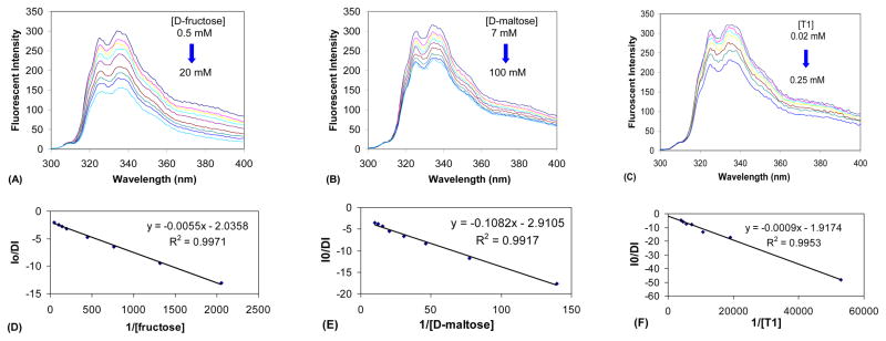
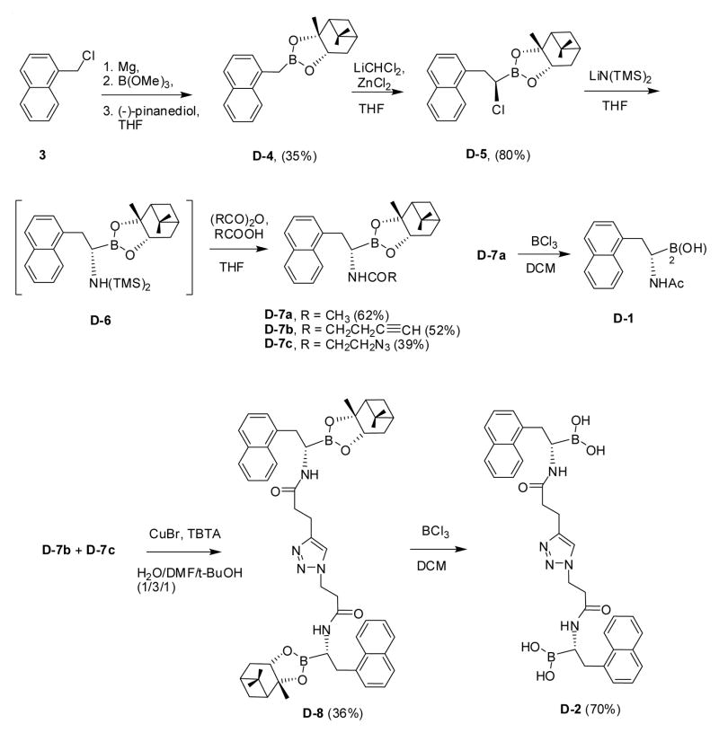
Fluorescent boronic acids are very useful for the design and synthesis of carbohydrate sensors. In an earlier communication, we first described the effort of developing water soluble fluorescent alpha-amidoboronic acids, which change fluorescence upon sugar binding. In this report, we describe a general method of functionalizing such boronic acids and their applications in the preparation of bis-alpha-amidoboronic acids with significantly enhanced binding for oligosaccharides as compared to their monoboronic acid counterparts. The advantages of good water solubility, easy modification to generate diversity, and modularity in synthesis will make alpha-amidoboronic acids very useful building blocks for future synthesis of boronic acid-based fluorescent sensors.
Published by Elsevier Ltd.
Figures


References
-
- Sugihara JM, Bowman CM. J Am Chem Soc. 1958;80:2443.
-
- Lorand JP, Edwards JO. J Org Chem. 1959;24:769.
-
- Yan J, Fang H, Wang B. Med Res Rev. 2005;25:490. - PubMed
-
- James TD, Shinkai S. Top Curr Chem. 2002;218:159.
Publication types
MeSH terms
Substances
Grants and funding
LinkOut - more resources
Full Text Sources
Other Literature Sources
Miscellaneous
