

Multiscale AM-FM methods for diabetic retinopathy lesion detection
- PMID: 20129850
- PMCID: PMC2825390
- DOI: 10.1109/TMI.2009.2037146
Multiscale AM-FM methods for diabetic retinopathy lesion detection
Abstract
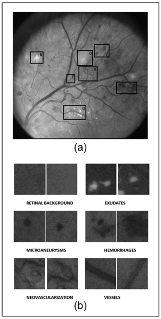
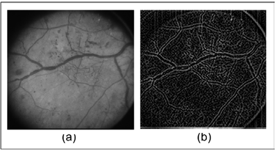
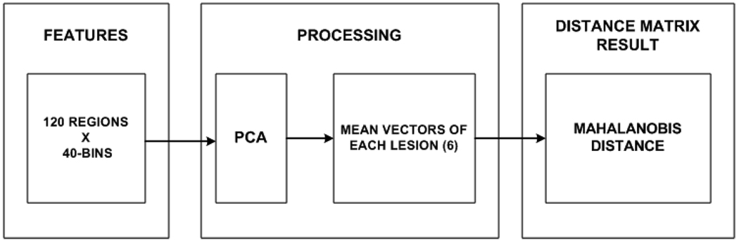
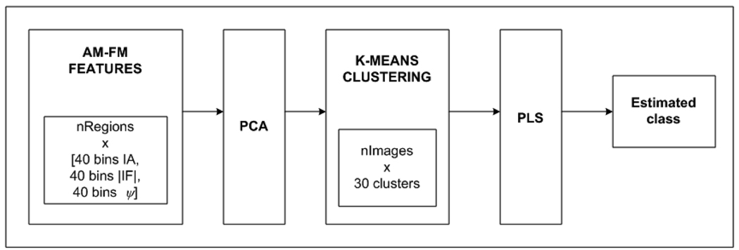
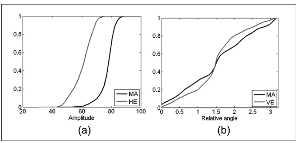
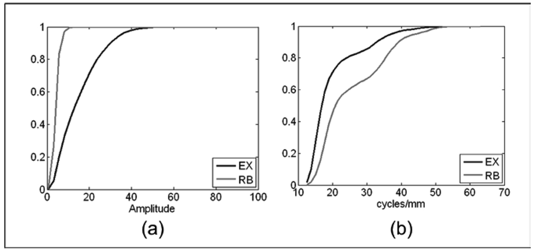
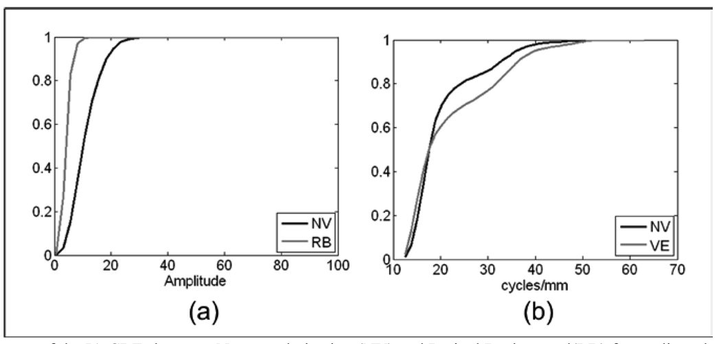
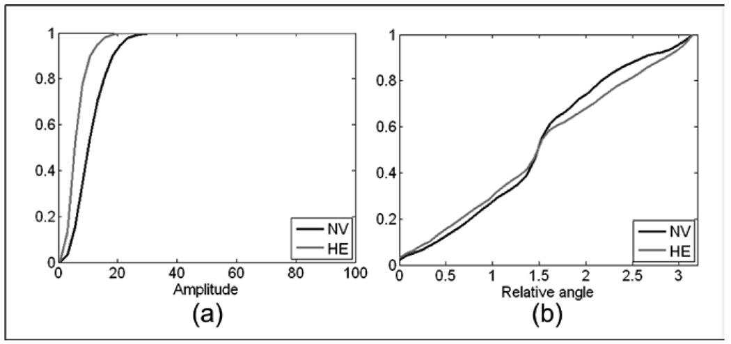
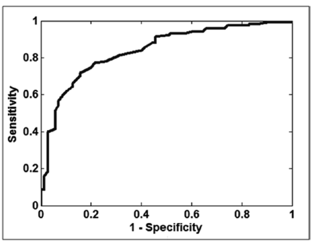
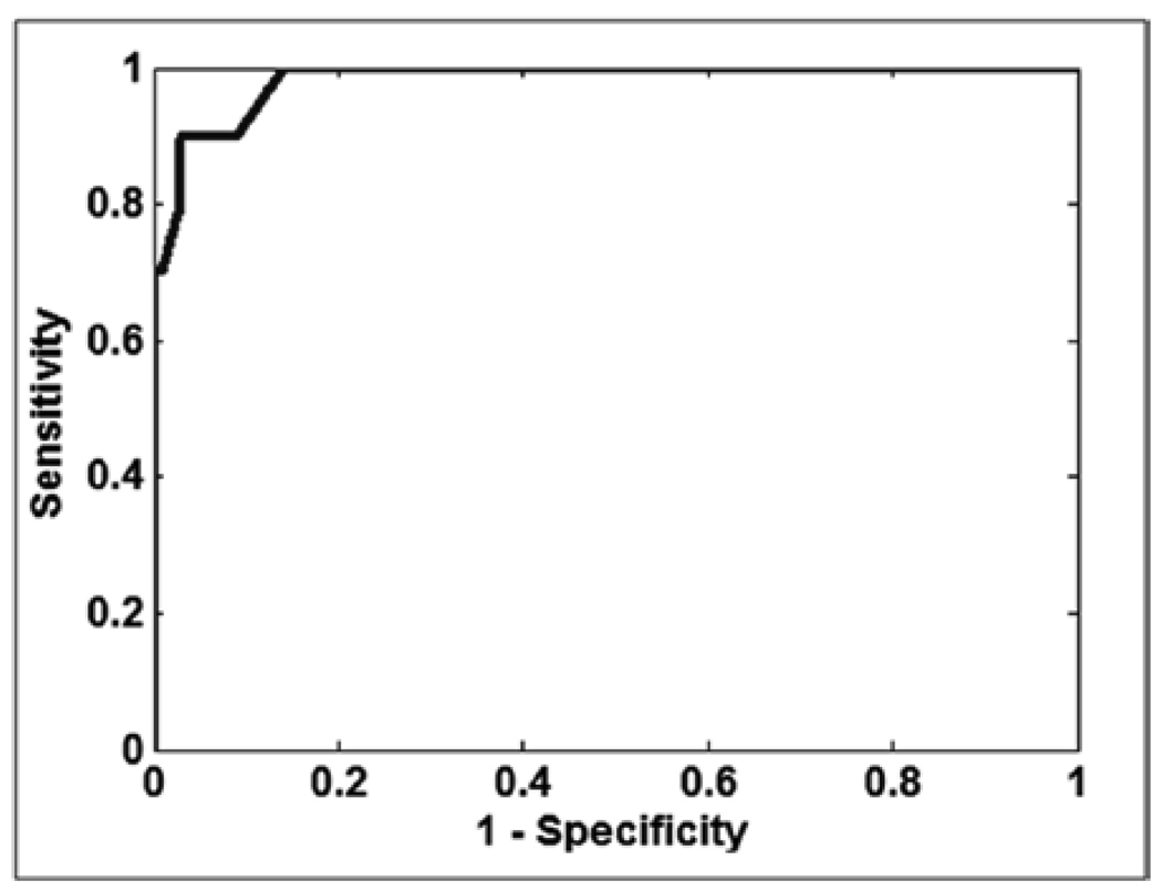
In this paper, we propose the use of multiscale amplitude-modulation-frequency-modulation (AM-FM) methods for discriminating between normal and pathological retinal images. The method presented in this paper is tested using standard images from the early treatment diabetic retinopathy study. We use 120 regions of 40 x 40 pixels containing four types of lesions commonly associated with diabetic retinopathy (DR) and two types of normal retinal regions that were manually selected by a trained analyst. The region types included microaneurysms, exudates, neovascularization on the retina, hemorrhages, normal retinal background, and normal vessels patterns. The cumulative distribution functions of the instantaneous amplitude, the instantaneous frequency magnitude, and the relative instantaneous frequency angle from multiple scales are used as texture feature vectors. We use distance metrics between the extracted feature vectors to measure interstructure similarity. Our results demonstrate a statistical differentiation of normal retinal structures and pathological lesions based on AM-FM features. We further demonstrate our AM-FM methodology by applying it to classification of retinal images from the MESSIDOR database. Overall, the proposed methodology shows significant capability for use in automatic DR screening.
Figures
Similar articles
-
Automated lesion detectors in retinal fundus images.Comput Biol Med. 2015 Nov 1;66:47-65. doi: 10.1016/j.compbiomed.2015.08.008. Epub 2015 Aug 18. Comput Biol Med. 2015. PMID: 26378502
-
Classification of diabetic retinopathy images using multi-class multiple-instance learning based on color correlogram features.Annu Int Conf IEEE Eng Med Biol Soc. 2012;2012:1462-5. doi: 10.1109/EMBC.2012.6346216. Annu Int Conf IEEE Eng Med Biol Soc. 2012. PMID: 23366177
-
Points of interest and visual dictionaries for automatic retinal lesion detection.IEEE Trans Biomed Eng. 2012 Aug;59(8):2244-53. doi: 10.1109/TBME.2012.2201717. Epub 2012 May 30. IEEE Trans Biomed Eng. 2012. PMID: 22665502
-
Pathology of diabetic retinopathy.Br Med Bull. 1970 May;26(2):137-42. doi: 10.1093/oxfordjournals.bmb.a070765. Br Med Bull. 1970. PMID: 4192644 Review. No abstract available.
-
Algorithms for the automated detection of diabetic retinopathy using digital fundus images: a review.J Med Syst. 2012 Feb;36(1):145-57. doi: 10.1007/s10916-010-9454-7. Epub 2010 Apr 6. J Med Syst. 2012. PMID: 20703740 Review.
Cited by
-
Discriminative Learning Approach Based on Flexible Mixture Model for Medical Data Categorization and Recognition.Sensors (Basel). 2021 Apr 2;21(7):2450. doi: 10.3390/s21072450. Sensors (Basel). 2021. PMID: 33918120 Free PMC article.
-
Automated analysis of diabetic retinopathy images: principles, recent developments, and emerging trends.Curr Diab Rep. 2013 Aug;13(4):453-9. doi: 10.1007/s11892-013-0393-9. Curr Diab Rep. 2013. PMID: 23686810 Review.
-
Fundus images analysis using deep features for detection of exudates, hemorrhages and microaneurysms.BMC Ophthalmol. 2018 Nov 6;18(1):288. doi: 10.1186/s12886-018-0954-4. BMC Ophthalmol. 2018. PMID: 30400869 Free PMC article.
-
Detection of Hard Exudates in Colour Fundus Images Using Fuzzy Support Vector Machine-Based Expert System.J Digit Imaging. 2015 Dec;28(6):761-8. doi: 10.1007/s10278-015-9793-5. J Digit Imaging. 2015. PMID: 25822397 Free PMC article.
-
Noninvasive temporal detection of early retinal vascular changes during diabetes.Sci Rep. 2020 Oct 15;10(1):17370. doi: 10.1038/s41598-020-73486-2. Sci Rep. 2020. PMID: 33060607 Free PMC article.
References
-
- Larsen M, Godt J, Larsen N, Lund-Andersen H, Sjolie AK, Agardh E, et al. Automated detection of fundus photographic red lesions in diabetic retinopathy. Invest Ophthalmol Vis Sci. 2003;44:761–766. - PubMed
-
- Niemeijer M, van Ginneken B, Staal J, Suttorp-Schulten MSA, Abràmoff MD. Automatic detection of red lesions in digital color fundus photographs. IEEE Transactions on Medical Imaging. 2005;24:584–592. - PubMed
-
- Sander B, Godt J, Lund-Andersen H, Grunkin M, Owens D, Larsen N, et al. Automatic detection of fundus photographic red lesions in diabetic retinopathy. ARVO. 2001 Poster No. 4338. - PubMed
-
- Murray V. Ph.D. dissertation. University of New Mexico; 2008. Sep, AM-FM methods for image and video processing.
Publication types
MeSH terms
Grants and funding
LinkOut - more resources
Full Text Sources
Other Literature Sources
Medical
