. 2010 Apr 2;285(14):10638-52.
doi: 10.1074/jbc.M110.108688. Epub 2010 Feb 3.

Activation of AMP-activated protein kinase by vascular endothelial growth factor mediates endothelial angiogenesis independently of nitric-oxide synthase

Affiliations

Activation of AMP-activated protein kinase by vascular endothelial growth factor mediates endothelial angiogenesis independently of nitric-oxide synthase

Nadine Stahmann et al. J Biol Chem. .

Abstract

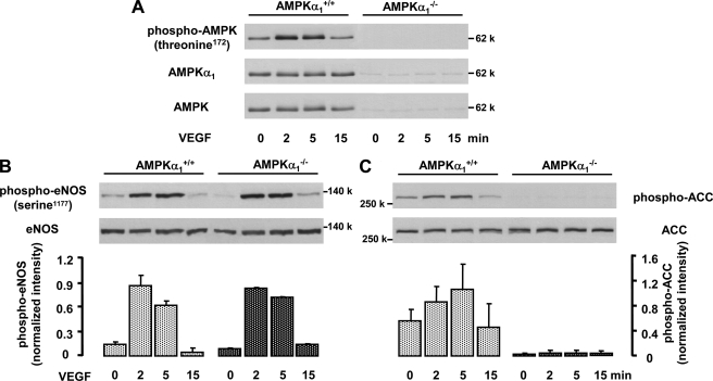
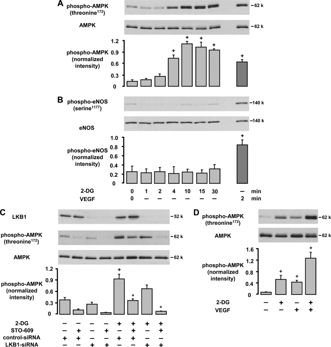
AMP-activated protein kinase (AMPK) is a sensor of cellular energy state and a regulator of cellular homeostasis. In endothelial cells, AMPK is stimulated via the upstream kinases LKB1 and Ca(2+)/calmodulin-dependent protein kinase kinase beta (CaMKKbeta). Previously, AMPK has been reported to activate endothelial nitric-oxide synthase (eNOS). Using genetic and pharmacological approaches, we show that vascular endothelial growth factor (VEGF) stimulates AMPK in human and mice endothelial cells via CaMKKbeta. VEGF-induced AMPK activation is potentiated under conditions of energy deprivation induced by 2-deoxyglucose. To investigate the role of AMPK in endothelial function, CaMKKbeta, AMPKalpha1, or AMPKalpha2 was down-regulated by RNA interference, and studies in AMPKalpha1(-/-) mice were performed. We demonstrate that AMPK does not mediate eNOS phosphorylation at serine residue 1177 or 633, NO- dependent cGMP generation, or Akt phosphorylation in response to VEGF. Using inhibitors of eNOS or soluble guanylyl cyclase and small interfering RNA against eNOS, we show that NO does not act upstream of AMPK. Taken together, these data indicate that VEGF-stimulated AMPK and eNOS pathways act independently of each other. However, acetyl-CoA carboxylase, a key enzyme in the regulation of fatty acid oxidation, was phosphorylated in response to VEGF in an AMPKalpha1- and AMPKalpha2-dependent manner. Our results show that AMPKalpha1 plays an essential role in VEGF-induced angiogenesis in vitro (tube formation and sprouting from spheroids) and in vivo (Matrigel plug assay). In contrast, AMPKalpha2 was not involved in VEGF-triggered sprouting. The data suggest that AMPKalpha1 promotes VEGF-induced angiogenesis independently of eNOS, possibly by providing energy via inhibition of acetyl-CoA carboxylase.


Figures

FIGURE 1.
FIGURE 1.
VEGF activates AMPK in endothelial cells via a CaMKKβ-mediated pathway. HUVEC were stimulated with 50 ng/ml VEGF for the indicated times (A) or for 5 min (B and C). B, cells were preincubated with STO-609 for 30 min. C, cells were pretreated with a transfection reagent (control), control-siRNA, or specific CaMKKβ-siRNA (1 μg/30-mm-diameter dish, 72 h). A–C, cell lysates were subjected to Western blot analysis using antibodies against phosphorylated AMPKα (Thr172) and total AMPKα. In parallel, AMPK activity was determined in AMPKα1 immune complexes using the SAMS peptide assay and calculated as nmol/min/mg lysate protein. C, CaMKKβ activity (nmol/min/mg AMPK) was measured in anti-CaMKKβ immune complexes by the activation of recombinant AMPK using the SAMS peptide assay to demonstrate down-regulation of the enzyme. Because Ca2+ and calmodulin were present in the test, the data may not reflect in vivo activation of CaMKKβ. A–C, representative blots, densitometric analyses, and enzyme activities are shown (mean ± S.E., n = 3). +, p < 0.05 versus unstimulated controls; *, p < 0.05 versus non-inhibitor-treated (B) or control-siRNA-treated (C) VEGF-stimulated cells.
FIGURE 2.
FIGURE 2.
CaMKKβ inhibition or down-regulation impairs VEGF-induced ACC phosphorylation but not eNOS phosphorylation. HUVEC were either preincubated for 30 min with STO-609, a CaMKK inhibitor, at the indicated concentrations (A, C, and E) or pretreated with CaMKKβ-specific or control-siRNA (1 μg/30-mm-diameter dish, 72 h) (B, D, and F) and subsequently stimulated with 50 ng/ml VEGF for 5 min (A, B, E, and F) or 20 min (C and D). A, B, E, and F, cells were lysed and subjected to immunoblotting using antibodies against phosphorylated eNOS (serine 1177) or total eNOS and against phosphorylated ACC or total ACC. Typical experiments and the densitometric analysis of three experiments for each staining are shown (mean ± S.E.). +, p < 0.05 versus unstimulated controls; *, p < 0.05 versus non-inhibitor-treated VEGF-stimulated cells. C and D, cells were denaturated with ethanol and processed for cGMP formation. cGMP was determined in ethanolic extracts by means of radioimmunoassay and calculated as pmol/mg cell protein, which was assayed in parallel samples (mean ± S.E., n = 4). +, p < 0.05 versus unstimulated controls.
FIGURE 3.
FIGURE 3.
AMPKα1 down-regulation inhibits VEGF-induced ACC phosphorylation but not eNOS or Akt phosphorylation. siRNA targeted to human AMPKα1 or containing an unrelated sequence (control-siRNA) was added to HUVEC for 72 h (0.5 μg/30-mm-diameter dish). Subsequently, HUVEC were stimulated with 50 ng/ml VEGF for the indicated times. Cells were lysed and subjected to immunoblotting using antibodies against phosphorylated AMPK (Thr172) or AMPKα1 (A); phosphorylated eNOS (serine 1177, serine 633) or total eNOS (B); phosphorylated Akt (serine 473) or total Akt (C); and phosphorylated or total ACC (D). Typical experiments and the densitometric analysis of four experiments for each staining are shown (mean ± S.E.). B, the gray and black columns represent eNOS phosphorylated at serine 1177 or serine 633, respectively. +, p < 0.05 versus unstimulated controls; *, p < 0.05 versus control-siRNA-pretreated VEGF-stimulated cells.
FIGURE 4.
FIGURE 4.
AMPKα2 down-regulation inhibits VEGF-induced ACC phosphorylation but not eNOS phosphorylation. siRNA targeted to human AMPKα2 or containing an unrelated sequence (control-siRNA) was added to HUVEC for 72 h (0.5 μg/30-mm-diameter dish). Subsequently, HUVEC were stimulated with 50 ng/ml VEGF for the indicated times. Cells were lysed and subjected to immunoblotting using antibodies against phosphorylated AMPK (Thr172) or AMPKα2 (A); phosphorylated eNOS (serine 1177, serine 633) or total eNOS (B); and phosphorylated or total ACC (C). Typical experiments and the densitometric analysis of four experiments for each staining are shown (mean ± S.E.). B, the gray and black columns represent eNOS phosphorylated at serine 1177 or serine 633, respectively. +, p < 0.05 versus unstimulated controls; *, p < 0.05 versus control-siRNA-pretreated VEGF-stimulated cells.
FIGURE 5.
FIGURE 5.
VEGF-induced ACC phosphorylation but not eNOS phosphorylation is inhibited in endothelial cells from AMPKα1−/− mice. MLEC from AMPKα1+/+ and AMPKα1−/− mice were stimulated with 50 ng/ml VEGF for the indicated times. Cell lysates were subjected to Western blot analysis using antibodies against phosphorylated AMPKα (Thr172), AMPKα1, or pan-AMPKα (A); phosphorylated (serine 1177) or total eNOS (B); and phosphorylated or total ACC (C). Representative blots and densitometric analysis from two experiments for each treatment are shown (mean ± S.E.). In addition to the kinetic studies shown here, three experiments with a single VEGF stimulation (5 min) were performed. Statistical analysis for the 5-min time points is given under “Results”. A significant inhibition of ACC but not of eNOS phosphorylation was observed.
FIGURE 6.
FIGURE 6.
VEGF-induced NO/cGMP formation is not required for VEGF-mediated AMPK activation. HUVEC were pretreated with l-NAME (0.5 mm, 30 min), eNOS-specific or control-siRNA duplexes (1 μg/30-mm-diameter dish, 72 h), or ODQ (10 μm, 30 min) and subsequently stimulated with VEGF (50 ng/ml, 5 min). Cells were lysed and subjected to immunoblotting using antibodies against AMPKα phosphorylated at Thr172 or total AMPKα. Down-regulation of eNOS was confirmed using anti-eNOS antibodies (middle panel). Typical experiments and densitometric analyses for each staining are shown (mean ± S.E., n = 5 for eNOS-siRNA experiments; n = 3 for others). +, p < 0.05 versus unstimulated controls.
FIGURE 7.
FIGURE 7.
2-Deoxyglucose activates AMPK via CaMKKβ- and LKB1-dependent pathways and acts synergistically with VEGF. A, B, and D, HUVEC were stimulated with 20 mm 2-deoxyglucose (2-DG) or 50 ng/ml VEGF for the indicated times (A and B) or with 2-DG for 4 min followed by VEGF for 2 min (D). Cell lysates were subjected to immunoblotting using antibodies against phosphorylated (Thr172) or total AMPKα (A and D) and against phosphorylated (serine 1177) or total eNOS (B). Representative blots and densitometric analyses are shown (mean ± S.E., n = 3). C, HUVEC were pretreated with LKB1-specific or control-siRNA (1 μg/30-mm-diameter dish, 72 h), additionally preincubated for 30 min with 20 μg/ml STO-609 as indicated, and subsequently stimulated with 2-deoxyglucose (20 mm, 4 min). Cell lysates were subjected to immunoblotting using antibodies against LKB1 and phosphorylated (threonine 172) or total AMPK. Representative blots and densitometric analyses are shown (mean ± S.E., n = 3). +, p < 0.05 versus unstimulated controls; *, p < 0.05 versus 2-deoxyglucose-stimulated, control-siRNA-pretreated cells.
FIGURE 8.
FIGURE 8.
AMPKα1 mediates tube formation of MLEC on Matrigel and in vivo angiogenesis in Matrigel plugs but is not involved in VEGF-mediated cell survival. A and B, MLEC from AMPKα1+/+ and AMPKα1−/− mice were grown in serum-depleted medium in the absence or presence of 50 ng/ml VEGF. Cells were counted (A) or subjected to cell cycle analysis (B) at the time of VEGF addition and 24 h later. Apoptotic cells were determined as cells with fragmented DNA (sub-G1 fraction) and are shown as a percentage of gated cells (B). Data are shown as mean ± S.E., n = 5 (A) or n = 4 (B); +, p < 0.05 versus initial values; *, p < 0.05 versus 24-h incubations without VEGF. C and D, MLEC from wild type and AMKα1 knock-out mice were seeded on a Matrigel matrix and grown in serum-depleted medium for 20 h in the absence or presence of 50 ng/ml VEGF, and tube formation was evaluated by light microscopy. Representative images (C) and an analysis of tube length (D) are shown. The average tube length per image was calculated from five independent pictures per condition in duplicates (mean ± S.E., n = 4). E and F, 500 μl of Matrigel mixed with 200 ng of VEGF plus 200 μg of heparin or with heparin alone was injected subcutaneously into wild type or AMPKα1 knock-out mice. Plugs were removed after 7 days, fixed in zinc fixative, and embedded in paraffin. Tissue sections were stained with anti-CD31 antibody and evaluated by fluorescence microscopy. Representative images (×400 (E)) and an analysis of the vascularized area (F) are shown. The average CD31-positive area was calculated from four images/section and two sections/plug using a standard imaging software (mean ± S.E.; n = 4 for control plugs, n = 13 or 14 for VEGF-containing plugs in wild type or AMPKα1 knock-out mice, respectively). D and F, +, p < 0.05 versus non-VEGF-treated controls; *, p < 0.05 versus the respective values in wild type cells (D) or mice (F).
FIGURE 9.
FIGURE 9.
AMPKα1 but not AMPKα2 mediates sprout formation from endothelial cell spheroids. Synthetic RNA duplexes targeted to human AMPKα1 or AMPKα2 or containing an unrelated sequence (control-siRNA) were added to HUVEC for 48 h (0.5 μg/30-mm-diameter dish). Subsequently, spheroids were generated, embedded in fibrin gels, and stimulated with 10 ng/ml VEGF for a further 48 h. Spheroids were viewed by light microscopy. Representative images (A) and an analysis of the number of sprouts per spheroid (B) are shown. The average sprout number per spheroid was calculated from five independent pictures per condition in duplicates. Means ± S.E. from five different experiments are shown; +, p < 0.05 versus unstimulated controls.

Similar articles

Cited by

References

    1. Carling D. (2004) Trends Biochem. Sci. 29, 18–24 - PubMed
    1. Hardie D. G. (2004) J. Cell Sci. 117, 5479–5487 - PubMed
    1. Kahn B. B., Alquier T., Carling D., Hardie D. G. (2005) Cell Metab. 1, 15–25 - PubMed
    1. Hardie D. G., Hawley S. A., Scott J. W. (2006) J. Physiol. 574, 7–15 - PMC - PubMed
    1. Towler M. C., Hardie D. G. (2007) Circ. Res. 100, 328–341 - PubMed

Publication types

MeSH terms

LinkOut - more resources