. 2010 Apr 2;285(14):10434-45.
doi: 10.1074/jbc.M109.078766. Epub 2010 Feb 2.

The fibroblast integrin alpha11beta1 is induced in a mechanosensitive manner involving activin A and regulates myofibroblast differentiation

Affiliations

The fibroblast integrin alpha11beta1 is induced in a mechanosensitive manner involving activin A and regulates myofibroblast differentiation

Sergio Carracedo et al. J Biol Chem. .

Abstract

Fibrotic tissue is characterized by an overabundance of myofibroblasts. Thus, understanding the factors that induce myofibroblast differentiation is paramount to preventing fibrotic healing. Previous studies have shown that mechanical stress derived from the integrin-mediated interaction between extracellular matrix and the cytoskeleton promotes myofibroblast differentiation. Integrin alpha11beta1 is a collagen receptor on fibroblasts. To determine whether alpha11beta1 can act as a mechanosensor to promote the myofibroblast phenotype, mouse embryonic fibroblasts and human corneal fibroblasts were utilized. We found that alpha11 mRNA and protein levels were up-regulated in mouse embryonic fibroblasts grown in attached three-dimensional collagen gels and conversely down-regulated in cells grown in floating gels. alpha11 up-regulation could be prevented by manually detaching the collagen gels or by cytochalasin D treatment. Furthermore, SB-431542, an inhibitor of signaling via ALK4, ALK5, and ALK7, prevented the up-regulation of alpha11 and the concomitant phosphorylation of Smad3 under attached conditions. In attached gels, TGF-beta1 was secreted in its inactive form but surprisingly not further activated, thus not influencing alpha11 regulation. However, inhibition of activin A attenuated the up-regulation of alpha11. To determine the role of alpha11 in myofibroblast differentiation, human corneal fibroblasts were transfected with small interfering RNA to alpha11, which decreased alpha-smooth muscle actin expression and myofibroblast differentiation. Our data suggest that alpha11beta1 is regulated by cell/matrix stress involving activin A and Smad3 and that alpha11beta1 regulates myofibroblast differentiation.


Figures
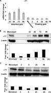
FIGURE 1.
FIGURE 1.
α11 levels are dynamically regulated inside collagen lattices. A, quantifications of α11 mRNA levels in wild type SV40 MEFs seeded in attached and floating collagen gels at different time points. mRNA quantifications under each condition were normalized to glyceraldehyde-3-phosphate dehydrogenase. Quantifications of mRNA levels in control cells (seeded in monolayer) were used for calibration/reference. The error bars represent asymmetric S.D. values for the respective conditions and were calculated as previously described (31). The specific values for each error bar are provided in supplemental Table S2. B, α11 protein levels in SV40 MEFs seeded in attached collagen gels at different time points. C, α11 protein levels in SV40 MEFs seeded in floating collagen gels at different time points. Band intensities in B and C were quantified, normalized to β-actin, and calibrated to the normalized value for cells in monolayer.
FIGURE 2.
FIGURE 2.
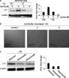
α11 levels inside attached collagen lattices depend on an intact actin cytoskeleton. A, effect of cytochalasin D at different concentrations on α11 protein levels in SV40 MEFs seeded in attached collagen lattices at 24 and 72 h. B, SV40 MEFs seeded in monolayer and treated with different concentrations of cytochalasin D (Cyt-D) for 72 h. C, α11 protein levels at 72 h in SV40 MEFs seeded in collagen lattices that were attached at all times (attached) or manually detached at 24 or 48 h in order to obtain floating conditions (Detached24 and Detached48, respectively). Band intensities in Western blots were quantified, normalized to β-actin, and calibrated to the normalized value corresponding to untreated cells at 24 h (A) or cells populating permanently attached gels (C).
FIGURE 3.
FIGURE 3.
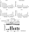
Involvement of the TGF-β superfamily in the regulation of α11 in attached collagen gels. A and B, quantifications of the mRNA levels for TGF-β superfamily ligands TGF-β1 and βA activin (A) and their respective receptors, ALK5 and ALK4 (B), at different time points in SV40 MEFs seeded in attached or floating collagen gels. mRNA quantifications for each target gene under each condition were normalized to glyceraldehyde-3-phosphate dehydrogenase. Quantifications of mRNA levels for each target gene in control cells (seeded in monolayer) were used for calibration/reference. The error bars represent asymmetric S.D. values for the respective conditions and were calculated as described previously (31). The specific value for each error bar is provided in supplemental Fig. S2. C, effect of the indicated concentrations of SB-431542 on α11 protein levels in SV40 MEFs seeded in attached gels at 96 h. Band intensities were quantified, normalized to β-actin, and calibrated to the normalized value corresponding to untreated cells at 24 h.
FIGURE 4.
FIGURE 4.
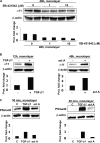
α11 levels in SV40 MEFs in monolayer are regulated by SB-431542, TGF-β1, and activin A. A, effect of the indicated concentrations of SB-431542 on α11 protein levels in SV40 MEFs seeded in monolayer at 48 h under serum free conditions. B, α11 protein levels in SV40 MEFs seeded in monolayer were serum-starved when stimulated (+) for the indicated time with 5 ng/ml of TGF-β1 or activin A (act A; 2 nm). C, phospho-Smad2 (PSmad2) and phospho-Smad3 (PSmad3) protein levels in SV40 MEFs seeded in monolayer and stimulated (+) for 30 min with 5 ng/ml TGF-β1 or activin A (act A; 2 nm). In A and B band intensities were quantified, normalized to β-actin, and calibrated to the normalized value for untreated cells. In C, the calibration was relative to the normalized band intensities corresponding to cells treated with TGF-β1.
FIGURE 5.
FIGURE 5.
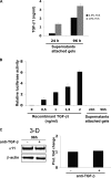
TGF-β1 is secreted but not activated by SV40 MEFs inside a collagen gel. A, amount of TGF-β in supernatants from attached collagen lattices containing SV40 MEFs determined by ELISA at the indicated time points. The analysis was performed in the presence of two different concentrations of serum. The bars show the average value for three independent experiments, whereas the error bars represent the S.D. for the corresponding average value. B, luciferase activity normalized to β-galactosidase activity in transiently transfected SV40 MEFs (see “Experimental Procedures”) cultured with supernatants similar to those used in A. Recombinant active TGF-β1 exogenously added was used as a control. Luciferase activities were calibrated to the normalized value for cells stimulated with 0.5 ng/ml recombinant active TGF-β1 exogenously added. C, α11 protein levels in SV40 MEFs seeded in attached collagen gels in the presence or absence of a 10 μg/ml concentration of a function-blocking antibody to TGF-β. Band intensities were quantified, normalized to β-actin, and calibrated to the normalized value corresponding to the sample obtained from cells without anti-TGF-β added.
FIGURE 6.
FIGURE 6.
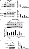
Activin A regulates α11 and Smad3 levels in MEFs cultured in attached collagen gels. A, α11 levels in SV40 MEFs seeded in attached collagen gels in the presence or absence of SB-431542 (SB; 10 μm), follistatin (Foll; 5 nm), or activin A function blocking antibody (anti-act A; 25 μg/ml). B, phospho-Smad3 (PSmad3) levels in SV40 MEFs seeded in attached collagen gels in the presence or absence of SB-431542 (10 μm), follistatin (5 nm), or activin A function-blocking antibody (25 μg/ml). C, α11 levels in primary MEFs seeded in attached collagen gels in the presence or absence of SB-431542 (10 μm), follistatin (5 nm), or activin A function-blocking antibody (25 μg/ml). D, α11 protein levels in wild type or Smad3−/− dermal fibroblasts seeded in attached lattices at the indicated time point. Band intensities were quantified and normalized to β-actin. Normalized bands were calibrated to the value corresponding to untreated cells (A and B), cells in monolayer (C), or Smad3+/+ cells (D).
FIGURE 7.
FIGURE 7.
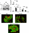
α11 levels and regulation correlate with a myofibroblastic phenotype. A, α11, α2, and α-SMA protein levels in SV40 MEFs in attached gels in the presence (+) or absence (−) of 20 ng/ml FGF-2 at the indicated time points. Band intensities were quantified, normalized to β-actin, and calibrated to the normalized value corresponding to untreated cells at 24 h. B, immunolocalization of α11 (red) at focal adhesions and α-SMA (green) in stress fibers (red) in primary MEFs, SV40 MEFs, and dermal fibroblasts isolated from an immortalized mouse (see “Experimental Procedures”).
FIGURE 8.
FIGURE 8.
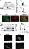
α11 influences myofibroblast differentiation in human corneal fibroblasts. A, α11 and α-SMA protein levels in corneal fibroblasts that remained unstimulated (−) or were treated (+) with 5 ng/ml of TGF-β1 or 2 nm activin A (act A) for 3 days. B, Immunolocalization of α11 (red) at focal adhesions and α-SMA (green) at stress fibers in corneal fibroblasts treated with 5 ng/ml TGF-β1 for 3 days. C, α11 and α-SMA protein levels in corneal fibroblasts that remained unstimulated (−) or were treated (+) for 3 days with 5 ng/ml TGF-β1 and siRNA (100 nm) to α11 or an off-target siRNA (mock) as a control. D, immunolocalization of α-SMA in corneal fibroblasts that remained untreated (upper left), were stimulated with 5 ng/ml TGF-β1 only (upper right), or were treated with 5 ng/ml TGF-β1 and siRNA (100 nm) to α11 (lower right) or an off-target siRNA (negative control (NC); lower left). The exposure time when acquiring pictures was identical in all conditions. In A and C, band intensities were quantified, normalized to β-actin, and calibrated to the normalized value corresponding to untreated cells. GAPDH, glyceraldehyde-3-phosphate dehydrogenase.

Similar articles

Cited by

References

    1. Hinz B. (2007) J. Invest. Dermatol. 127, 526–537 - PubMed
    1. Tomasek J. J., Gabbiani G., Hinz B., Chaponnier C., Brown R. A. (2002) Nat. Rev. Mol. Cell Biol. 3, 349–363 - PubMed
    1. Wipff P. J., Rifkin D. B., Meister J. J., Hinz B. (2007) J. Cell Biol. 179, 1311–1323 - PMC - PubMed
    1. Desmoulière A., Guyot C., Gabbiani G. (2004) Int. J. Dev. Biol. 48, 509–517 - PubMed
    1. Hinz B., Dugina V., Ballestrem C., Wehrle-Haller B., Chaponnier C. (2003) Mol. Biol. Cell 14, 2508–2519 - PMC - PubMed

Publication types

MeSH terms