. 2010 Jun;38(10):3351-61.
doi: 10.1093/nar/gkq033. Epub 2010 Feb 2.

Arginine methylation of REF/ALY promotes efficient handover of mRNA to TAP/NXF1

Affiliations

Arginine methylation of REF/ALY promotes efficient handover of mRNA to TAP/NXF1

Ming-Lung Hung et al. Nucleic Acids Res. 2010 Jun.

Abstract

The REF/ALY mRNA export adaptor binds TAP/NXF1 via an arginine-rich region, which overlaps with its RNA-binding domain. When TAP binds a REF:RNA complex, it triggers transfer of the RNA from REF to TAP. Here, we have examined the effects of arginine methylation on the activities of the REF protein in mRNA export. We have mapped the arginine methylation sites of REF using mass spectrometry and find that several arginines within the TAP and RNA binding domains are methylated in vivo. However, arginine methylation has no effect on the REF:TAP interaction. Instead, arginine methylation reduces the RNA-binding activity of REF in vitro and in vivo. The reduced RNA-binding activity of REF in its methylated state is essential for efficient displacement of RNA from REF by TAP in vivo. Therefore, arginine methylation fine-tunes the RNA-binding activity of REF such that the RNA-protein interaction can be readily disrupted by export factors further down the pathway.


Figures
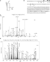
Figure 1.
Figure 1.
Identification of REF methylation sites. (A) Extract from 293T cells transfected with FLAG-Myc tagged REF was subjected to α-FLAG immunopurification in stringent conditions. Purified protein was analysed on SDS-PAGE stained with Coomassie blue. (B) Dimethylation (R) and both mono or dimethylation (R) sites are mapped on the primary sequence of REF together with residues involved in the interaction with TAP (closed circle) and RNA (open triangle). The underlined sequence corresponds to the RRM domain of REF. (C) CID MS/MS fragmentation spectra of the methylated peptide N(dmeR)PAIA(dimeR)GGR [M+2H]2+. (D) ETD MS/MS fragmentation spectra of the methylated peptide S(dimeR)GSGGFGG(dimeR)GSQG(dimeR)G(dimeR)GTGR [M+3H]3. The prominent y, b, c′, z′• ions and the characteristic neutral losses/precursor ion associated with asymmetric dimethylation (dimethylamine) are highlighted.
Figure 2.
Figure 2.
PRMT1 interacts with REF. (A) Co-immunoprecipitation of FLAG-Myc-REF with PRMT1. The left two panels show western blots with the PRMT1 antibody. The right panel is a Coomassie stained gel of the immunoprecipitations. FLAG-Myc-REF is expressed at high levels and clearly visible after immunopuriifcation. (B) Pull-down assays with GST-PRMT1 and in vitro translated REF. The left panel shows the Coomassie stained proteins following the pull-down and the right panels show phosphoimages of the radiolabelled REF input sample and pull-downs.
Figure 3.
Figure 3.
Arginine methylation of REF does not affect the binding of TAP. (A) Pull-down assay. GST or GST-TAP-p15 were first immobilized onto glutathione-coated beads before total extracts from +/– AdOx-treated 293T cells transfected with FLAG-Myc-REF were added to the binding reactions. Eluted proteins were analysed by SDS-PAGE stained with Coomassie blue (left) and western blotting (right). A 293T cell extract expressing FLAG-tagged BAP was used as a control (B) 293T cells co-transfected with either a FLAG control or a FLAG-Myc-tagged REF and either a 13Myc-tagged TAP or a Myc-control vector were cultured +/– AdOx. Input samples are shown in the top panel following western blotting with α-Myc antibody. Extracts were then subjected to α-FLAG immunoprecipitation (IP) and eluted proteins were analysed by western blotting (WB) with α-Myc antibody (WB) (bottom). (C) 293T cells co-transfected with either a FLAG control or a FLAG-Myc-tagged REF were cultured +/– AdOx. Extracts were then subjected to α-FLAG immunoprecipitation (IP) and eluted proteins were analysed by western blotting with α-FLAG (middle) and α-TAP (bottom).
Figure 4.
Figure 4.
Arginine methylation decreases the mRNA-binding activity of REF and human REF/ALY. (A) In vitro protein:RNA UV cross-linking assay. Immuno-purified FLAG or FLAG-Myc-REF from transfected 293T cells treated (+) or not (–) with AdOx was UV-cross linked (+) or not (–) with a 32P-radiolabelled RNA oligonucleotide. Resulting complexes were analysed by SDS-PAGE stained with Coomassie blue (lower panel) and Phosphoimage (Upper panel). (B) RNA-binding affinities were measured for immunopurified FLAG-REF from ADOX-treated or untreated 293T cells and a 32P-labelled 21-mer oligoribonucleotide as detailed in ‘Materials and Methods’ section. Standard error bars were calculated from two independent experiments. (C) mRNP capture assay. Poly(A)+ RNA from 293T cells +/– AdOx transfected with FLAG, FLAG-Myc-REF (REF) or FLAG-Myc-BAP was purified on oligo-dT or IgG Sepharose beads as indicated in denaturing conditions after UV cross-linking (+) or not (–). Total extract (1% of input) and eluted proteins were analysed by western blotting (WB) with α-FLAG antibody. (D) Same experiment as in (C) using untransfected 293T cells and α-ALY antibody to detect the endogenous REF/ALY protein.
Figure 5.
Figure 5.
Arginine methylation of REF facilitates the RNA handover from REF to TAP-p15 during mRNA export. (A) In vivo competition assay. 293T cells transfected with either a FLAG control (lanes 1 and 7) or FLAG-Myc-REF (lanes 2–6 and 8–12) and increasing amounts of 13-Myc-TAP and p15 expression plasmids, were treated +/– AdOx before UV cross-linking. Extracts were subjected to α-FLAG immunoprecipitation and REF:RNA complexes were treated with RNase. End-restricted mRNAs cross-linked to REF were radiolabelled using polynucleotide kinase before phosphoimaging (Top). The increasing expression of TAP was confirmed in total extracts by α-Myc western blotting (WB) (Middle) and specific immunoprecipitation of FLAG-Myc-REF was confirmed by α-FLAG WB (Bottom). (B) Quantification of the radiolabelled RNA detected on the phosphoimage of the competition assay. Error bars represent the standard error of the mean from three independent experiments.

References

    1. Kohler A, Hurt E. Exporting RNA from the nucleus to the cytoplasm. Nat. Rev. Mol. Cell. Biol. 2007;8:761–773. - PubMed
    1. Farny NG, Hurt JA, Silver PA. Definition of global and transcript-specific mRNA export pathways in metazoans. Genes Dev. 2008;22:66–78. - PMC - PubMed
    1. Carmody SR, Wente SR. mRNA nuclear export at a glance. J. Cell. Sci. 2009;122:1933–1937. - PMC - PubMed
    1. Johnson SA, Cubberley G, Bentley DL. Cotranscriptional recruitment of the mRNA export factor Yra1 by direct interaction with the 3′ end processing factor Pcf11. Mol. Cell. 2009;33:215–226. - PMC - PubMed
    1. Yoh SM, Cho H, Pickle L, Evans RM, Jones KA. The Spt6 SH2 domain binds Ser2-P RNAPII to direct Iws1-dependent mRNA splicing and export. Genes Dev. 2007;21:160–174. - PMC - PubMed

Publication types

MeSH terms