. 2010 Apr;84(8):4041-9.
doi: 10.1128/JVI.02471-09. Epub 2010 Feb 3.

Direct evidence from single-cell analysis that human {alpha}-defensins block adenovirus uncoating to neutralize infection

Affiliations

Direct evidence from single-cell analysis that human {alpha}-defensins block adenovirus uncoating to neutralize infection

Emily K Nguyen et al. J Virol. 2010 Apr.

Abstract

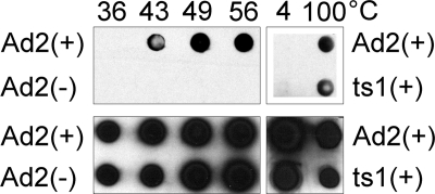
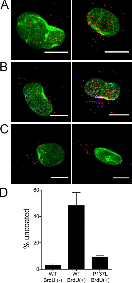
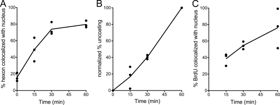
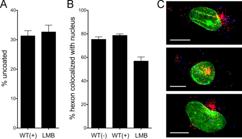
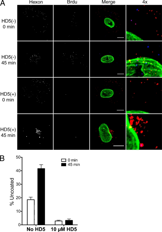
Human alpha-defensins are evolutionarily conserved effectors of the innate immune response with broadly acting antibacterial activity. Their role in antiviral immunity is less well understood. We previously showed that these antimicrobial peptides are potent inhibitors of human adenovirus infection. Based on biochemical studies and indirect evidence from confocal microscopy, we proposed that defensins bind to and stabilize the virus capsid and neutralize infection by preventing the release of the endosomalytic protein VI. To determine whether defensin action also restricts exposure of the viral genome, we developed a system to evaluate adenovirus uncoating during cell entry by monitoring the exposure of BrdU-labeled viral genomes. This assay allowed us to determine the kinetics of uncoating of virus particles in single cells. Using this assay, we now provide direct evidence that human alpha-defensins block adenovirus infection by preventing uncoating during cell entry.


Figures

FIG. 1.
FIG. 1.
BrdU-labeled genomes are exposed upon capsid disassembly. HAdV-2 (Ad2) or HAdV-2ts1 (ts1), labeled (+) or unlabeled (−) with BrdU, was incubated at the indicated temperatures before being analyzed by immunoblotting for BrdU (upper panels) or HAdV-2 proteins (lower panels).
FIG. 2.
FIG. 2.
Detection of BrdU in labeled, uncoated virus particles in infected cells. Representative images, which are stained for hexon (red), BrdU (blue), and lamin (green), are shown for unlabeled HAdV-5 wild type (WT) (A), labeled HAdV-5 WT (B), and labeled HAdV-5P137L (C) viruses. Scale bars are 10 μm. (D) Percent uncoating was determined for HAdV-5 (WT) or HAdV-5P137L (5P137L) vectors, labeled (+) or unlabeled (−) with BrdU, at 45 min postinfection in A549 cells. Data are the means and standard errors of the means of the results from three independent experiments in which ∼30 cells were evaluated for each condition. Note that the low value for the unlabeled WT virus in this assay does not indicate that the virus failed to uncoat but reflects the absence of BrdU in its genome.
FIG. 3.
FIG. 3.
Kinetics of hexon trafficking and genome exposure and translocation in infected cells. A549 cells were infected with BrdU-labeled HAdV-5 and analyzed for hexon colocalization with the nucleus (A), normalized percent uncoating (B), and BrdU colocalization with the nucleus (C) at the indicated times postinfection. Points represent the mean values from each of three independent experiments in which ∼30 cells were evaluated at each time point. Lines connect the averages of these mean values. In panel C, no value is given for the zero time point, as no uncoating occurred.
FIG. 4.
FIG. 4.
BrdU labeling does not alter virus trafficking to the MTOC. A549 cells infected with HAdV-5 (WT), labeled (+) or unlabeled (−) with BrdU, or with BrdU-labeled HAdV-5 in the presence of 20 nM LMB (LMB) were evaluated at 45 min postinfection for percent uncoating (A) and hexon colocalization with the nucleus (B). Data are the mean values and standard errors for ∼30 cells for each condition. Note that the WT(−) and WT(+) controls from this experiment were included as one of the three experiments averaged for Fig. 2. (C) Three cells representative of the LMB phenotype, colored as in Fig. 2, are shown. Scale bar is 10 μm.
FIG. 5.
FIG. 5.
The α-defensin HD5 blocks genome exposure during cell entry. A549 cells were infected with BrdU-labeled HAdV-5 in the presence (+) or absence (−) of 10 μM HD5. (A) Representative cells, separated by color channel as indicated or merged and colored as in Fig. 2, are shown for the indicated time points. A section of the merged image for each condition was enlarged fourfold (4x). Scale bar, 10 μm. (B) Percent uncoating was evaluated at 0 and 45 min postinfection. Data are the mean values and standard errors for ∼30 cells for each condition from one experiment representative of three independent experiments.

Similar articles

Cited by

References

    1. Arnberg, N. 2009. Adenovirus receptors: implications for tropism, treatment and targeting. Rev. Med. Virol. 19:165-178. - PubMed
    1. Bailey, C. J., R. G. Crystal, and P. L. Leopold. 2003. Association of adenovirus with the microtubule organizing center. J. Virol. 77:13275-13287. - PMC - PubMed
    1. Berns, K. I., and S. Adler. 1972. Separation of two types of adeno-associated virus particles containing complementary polynucleotide chains. J. Virol. 9:394-396. - PMC - PubMed
    1. Berns, K. I., and J. A. Rose. 1970. Evidence for a single-stranded adenovirus-associated virus genome: isolation and separation of complementary single strands. J. Virol. 5:693-699. - PMC - PubMed
    1. Cerullo, V., M. P. Seiler, V. Mane, N. Brunetti-Pierri, C. Clarke, T. K. Bertin, J. R. Rodgers, and B. Lee. 2007. Toll-like receptor 9 triggers an innate immune response to helper-dependent adenoviral vectors. Mol. Ther. 15:378-385. - PubMed

Publication types

LinkOut - more resources