. 2010 Apr;84(8):3993-4001.
doi: 10.1128/JVI.02289-09. Epub 2010 Feb 3.

Killing of avian and Swine influenza virus by natural killer cells

Affiliations

Killing of avian and Swine influenza virus by natural killer cells

Hagit Achdout et al. J Virol. 2010 Apr.

Abstract

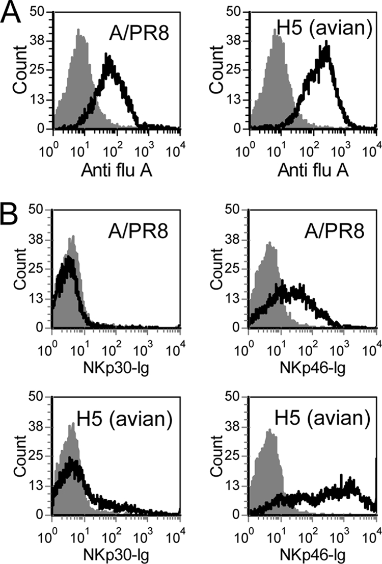
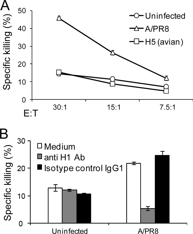
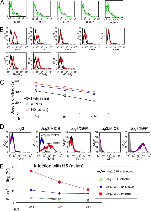
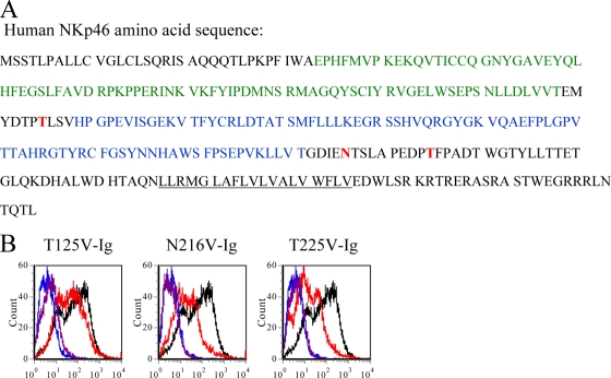
Today, global attention is focused on two influenza virus strains: the current pandemic strain, swine origin influenza virus (H1N1-2009), and the highly pathogenic avian influenza virus, H5N1. At present, the infection caused by the H1N1-2009 is moderate, with mortality rates of less <1%. In contrast, infection with the H5N1 virus resulted in high mortality rates, and ca. 60% of the infected patients succumb to the infection. Thus, one of the world greatest concerns is that the H5N1 virus will evolve to allow an efficient human infection and human-to-human transmission. Natural killer (NK) cells are one of the innate immune components playing an important role in fighting against influenza viruses. One of the major NK activating receptors involved in NK cell cytotoxicity is NKp46. We previously demonstrated that NKp46 recognizes the hemagglutinin proteins of B and A influenza virus strains. Whether NKp46 could also interact with H1N1-2009 virus or with the avian influenza virus is still unknown. We analyzed the immunological properties of both the avian and the H1N1-2009 influenza viruses. We show that NKp46 recognizes the hemagglutinins of H1N1-2009 and H5 and that this recognition leads to virus killing both in vitro and in vivo. However, importantly, while the swine H1-NKp46 interactions lead to the direct killing of the infected cells, the H5-NKp46 interactions were unable to elicit direct killing, probably because the NKp46 binding sites for these two viruses are different.


Figures

FIG. 1.
FIG. 1.
NKp46 recognition of influenza virus-infected Jeg3 cells. fluorescence-activated cell sorting (FACS) staining of uninfected Jeg3 cells (gray filled histogram) or Jeg3 cells infected with A/PR8 or H5 (avian) viruses (black line). Staining was performed with anti-influenza virus A (Anti-flu A), NKp30-Ig, and NKp46-Ig fusion proteins (B) (indicated in the x axis of the figure). The figure shows the results for one representative experiment out of four performed.
FIG. 2.
FIG. 2.
Jeg3 cells infected with avian influenza virus are not killed by NK cells. (A) Killing assay. Jeg3 cells, either uninfected or infected with A/PR8 or H5 (avian) influenza viruses, were tested in killing assays by using bulk NK cells. The effector to target ratio (E:T) is indicated in the figure. (B) The killing of Jeg3 cells is mediated by the interaction with HA. Jeg3 cells infected or not with A/PR8 influenza virus were preincubated with anti-H1 MAb, H28-E23 or with anti-CD99 MAb (negative control) and were tested in killing assays by bulk NK cells at an E:T ratio of 10:1. The figure shows the results for one representative experiment of three performed. Error bars indicate the standard deviations.
FIG. 3.
FIG. 3.
The killing of the avian-infected cells is possible when several NK activating receptors are engaged. (A) FACS staining of Jeg3 cells for NKG2D ligands. Staining was performed with anti-MICA, MICB, and ULBP1-3 MAbs. (B) FACS staining of Hep3b cells for NKG2D and NCR ligands. Staining was performed with anti-MICA, MICB, ULBP1-3 MAbs, NKp30-Ig, NKp44-Ig, and NKp46-Ig fusion proteins (indicated in the x axis of the figure). (C) Killing experiment of Hep3b cells infected or not with A/PR8 or with H5N1 viruses by bulk NK cells. The various E:T ratios are indicated in the figure. (D) FACS staining of different Jeg3 cells transfected with MICB or with GFP as control. In the three left histograms, staining was performed with an anti-MICB MAb (red line) and with either a negative control MAb (blue line) or with the secondary MAb (black line). In the two right histograms the GFP intensity of the indicated cells is shown. (E) Killing experiment of H5N1-infected Jeg3/GFP and Jeg3/MICB cells by bulk NK cells. The E:T ratio is indicated in the figure. The figure shows the results for one representative experiment out of three performed. Error bars indicate standard deviations.
FIG. 4.
FIG. 4.
Glycosylation at amino acids Asp216 and Thr225 of NKp46 are involved in its binding to H5-infected cells. (A) Amino acid sequences of human NKp46. The first Ig and the second Ig domains are indicated in green and in blue, respectively. The transmembrane domain is underlined, and the three putative glycosylation sites are indicated in bold red. (B) H5N1 (avian) influenza virus-infected or noninfected Jeg3 cells were stained with NKp46-Ig (black and blue lines, respectively) and with the indicated mutated proteins (uninfected [purple] and infected [red]). The figure shows the results of one representative experiment of three performed.
FIG. 5.
FIG. 5.
The binding sites of H1 and H5 to NKp46 are distinct. Jeg3/MICB cells were infected with A/PR8 or with H5 (avian) viruses, and their killing by bulk NK cells was tested in the presence or absence of the indicated anti-NKp46 MAbs. The E:T ratio is 60:1. The figure shows the results for one representative experiment of three performed. Error bars indicate standard deviations.
FIG. 6.
FIG. 6.
The H1N1-2009-NKp46 interaction is similar to other human origin influenza virus HAs. (A) Jeg3 cells infected or not infected with 19 different isolations of H1N1-2009 (the isolation number is indicated at the x axis) that were obtained from different Israeli patients. Jeg3 cells were stained either with NKp46-Ig or with T225V-Ig (white and gray bars, respectively). (B) The cells in panel A were tested for killing by NK cells in E:T ratio of 25:1. Error bars indicate standard deviations.
FIG. 7.
FIG. 7.
Blocking of NKG2D activity during infection. (A) Redirected killing assay. 35S-labeled P815 cells were infected or not with H5 (avian virus) or H3N2 (human influenza virus) and then assayed in a redirected killing assay with human NK cells at E:T of 3:1 in the presence of either anti-NKG2D MAb or control IgG1 MAb. (B) Bulk NK cells were preincubated with anti-NKG2D MAb or with anti-CD99 MAb (negative control) and were tested in killing assays at an E:T ratio of 10:1 against Jeg3/MICB cells that were infected or not infected with A/PR8 or H5N1 influenza viruses. (C) Anti-NKG2D receptor antibody was injected or not injected into C57BL/6 mice (WT) or C57BL/6 NCR1gfp/gfp mice (KO) 6 h prior to their intranasal inoculation with 16 TCID50 of the H1N1-2009 virus (isolation number 926)/mouse or with 1.6 TCID50 of the H5N1 virus/mouse. Five or six days (for avian and H1N1-2009 infections, respectively) after infection the mice were sacrificed, their lungs were homogenized, RNA was extracted, and the presence of virus in the infected lungs was detected by specific primers using real-time RT-PCR. The virus titer was determined based on the viral genome copy numbers.

Similar articles

Cited by

References

    1. Achdout, H., T. I. Arnon, G. Markel, T. Gonen-Gross, G. Katz, N. Lieberman, R. Gazit, A. Joseph, E. Kedar, and O. Mandelboim. 2003. Enhanced recognition of human NK receptors after influenza virus infection. J. Immunol. 171:915-923. - PubMed
    1. Achdout, H., I. Manaster, and O. Mandelboim. 2008. Influenza virus infection augments NK cell inhibition through reorganization of major histocompatibility complex class I proteins. J. Virol. 82:8030-8037. - PMC - PubMed
    1. Arnon, T. I., H. Achdout, O. Levi, G. Markel, N. Saleh, G. Katz, R. Gazit, T. Gonen-Gross, J. Hanna, E. Nahari, A. Porgador, A. Honigman, B. Plachter, D. Mevorach, D. G. Wolf, and O. Mandelboim. 2005. Inhibition of the NKp30 activating receptor by pp65 of human cytomegalovirus. Nat. Immunol. 6:515-523. - PubMed
    1. Arnon, T. I., H. Achdout, N. Lieberman, R. Gazit, T. Gonen-Gross, G. Katz, A. Bar-Ilan, N. Bloushtain, M. Lev, A. Joseph, E. Kedar, A. Porgador, and O. Mandelboim. 2004. The mechanisms controlling the recognition of tumor- and virus-infected cells by NKp46. Blood 103:664-672. - PubMed
    1. Arnon, T. I., M. Lev, G. Katz, Y. Chernobrov, A. Porgador, and O. Mandelboim. 2001. Recognition of viral hemagglutinins by NKp44 but not by NKp30. Eur. J. Immunol. 31:2680-2689. - PubMed

Publication types

MeSH terms

Substances