. 2010 Apr 1;21(7):1153-65.
doi: 10.1091/mbc.e09-10-0910. Epub 2010 Feb 3.

Evasion of endoplasmic reticulum surveillance makes Wsc1p an obligate substrate of Golgi quality control

Affiliations

Evasion of endoplasmic reticulum surveillance makes Wsc1p an obligate substrate of Golgi quality control

Songyu Wang et al. Mol Biol Cell. .

Abstract

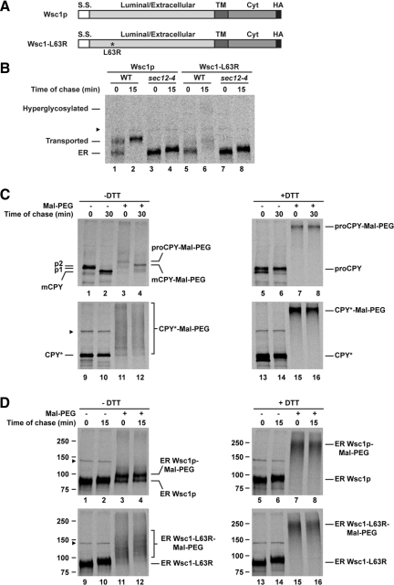
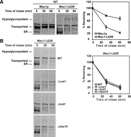
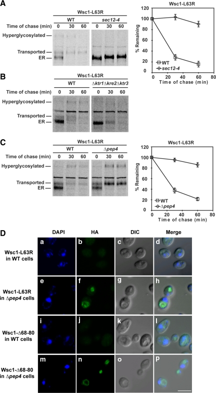
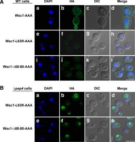
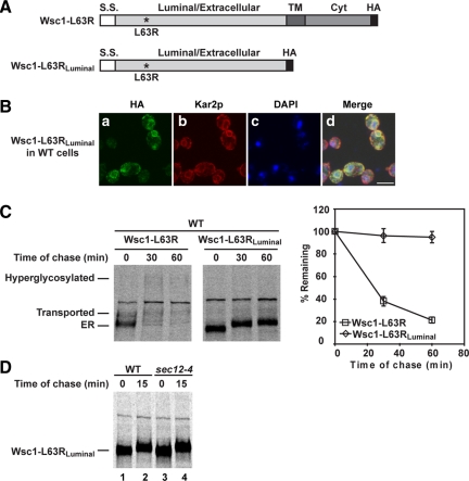
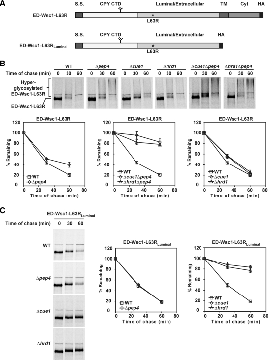
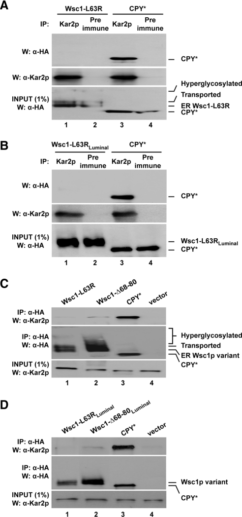
In the endoplasmic reticulum (ER), most newly synthesized proteins are retained by quality control mechanisms until folded. Misfolded molecules are sorted to ER-associated degradation (ERAD) pathways for disposal. Reports of mutant proteins degraded in the vacuole/lysosome suggested an independent Golgi-based mechanism also at work. Although little is understood of the post-ER pathway, the growing number of variants using it suggests a major role in quality control. Why seemingly redundant mechanisms in sequential compartments are needed is unclear. To understand their physiological relationship, the identification of endogenous pathway-specific substrates is a prerequisite. With ERAD substrates already well characterized, the discovery of Wsc1p as an obligate substrate of Golgi quality control enabled detailed cross-pathway analyses for the first time. By analyzing a panel of engineered substrates, the data show that the surveillance mode is determined by each polypeptide's intrinsic design. Although most secretory pathway proteins can display ERAD determinants when misfolded, the lack thereof shields Wsc1p from inspection by ER surveillance. Additionally, a powerful ER export signal mediates transport whether the luminal domain is folded or not. By evading ERAD through these passive and active mechanisms, Wsc1p is fully dependent on the post-ER system for its quality control.


Figures

Figure 1.
Figure 1.
A misfolded variant of Wsc1p. (A) Schematic representation of HA-tagged Wsc1p and Wsc1-L63R. The asterisk marks the position of the L63R mutation. S.S., signal sequence; TM, transmembrane domain; Cyt, cytoplasmic domain. (B) Wild-type (WT) and sec12-4 cells expressing Wsc1p or Wsc1-L63R were grown to log phase at 23°C and shifted to 37°C for 20 min before a 5-min pulse-label with [35S]methionine/cysteine and a 15-min chase. Wsc1p and Wsc1-L63R were immunoprecipitated from detergent lysates using anti-HA antibody and resolved by SDS-PAGE. (C) Wild-type cells incubated in the presence or absence of 10 mM DTT were pulse-labeled for 5 min and chased for 0 and 30 min at 30°C. Proteins were incubated with or without Mal-PEG after TCA precipitation. CPY and CPY* were immunoprecipitated from detergent lysates using anti-CPY and anti-HA antisera, respectively. The positions of ER (p1), Golgi (p2), and mature vacuolar form (mCPY) of CPY are indicated. (D) The sec12-4 strain expressing Wsc1p or Wsc1-L63R was labeled for 5 min and chased for 15 min as described in B. PEGylation-based folding assay was performed as described in C. Immunoprecipitated Wsc1p and Wsc1-L63R were resolved by SDS-PAGE. The arrowhead denotes the position of a nonspecific protein that cross-reacts with the anti-HA antibody.
Figure 2.
Figure 2.
Wsc1-L63R is degraded rapidly using an ERAD-independent pathway. (A) Wild-type cells expressing Wsc1p or Wsc1-L63R were pulse-labeled at 30°C with [35S]methionine/cysteine for 10 min followed by a cold chase for times indicated. Proteins were resolved by SDS-PAGE and quantified by phosphorimager analysis. The data plotted reflect three independent experiments with mean ± SD indicated. (B) Wsc1-L63R turnover was measured in wild-type, Δcue1, Δhrd1, and Δdoa10 strains by pulse-chase analysis as described in A.
Figure 3.
Figure 3.
Wsc1-L63R is transported to the vacuole via the Golgi for degradation. (A) Stability of Wsc1-L63R was examined in wild-type and sec12-4 cells using pulse-chase analysis as described in Figure 2A, except both strains were grown to log phase at 23°C and shifted to 37°C for 20 min before labeling. (B and C) Pulse-chase analysis was performed in wild-type, Δktr1Δkre2Δktr3, and Δpep4 cells expressing Wsc1-L63R as described in Figure 2A. (D) Wild-type and Δpep4 cells expressing Wsc1-L63R or Wsc1-Δ68-80 were fixed and permeabilized. Staining was performed using anti-HA primary antibodies followed by Alexa Fluor 488 goat anti-mouse secondary antibodies. DAPI staining marks the position of nuclei. Cells were visualized by confocal and DIC microscopy. Scale bar, 5 μm.
Figure 4.
Figure 4.
Misfolded Wsc1p is degraded by Golgi QC. (A and B) Wild-type and Δpep4 cells expressing Wsc1-AAA, Wsc1-L63R-AAA, or Wsc1-Δ68-80-AAA proteins driven by the GAS1 promoter were processed as described in Figure 3D. Staining was performed using anti-HA primary antibodies and Alexa Fluor 488 goat anti-mouse secondary antibodies. Images were captured by confocal and DIC microscopy. Scale bars, 5 μm.
Figure 5.
Figure 5.
Wsc1-L63R lacks an ERAD determinant in its luminal domain. (A) A schematic diagram of Wsc1-L63R and Wsc1-L63RLuminal. Asterisk indicates the L63R mutation. S.S., signal sequence; TM, transmembrane domain; Cyt, cytoplasmic domain. (B) Wild-type cells expressing Wsc1-L63RLuminal were processed as in Figure 3D. The cells were stained with anti-HA and anti-Kar2p primary antibodies followed by Alexa Fluor 488 goat anti-mouse and Alexa Fluor 594 goat anti-rabbit secondary antibodies. Cells were visualized by confocal microscopy. Scale bar, 5 μm. (C) The turnover of Wsc1-L63R and Wsc1-L63RLuminal in the wild-type strain was measured by pulse-chase analysis as described in Figure 2A. (D) Pulse-chase analysis was performed in wild-type and sec12-4 cells expressing Wsc1-L63RLuminal as described in Figure 1B.
Figure 6.
Figure 6.
ED-Wsc1-L63R and ED-Wsc1-L63RLuminal are ERAD substrates. (A) Schematic representations of ED-Wsc1-L63R and ED-Wsc1-L63RLuminal. S.S., signal sequence from Kar2p; CPY CTD, CPY C-terminal domain; TM, transmembrane domain; Cyt, Wsc1p cytoplasmic domain; HA, hemagglutinin tag. (B) The turnover of ED-Wsc1-L63R was examined in the wild-type and mutant strains using pulse-chase analysis as described in Figure 2A. (C) Degradation rates of ED-Wsc1-L63RLuminal were determined in wild-type and mutant cells by pulse-chase analysis as in Figure 2A.
Figure 7.
Figure 7.
Misfolded Wsc1p is not recognized by the major ER chaperone BiP/Kar2p. (A and B) Kar2p was immunoprecipitated from wild-type cells expressing Wsc1-L63R, Wsc1-L63RLuminal, or CPY* under native conditions. Protein complexes were resolved by SDS-PAGE and analyzed on immunoblots. Blots were probed using anti-HA antibody for substrates (top), stripped, and reprobed with anti-Kar2p antiserum as a control (middle). One percent of each lysate was loaded and analyzed in parallel to measure the relative amounts of input substrate used for each experiment (bottom). (C and D) Lysates from wild-type cells expressing the indicated substrates were subjected to native immunoprecipitation with anti-HA resin and Western blot analysis. Coimmunoprecipitated Kar2p was detected by anti-Kar2p immunoblots (top). The same blots were reprobed with anti-HA antibody as a control (middle). 1% of the lysates was analyzed in parallel and probed with anti-Kar2p antibody as an input control (bottom).
Figure 8.
Figure 8.
Misfolded Wsc1p fused with an ERAD determinant binds Kar2p efficiently. Lysates from wild-type cells expressing the indicated substrates were subjected to immunoprecipitation with anti-Kar2p antibody (A and B) or anti-HA resin (C and D) followed by Western blotting as described in Figure 7.

Similar articles

Cited by

References

    1. Ammerer G., Hunter C. P., Rothman J. H., Saari G. C., Valls L. A., Stevens T. H. PEP4 gene of Saccharomyces cerevisiae encodes proteinase A, a vacuolar enzyme required for processing of vacuolar precursors. Mol. Cell. Biol. 1986;6:2490–2499. - PMC - PubMed
    1. Anelli T., Sitia R. Protein quality control in the early secretory pathway. EMBO J. 2008;27:315–327. - PMC - PubMed
    1. Ashok A., Hegde R. S. Selective processing and metabolism of disease-causing mutant prion proteins. PLoS Pathog. 2009;5:e1000479. - PMC - PubMed
    1. Barlowe C. Signals for COPII-dependent export from the ER: what's the ticket out? Trends Cell Biol. 2003;13:295–300. - PubMed
    1. Barlowe C., Schekman R. SEC12 encodes a guanine-nucleotide-exchange factor essential for transport vesicle budding from the ER. Nature. 1993;365:347–349. - PubMed

Publication types

MeSH terms

Substances