. 2010 Feb 3;30(5):1582-94.
doi: 10.1523/JNEUROSCI.4515-09.2010.

Genetic fate mapping reveals that the caudal ganglionic eminence produces a large and diverse population of superficial cortical interneurons

Affiliations

Genetic fate mapping reveals that the caudal ganglionic eminence produces a large and diverse population of superficial cortical interneurons

Goichi Miyoshi et al. J Neurosci. .

Abstract

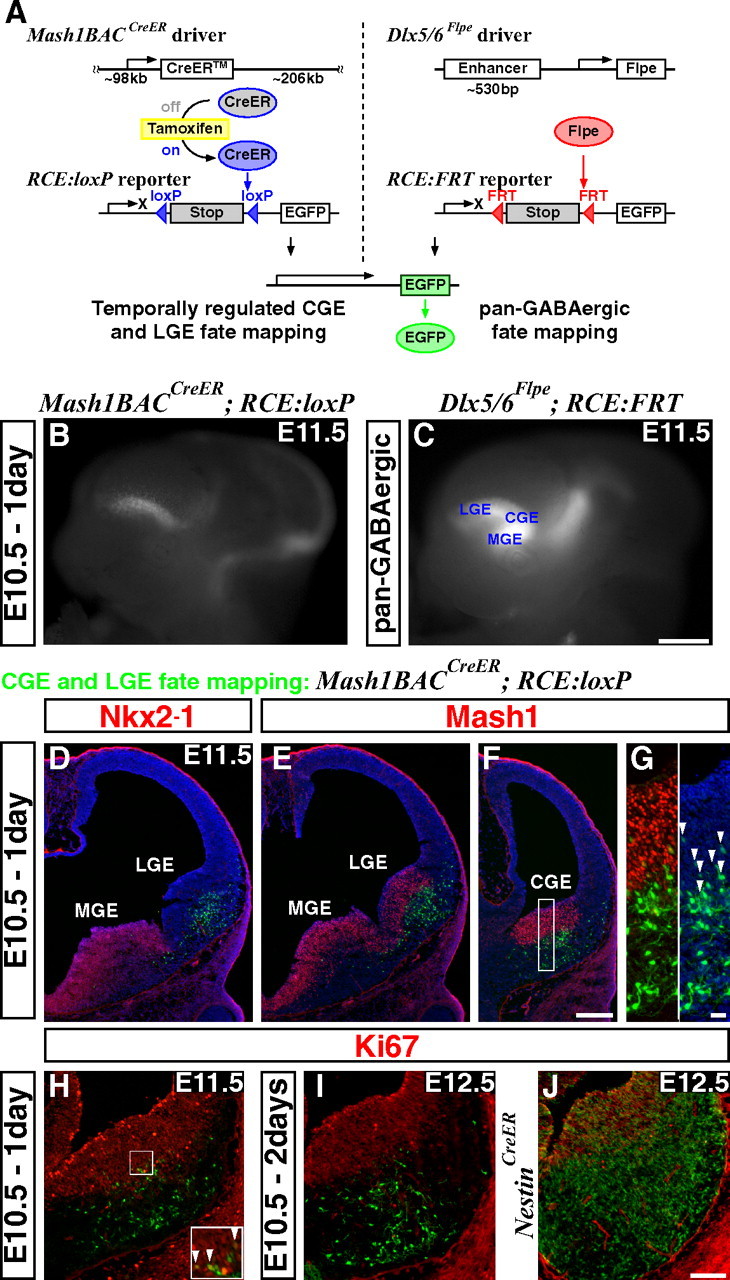
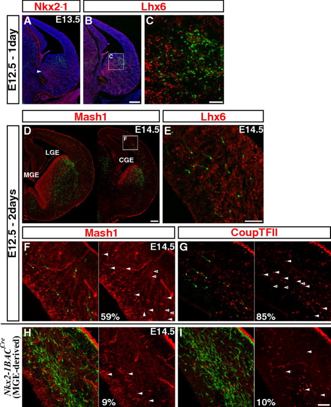
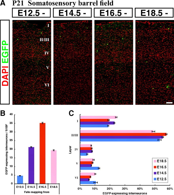
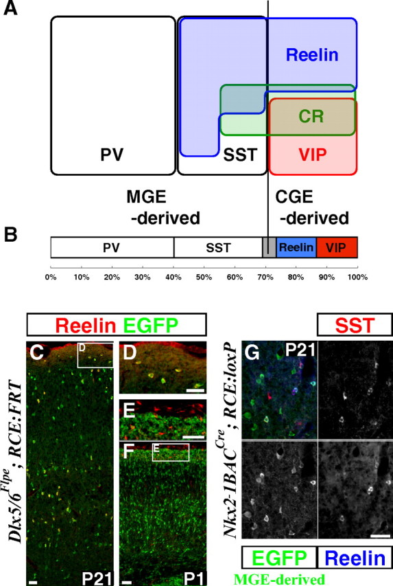
By combining an inducible genetic fate mapping strategy with electrophysiological analysis, we have systematically characterized the populations of cortical GABAergic interneurons that originate from the caudal ganglionic eminence (CGE). Interestingly, compared with medial ganglionic eminence (MGE)-derived cortical interneuron populations, the initiation [embryonic day 12.5 (E12.5)] and peak production (E16.5) of interneurons from this embryonic structure occurs 3 d later in development. Moreover, unlike either pyramidal cells or MGE-derived cortical interneurons, CGE-derived interneurons do not integrate into the cortex in an inside-out manner but preferentially (75%) occupy superficial cortical layers independent of birthdate. In contrast to previous estimates, CGE-derived interneurons are both considerably greater in number (approximately 30% of all cortical interneurons) and diversity (comprised by at least nine distinct subtypes). Furthermore, we found that a large proportion of CGE-derived interneurons, including the neurogliaform subtype, express the glycoprotein Reelin. In fact, most CGE-derived cortical interneurons express either Reelin or vasoactive intestinal polypeptide. Thus, in conjunction with previous studies, we have now determined the spatial and temporal origins of the vast majority of cortical interneuron subtypes.


Figures

Figure 1.
Figure 1.
An inducible genetic strategy for fate mapping temporally distinct interneuron cohorts derived from the CGE. A, Schematics of an inducible genetic fate mapping strategy for CGE- and LGE-derived cells (left), and a method for fate mapping the entire GABAergic population in the forebrain (right). Left, By combining a Mash1BAC-CreER driver with an RCE:loxP reporter, Mash1-expressing cells in the CGE and LGE can be labeled at the desired time point by administration of tamoxifen. Within 6–24 h of tamoxifen administration, CreER is activated and removes the stop cassette in the RCE:loxP reporter by recombining the flanking loxP sites, resulting in permanent EGFP labeling of the cells expressing Mash1 in the CGE and LGE at this time. Right, Our Dlx5/6-Flpe driver expresses the site-specific recombinase Flpe under the regulation of the intergenic enhancer between Dlx5 and Dlx6 (id6/id5). By combining this line with the RCE:FRT reporter, which possesses a stop cassette flanked by FRT sites, the entire forebrain GABAergic population is labeled with EGFP. Note that both RCE reporters (RCE:loxP and RCE:FRT) were generated from a single dual-stop reporter (RCE:dual; see supplemental Fig. 5, available at www.jneurosci.org as supplemental material). B, EGFP expression in E11.5 whole mount brains after E10.5 tamoxifen administration. Note that EGFP expression is excluded from the MGE (compare with C). In addition to the labelings in the LGE and CGE, EGFP expression was observed in the thalamus and mid/hindbrain regions. C, Dlx5/6-Flpe directed pan-GABAergic EGFP labeling in the forebrain region is observed throughout the MGE, CGE and LGE, as well as in the thalamus at E11.5. D–G, Coronal telencephalic sections of B. D, As suggested by whole mount analysis (B), EGFP labeling is excluded from the MGE, which is marked by Nkx2–1 expression. E, F, Although Mash1 is normally expressed throughout the entire ventral forebrain progenitor zone (E, F), this particular founder does not recombine the reporter in the MGE. This allowed us to specifically label the cells in the CGE (F) and the LGE (E). G, Higher magnification of the CGE progenitor domain of F. All of the EGFP-positive cells coexpressing Mash1 (arrowheads) show only low levels of EGFP expression, suggesting that the removal of the stop cassette in the reporter has occurred only recently in these cells. H, A section adjacent to F. Very few proliferating cells (Ki67-positive) are labeled 1 d after tamoxifen administration (arrowheads in inset). I, Two days after tamoxifen administration, no fate-mapped cells coexpress Ki67. J, By comparison, many of the cells fate mapped using a Nestin-CreER driver line continue to proliferate 2 d after tamoxifen administration. Scale bars: B, C, 500 μm; D–F, 200 μm; G, 20 μm; H–J, 100 μm.
Figure 2.
Figure 2.
CGE-derived cortical interneurons are first found at E12.5 fate mapping and express Mash1 and CoupTFII during tangential migration. A–C, To assess the initial labeling, coronal sections through the E13.5 telencephalon were examined 1 d after tamoxifen administration (E12.5). A, Labeling occurs in the CGE but not in the MGE where Nkx2–1 is expressed (arrowhead: caudal portion of the MGE). B, C, The genetically fate mapped cells in the CGE do not express Lhx6, a gene maintained within the MGE-lineages. D, E, Two days after tamoxifen administration at E12.5, fate mapped cells are found migrating through the cortex. Cells migrating in the cortex are first found in the caudal regions (D), and as before, they do not express MGE-lineage marker Lhx6 (E). F, G, Many of the CGE-derived interneurons migrating through cortex express low levels of Mash1 (F) and CoupTFII (G). H, I, In contrast to CGE-derived interneurons, only very few of the MGE-derived cells (labeled using a combination of the Nkx2–1BAC-Cre driver and RCE:loxP reporter lines) express Mash1 (H) or CoupTFII (I). Scale bars: A, B, D, 200 μm; C, E–I, 50 μm.
Figure 3.
Figure 3.
CGE-derived interneurons integrate preferentially into superficial cortical layers independent of their birthdates with peak production occurring at E16.5. A, Representative pictures within the P21 somatosensory cortex showing fate-mapped interneurons from four embryonic time points (E12.5, E14.5, E16.5 and E18.5). Cortical layering was revealed by DAPI nuclear counterstaining (pseudo colored in red). B, A quantification of the cell numbers at P21 from four embryonic fate-mapping time points. The largest numbers of cortical interneurons were fate mapped from the E16.5 time point. C, The laminar distributions of fate-mapped interneurons from four embryonic time points (E12.5, E14.5, E16.5, and E18.5) were analyzed. At all time points examined, CGE-derived interneurons were found to preferentially occupy superficial layers in the P21 cortex. Error bars, SEM. Scale bar, 100 μm.
Figure 4.
Figure 4.
A large proportion of interneurons is derived from the CGE and express either Reelin or VIP. A, Schematic showing the molecular expression profiles of GABAergic interneurons in the P21 somatosensory mouse barrel cortex. PV-, SST-, and VIP-immunopositive populations constitute mutually exclusive interneuron subtypes. PV- and SST-expressing populations are exclusively derived from the Lhx6-expressing MGE lineage (Fogarty et al., 2007). All VIP-expressing interneurons are derived from the CGE (see also Table 1). The MGE gives rise to a population of interneurons that coexpress Reelin and SST, while those Reelin expressing interneurons lacking SST-expression are CGE-derived (Table 3). CR is coexpressed with all markers with the exception of PV. In addition, there appears to be a small population that is solely CR-positive within the CGE-lineage. B, Within the total cortical interneuron population labeled with Dlx5/6-Flpe driver and RCE:FRT reporter, at least 69.1% (PV: 40.3% ± 0.90% and SST: 28.8%% ± 1.82%) are derived from the MGE and at least 26.4% (Reelin: 13.1% ± 1.03% and VIP: 13.3% ± 0.28%) are derived from the CGE. The gray region represents the ∼5% of all cortical interneurons for which we presently lack any marker. C–F, Reelin expression in the P21 (C, D) and P1 (E, F) mouse somatosensory cortex. The Reelin-expressing cells are GABAergic interneurons that are marked by EGFP expression from the Dlx5/6-Flpe driver and RCE:FRT reporter combination. Most of the interneurons in layer I coexpress Reelin (C, D). In contrast at P1, none of the Reelin-expressing cells in layer I are GABAergic, but instead are glutamatergic Cajal-Retzius cells (E, F). G, All of the Reelin-expressing cells derived from the MGE (marked with Nkx2–1BAC-Cre and RCE:loxP) coexpress SST, suggesting that Reelin-expressing SST-negative interneurons are exclusively derived from the CGE Table 3. Scale bars, 50 μm.
Figure 5.
Figure 5.
Reelin- and VIP-expressing interneurons originate from the CGE in relatively similar proportions between E12.5 and E18.5. A–D, Representative examples of Reelin-expressing multipolar cells (A, B) and VIP-expressing bipolar (C) and multipolar (D) cells. E, Histogram quantification of the percentage of CGE-derived interneurons at P21 fate mapped from E12.5, E14.5, E16.5 and E18.5 that express: Reelin, VIP, PV and SST. Reelin- and VIP-expressing interneurons are generated in a similar ratio (∼40% of production) from all developmental time points except at E14.5 (p = 0.001). At all time points examined, very few MGE-derived interneurons (PV- and SST-expressing) were observed. This suggests that most of the Reelin-expressing cells fate mapped do not coexpress SST. Error bars, SEM. F, The proportion of cortical interneuron subtypes generated at the onset (E12.5) and peak (E16.5) of the fate mapping of CGE-derived cortical interneurons. The sIB subtype was only found within the E12.5 fate mapped pool, and the fAD subtype was more prevalent following E16.5 labeling. Scale bar: A–D, 50 μm.
Figure 6.
Figure 6.
Nine subtypes of cortical interneurons with distinct intrinsic electrophysiological properties and morphologies are CGE-derived. CGE-derived cortical interneurons were characterized based on intrinsic electrophysiological properties and morphologies, and correlated with post hoc Reelin or VIP immunoreactivity. Top, Responses to 500 ms threshold (black trace) and below threshold (red trace) current injections (middle) responses to a hyperpolarizing current injection protocol (bottom) responses to a near maximum current injection. The LS1 (late-spiking) subtype shows a typical delayed threshold spike, a delay that persists during the firing of multiple spikes (arrows). The LS2 subtype often shows a delay to the threshold spike (top) but this delay is always eliminated at firing of more than one spike (arrow). The dIB (delayed intrinsic-bursting) subtype shows a bursting firing pattern from threshold with an AHP that typically goes below resting membrane potential of the cell (arrow). This subtype has a significant burst nonadapting (bNA) firing pattern, which persists at suprathreshold current injections (bottom). The sIB (sigmoid intrisic-bursting) subtype fires a 4–6 spike burst from threshold after a pronounced delay. The bNA1 (burst nonadapting) subtype shows a clear hump at near threshold levels early in the current injection (red arrow) and a biphasic AHP. The typical bNA pattern is seen at intermediate current injection (black arrow) of bNA1 subtype. The bNA2 subtype shows a pattern similar to that of bNA1, except for no hump in the beginning of the near threshold current injection (red arrow) and that the bNA-pattern typically persists at near maximum firing. The dNFS3 (delayed non-fast spiking) subtype shows a delay to the initial spike with a sigmoid shape and a sharp AHP (arrow in the top panel). All dNFS3 interneurons display rectification between hyperpolarizing steps (arrow in the middle panel) and a regular firing pattern with slight intraspike interval adaptation during intermediate current injections. The firing pattern of IS (irregular-spiking) subtype reveals a delay to the initial spike with subthreshold oscillations but it is not preceded by a ramp or a sigmoid shape. The shape of AHP is typically rounded (arrow in the top panel). The IS typically shows irregular firing during intermediate current injections (arrow in the middle panel) and a pronounced spike height adaptation throughout the suprathreshold current injection (arrow in the bottom panel). The fAD (fast-adapting) subtype fails to fire throughout the 500 ms current injection and show a hyperpolarization after the end of current injection (arrow in the middle panel). Note the slope of the trace after the spike train at intermediate (middle panel) and suprathreshold current injections (arrow in the bottom panel) for the fAD subtype. Bottom, Examples of morphological reconstructions by neurolucida tracing for each interneuron subtype. Dendrites are shown in blue and axon in red. The LS1 subtype exhibited the typical highly arborized dendritic pattern of neurogliaform cells while the LS2 subtype is generally larger and extends longer dendrites. The multipolar dIB subtypes were mainly found in superficial layers and often extended an axon toward deeper layers (similar to the bNA1). The bipolar sIB subtypes were found in deeper layers. The bNA2 subtypes have bipolar morphologies and were only recorded in layers II/III. Note the dNFS3 subtype showing a typical “axon arcade” morphology with an axon that starts out toward superficial layers from soma but the branches form arches when they project downward (arrows). The IS subtypes are bitufted with one dendritic branch reaching the pia and an axon extending deep while the fAD subtypes are bipolar/tripolar with an axon projecting locally as well as toward deeper layers. Numbers denote total cells recorded.

Comment in

References

    1. Alcántara S, Ruiz M, D'Arcangelo G, Ezan F, de Lecea L, Curran T, Sotelo C, Soriano E. Regional and cellular patterns of reelin mRNA expression in the forebrain of the developing and adult mouse. J Neurosci. 1998;18:7779–7799. - PMC - PubMed
    1. Anderson SA, Eisenstat DD, Shi L, Rubenstein JL. Interneuron migration from basal forebrain to neocortex: dependence on Dlx genes. Science. 1997;278:474–476. - PubMed
    1. Angevine JB, II, Sidman RL. Autoradiographic study of cell migration during histogenesis of cerebral cortex in the mouse. Nature. 1961;192:766–768. - PubMed
    1. Ascoli GA, Alonso-Nanclares L, Anderson SA, Barrionuevo G, Benavides-Piccione R, Burkhalter A, Buzsáki G, Cauli B, Defelipe J, Fairén A, Feldmeyer D, Fishell G, Fregnac Y, Freund TF, Gardner D, Gardner EP, Goldberg JH, Helmstaedter M, Hestrin S, Karube F, et al. Petilla terminology: nomenclature of features of GABAergic interneurons of the cerebral cortex. Nat Rev Neurosci. 2008;9:557–568. - PMC - PubMed
    1. Bai CB, Auerbach W, Lee JS, Stephen D, Joyner AL. Gli2, but not Gli1, is required for initial Shh signaling and ectopic activation of the Shh pathway. Development. 2002;129:4753–4761. - PubMed

Publication types

MeSH terms

LinkOut - more resources