. 2010 Feb 3;30(5):1637-47.
doi: 10.1523/JNEUROSCI.4872-09.2010.

ERK1/2 mitogen-activated protein kinase phosphorylates sodium channel Na(v)1.7 and alters its gating properties

Affiliations

ERK1/2 mitogen-activated protein kinase phosphorylates sodium channel Na(v)1.7 and alters its gating properties

Severine Stamboulian et al. J Neurosci. .

Abstract

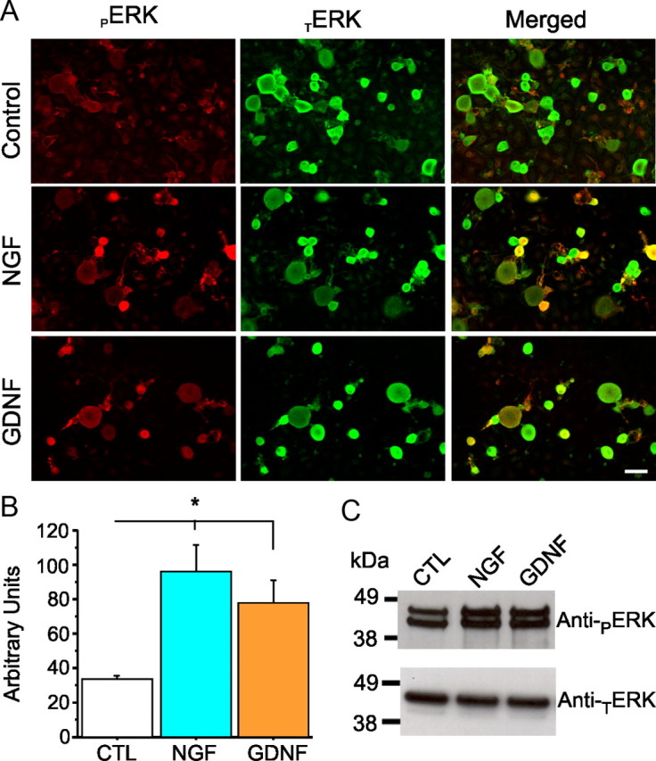
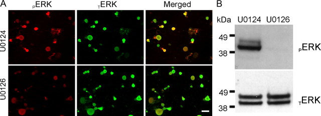
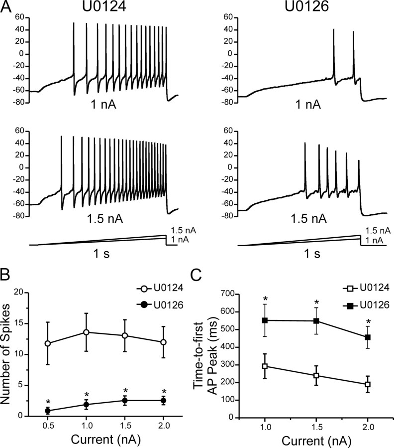
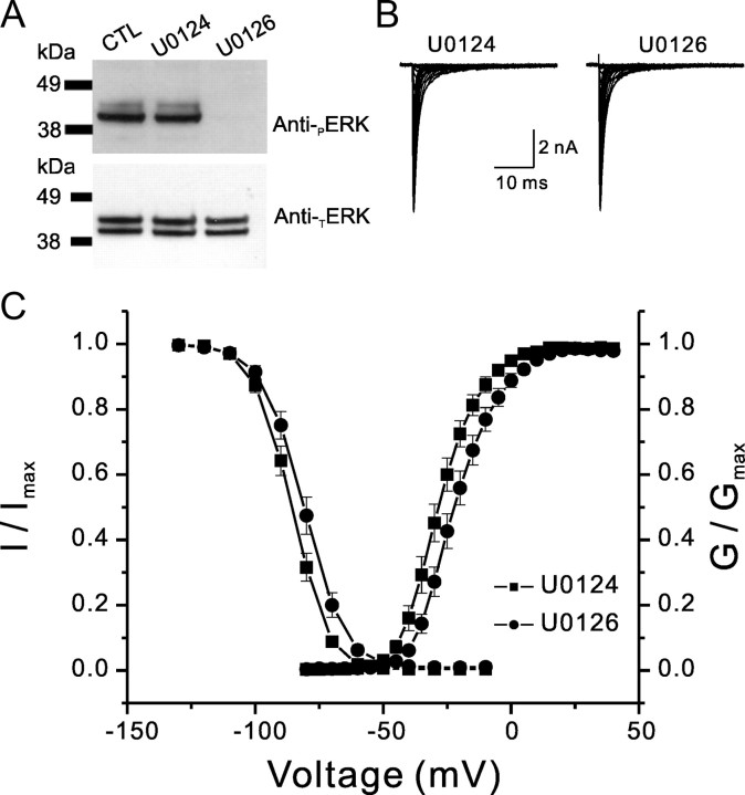
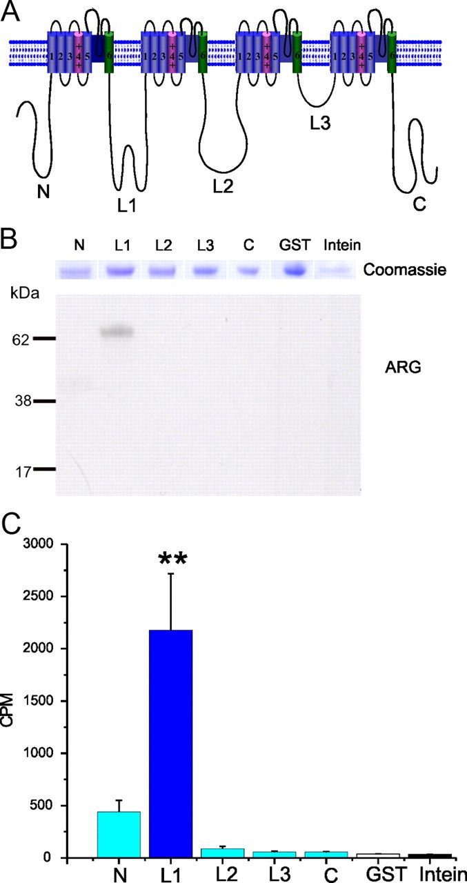
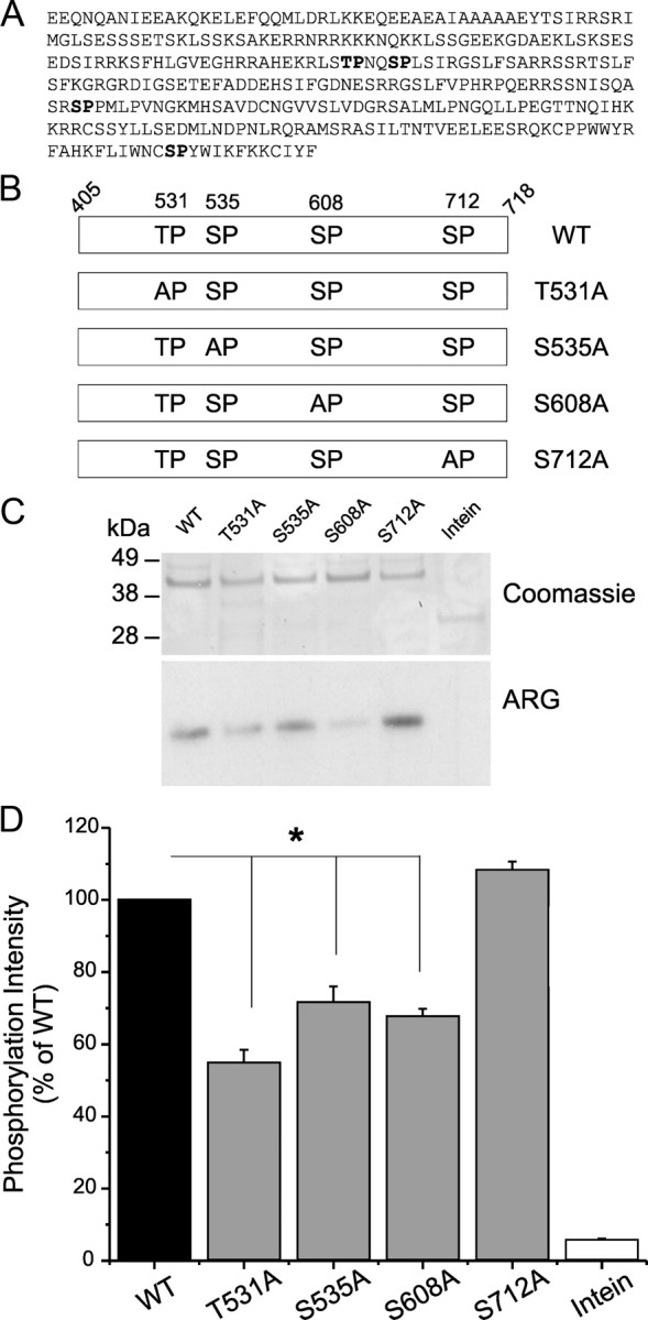
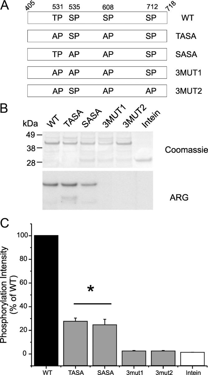
Na(v)1.7 sodium channels can amplify weak stimuli in neurons and act as threshold channels for firing action potentials. Neurotrophic factors and pro-nociceptive cytokines that are released during development and under pathological conditions activate mitogen-activated protein kinases (MAPKs). Previous studies have shown that MAPKs can transduce developmental or pathological signals by regulating transcription factors that initiate a gene expression response, a long-term effect, and directly modulate neuronal ion channels including sodium channels, thus acutely regulating dorsal root ganglion (DRG) neuron excitability. For example, neurotrophic growth factor activates (phosphorylates) ERK1/2 MAPK (pERK1/2) in DRG neurons, an effect that has been implicated in injury-induced hyperalgesia. However, the acute effects of pERK1/2 on sodium channels are not known. We have shown previously that activated p38 MAPK (pp38) directly phosphorylates Na(v)1.6 and Na(v)1.8 sodium channels and regulates their current densities without altering their gating properties. We now report that acute inhibition of pERK1/2 regulates resting membrane potential and firing properties of DRG neurons. We also show that pERK1 phosphorylates specific residues within L1 of Na(v)1.7, inhibition of pERK1/2 causes a depolarizing shift of activation and fast inactivation of Na(v)1.7 without altering current density, and mutation of these L1 phosphoacceptor sites abrogates the effect of pERK1/2 on this channel. Together, these data are consistent with direct phosphorylation and modulation of Na(v)1.7 by pERK1/2, which unlike the modulation of Na(v)1.6 and Na(v)1.8 by pp38, regulates gating properties of this channel but not its current density and contributes to the effects of MAPKs on DRG neuron excitability.


Figures

Figure 1.
Figure 1.
ERK1/2 is widely expressed in DRG neurons and colocalized with Nav1.7 sodium channels. A, Immunofluorescence labeling of adult rat DRG sections probed with anti-TERK1/2 and anti-PGP9.5 antibodies. Merged image (yellow) shows that TERK1/2 (red) and PGP9.5 (green) immunofluorescent signals colocalize within small- and medium-diameter DRG neurons. Large-diameter DRG neurons generally do not express TERK1/2. Scale bar, 50 μm. B, Immunostaining of TERK1/2 (green) and Nav1.7 (red) in DRG neurons in culture. Merged image (yellow) shows that TERK1/2 and Nav1.7 colocalize in small- and medium-diameter DRG neurons. Scale bar, 50 μm.
Figure 2.
Figure 2.
ERK1/2 are widely expressed in DRG neurons in culture containing NGF or GDNF. A, Immunostaining of ERK1/2 in adult rat DRG neurons in culture (24 h). In the morning, DRG neurons were treated for 30 min with NGF (50 μg/ml) or GDNF (50 μg/ml), and levels of TERK1/2 and pERK1/2 were examined using anti-TERK1/2 (green) and anti-pERK1/2 (red). Scale bar, 50 μm. TERK1/2 signal is present in DRG neurons in culture, and a 20 min treatment with NGF or GDNF increases the signal of pERK1/2 (merged image). B, Histogram showing pERK1/2 expression level for each culture condition. Treatment with NGF or GDNF elicits a significant increase of pERK1/2 compared with the control condition (CTL). Data are mean ± SEM. *p < 0.05, Student's t test. C, Western blot using anti-pERK1/2 antibody to assess activation of ERK1/2 in DRG neurons in culture after 30 min treatment with NGF (50 μg/ml) or GDNF (50 μg/ml). Basal levels of pERK1/2 are detected in these cultures (which contain Schwann cells and fibroblasts in addition to neurons), and levels of pERK1/2 are increased acutely by the presence of NGF or GDNF in the culture media.
Figure 3.
Figure 3.
Activation of ERK1/2 in DRG neurons is inhibited by acute treatment with U0126. A, Immunostaining of ERK1/2 in adult rat DRG neurons in culture (24 h) containing NGF (50 ng/ml) and GDNF (50 ng/ml). ERK1/2 activation inhibitor U0126 (10 μm) or its inactive analog U0124 (10 μm) were added to the media on the morning of the experiment for 20 min before cells are fixed and immunolabeled for TERK1/2 (green) and pERK1/2 (red). Scale bar, 50 μm. B, Western blot using anti-pERK1/2 (top) and anti-TERK1/2 (bottom) to assess effect of U0126 treatment on DRG neurons in culture (24 h) in the presence of NGF (50 ng/ml) and GDNF (50 ng/ml). U0126 (10 μm) or the inactive analog U0124 (10 μm) were added to the cultures for 20 min before cell lysis. Treatment with U0126 abolishes ERK1/2 activation in DRG cultures.
Figure 4.
Figure 4.
Inhibition of pERK1/2 reduces excitability of DRG neurons. Whole-cell current-clamp recordings were performed in DRG neurons pretreated for 20 min with U0124 or U0126. Action potentials were elicited by 1 s ramp current injection ranging from 0.5 to 2 nA in 0.5 nA increments from resting membrane potential. A, Top row shows representative trains of action potentials in response to a 1 nA (top traces) and 1.5 nA (bottom traces) ramp stimuli. DRG neurons treated with U0126 show reduced numbers of action potentials and longer time-to-peak for the first action potential spike compared with neurons treated with the inactive analog U0124. B, The mean number of action potentials (defined as action potentials that overshoot 0 mV) for the population of DRG neurons studied was significantly reduced for neurons pretreated with U0126 (filled symbols; n = 20) compared with U0124 (open symbols; n = 20). C, For the population of DRG neurons studied, the mean time-to-peak for the first action potential (AP) spike was significantly (*p < 0.05) longer in neurons treated with U0126 (filled symbols; n = 20) compared with U0124 (open symbols; n = 20). Data are expressed as means ± SEM.
Figure 5.
Figure 5.
pERK1/2 inhibitor U0126 shifts activation and fast inactivation of Nav1.7. A, Western blot using anti-pERK1/2 antibody (top panel) and anti-TERK1/2 antibody (bottom panel) showing the effect of treatment with U0124 (10 μm) or U0126 (10 μm) on ERK1/2 activation in HEK 293 cells transiently transfected with hNav1.7 together with β1 and β2 subunits. At 24–36 h after transfection, HEK 293 cells were incubated with 10 μm of the drug for 20 min at 37°C. Cells were then washed with ice-cold PBS, lysed, and resuspended in 2× sample buffer. Untreated [control (CTL)] or U0124-treated cells show pERK1/2 signal, whereas treatment with U0126 abolishes the pERK1/2 signal. B, Representative sodium current traces were recorded from HEK 293 cells transiently expressing hNav1.7 channels together with hβ1 and hβ2. Cells were treated with either U0124 or U0126 for 20 min before recording. Peak hNav1.7 current amplitude was comparable in cells treated with U0124 (n = 14) or U0126 (n = 13). C, For activation (right curves), whole-cell Na+ currents were elicited by 50 ms test pulses to potentials between −80 and +40 mV in steps of 5 mV from a holding potential of −120 mV. For steady-state fast inactivation (left curves), currents were elicited with test pulses to −10 mV after 500 ms conditioning pulses. The V1/2 for activation and steady-state fast inactivation were measured from curves that were fitted by Boltzmann distribution equations. For activation, V1/2,Act = −27.5 ± 1.8 mV and k = 7.0 ± 0.4 mV for U0124 (■; n = 14); V1/2,Act = −20.9 ± 1.8 mV and k = 7.8 ± 0.4 mV for U0126 (●; n = 13). For steady-state fast inactivation, V1/2,Fast = −85.8 ± 1.4 mV and k = 6.4 ± 0.1 mV for U0124; V1/2,Fast = −81.0 ± 1.7 mV and k = 7.2 ± 0.3 mV for U0126. Both activation and steady-state fast inactivation curves for the cells pretreated with U0126 were significantly different from those for U0124 (p < 0.05).
Figure 6.
Figure 6.
The first intracellular loop of the sodium channel Nav1.7 is phosphorylated by pERK1. A, Schematic of a sodium channel polypeptide showing the organization in four domains (DI–DIV) joined by three intracellular loops (L1–L3) and the intracellular N and C termini. The N, L2, L3, and C intracellular regions were produced as GST fusion proteins. The L1 intracellular region was produced as an Intein fusion protein. B, L1 is the only substrate for pERK1. Comparable amounts of fusion proteins, as determined by Coomassie blue staining (top panel), were used in the kinase assay. ARG shows that only L1 is phosphorylated by pERK1 in this assay. C, Histogram showing the Cerenkov counts of incorporated [32P]ATP in the different fusion proteins, after the kinase assay. **p < 0.01. Data are expressed as mean ± SEM; n = 3.
Figure 7.
Figure 7.
Identification of the pERK1 phosphorylation sites in L1 of Nav1.7. A, Four putative SP/TP phosphoacceptor sites (bold type) are identified within Nav1.7/L1. B, Schematic showing the replacement of S/T residues by the nonphosphorylatable alanine (T531A, S535A, S608A, S712A) to determine the contribution of each of these sites to pERK1/2 phosphorylation of Nav1.7/L1. C, Comparable amounts of WT and mutant fusion proteins, as determined by Coomassie blue staining (top), were used in the kinase assay. ARG shows that T531A, S535A, and S608A reduce [32P]ATP incorporation compared with WT, indicating that they contribute to the total phosphorylation of L1 (bottom). D, The [32P]ATP incorporation into WT L1 was corrected by the intensity of Coomassie blue staining of the band (top) and was set as 100% to measure the relative effect of individual replacement of the putative phosphorylation sites in L1. Histogram of the corrected [32P]ATP incorporation (Cerenkov count/Coomassie blue intensity) shows significant decrease for T531A, S535A, and S608A, but not S712A, mutant proteins compared with WT. *p < 0.05. Data are expressed as mean ± SEM; n = 4.
Figure 8.
Figure 8.
Triple substitutions are required to abolish phosphorylation of L1. A, Schematic diagram illustrating several double and triple replacements of phosphorylation residues by alanine [WT, TASA (T531S535/AA), SASA (S535S608/AA), 3MUT1 (T531S535S608/AAA), and 3MUT2 (T531S608S712/AAA)] that were tested as substrates for pERK1. B, Comparable amounts of WT and mutant fusion proteins, as determined by Coomassie blue staining (top), were used in the kinase assay. ARG shows that TASA and SASA are substrates for pERK1, whereas both triple mutants 3MUT1 and 3MUT2 are no longer phosphorylated by pERK1, indicating that they contribute to the total phosphorylation of L1 (bottom). C, The [32P]ATP incorporation into WT L1 was corrected by the intensity of Coomassie blue staining of the band (top) and was set as 100% to measure the relative efficiency of several double- and triple-mutant derivatives as pERK1 substrates. Histogram of the corrected [32P]ATP incorporation (Cerenkov count/Coomassie blue intensity) shows significant decrease for TASA and SASA mutant proteins, whereas 3MUT1 and 3MUT2 triple-mutant proteins reach only background levels (same as the control Intein tag), indicating that S712 may contribute to phosphorylation of L1. *p < 0.05. Data are expressed as mean ± SEM; n = 3.

Similar articles

Cited by

References

    1. Adams JP, Anderson AE, Varga AW, Dineley KT, Cook RG, Pfaffinger PJ, Sweatt JD. The A-type potassium channel Kv4.2 is a substrate for the mitogen-activated protein kinase ERK. J Neurochem. 2000;75:2277–2287. - PubMed
    1. Agthong S, Kaewsema A, Tanomsridejchai N, Chentanez V. Activation of MAPK ERK in peripheral nerve after injury. BMC Neurosci. 2006;7:45. - PMC - PubMed
    1. Averill S, Delcroix JD, Michael GJ, Tomlinson DR, Fernyhough P, Priestley JV. Nerve growth factor modulates the activation status and fast axonal transport of ERK 1/2 in adult nociceptive neurones. Mol Cell Neurosci. 2001;18:183–196. - PubMed
    1. Binshtok AM, Wang H, Zimmermann K, Amaya F, Vardeh D, Shi L, Brenner GJ, Ji RR, Bean BP, Woolf CJ, Samad TA. Nociceptors are interleukin-1β sensors. J Neurosci. 2008;28:14062–14073. - PMC - PubMed
    1. Black JA, Dib-Hajj S, McNabola K, Jeste S, Rizzo MA, Kocsis JD, Waxman SG. Spinal sensory neurons express multiple sodium channel alpha-subunit mRNAs. Mol Brain Res. 1996;43:117–131. - PubMed

Publication types

MeSH terms

Substances

LinkOut - more resources