. 2010 Feb 3;30(5):1766-76.
doi: 10.1523/JNEUROSCI.4962-08.2010.

The N-glycanase png-1 acts to limit axon branching during organ formation in Caenorhabditis elegans

Affiliations

The N-glycanase png-1 acts to limit axon branching during organ formation in Caenorhabditis elegans

Nasrin Habibi-Babadi et al. J Neurosci. .

Abstract

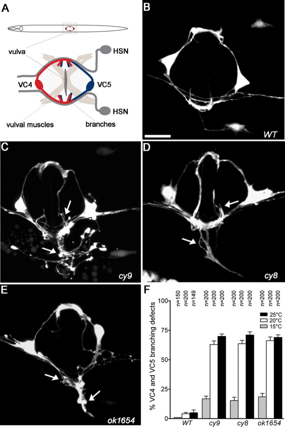
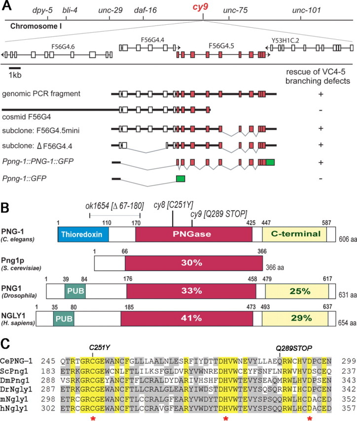
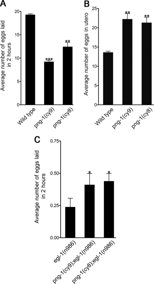
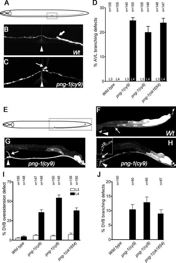
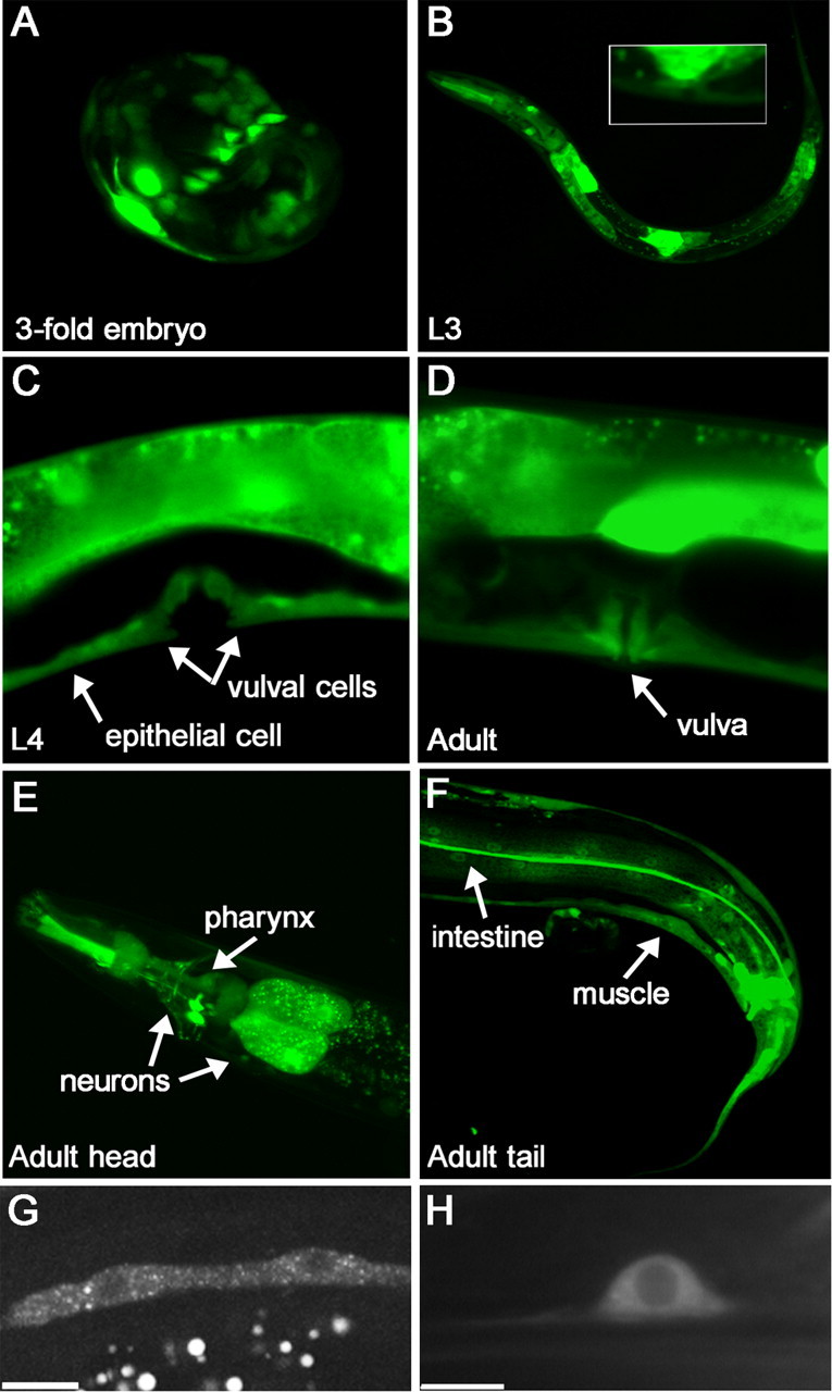
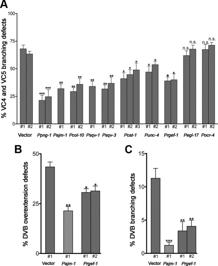
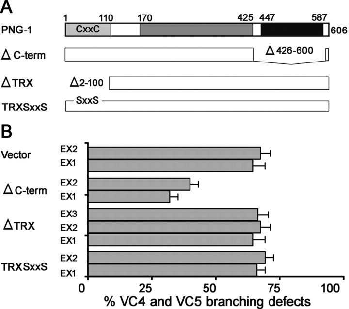
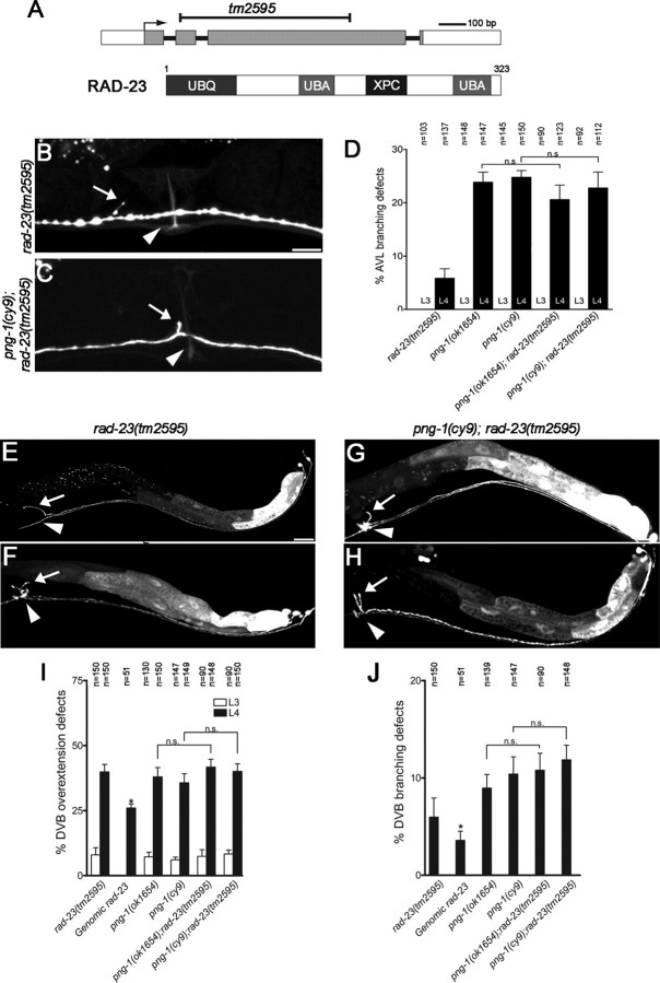
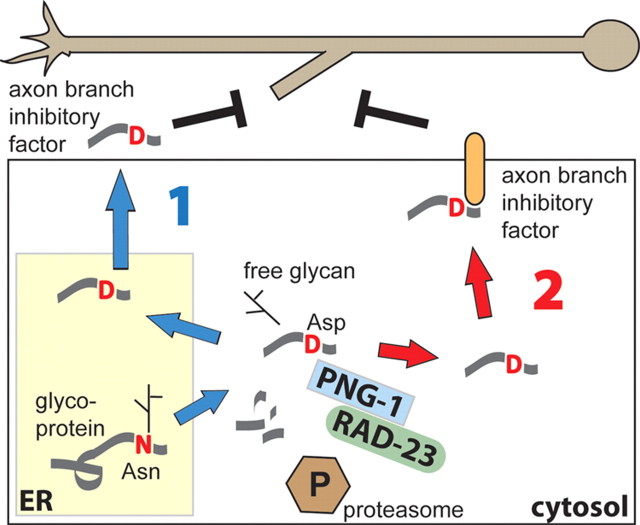
Peptide:N-glycanases (PNGases) are cytoplasmic de-N-glycosylation enzymes that have been shown in cultured cells to facilitate the degradation of misfolded glycoproteins during endoplasmic reticulum-associated degradation and in the processing of major histocompatibility complex class I antigens for proper cell-surface presentation. The gene encoding PNGase activity was initially described in budding yeast (Png1p) and shown to be highly conserved from yeast to humans, but physiological roles in higher organisms have not been elucidated. Here we describe peripheral nervous system defects associated with the first loss-of-function mutations in an animal PNGase. Mutations in png-1, the Caenorhabditis elegans PNGase ortholog, result in an increase in axon branching during morphogenesis of the vulval egg-laying organ and egg-laying behavior changes. Neuronal defects include an increase in the branched morphology of the VC4 and VC5 egg-laying neurons as well as inappropriate branches from axons that run adjacent to the vulva but would normally remain unbranched. We show that png-1 is widely expressed and can act from both neurons and epithelial cells to restrict axon branching. A deletion allele of the DNA repair gene rad-23, orthologs of which are known to physically interact with PNGases in yeast and mammals, displays similar axon branching defects and genetic interactions with png-1. In summary, our analysis reveals a novel developmental role for a PNGase and Rad-23 in the regulation of neuronal branching during organ innervation.


Figures

Figure 1.
Figure 1.
Mutants displaying excessively branched VC4 and VC5 axons. A, Schematic of a worm (ventral view) showing the positions of VC4 and VC5 and the vulval egg-laying organ innervated by the HSN and VC motor neurons. B–E, Ventral view of VC4 and VC5 neurons in adult wild-type (B; WT) and cy9, cy8, and ok1654 (C–E) mutants visualized using a cyIs4[Pcat-1::GFP] reporter. Arrows indicate axon branching defects. F, Quantification of VC4-5 excess branching defects in wild-type and mutant animals. Branching defects decrease when animals are grown at lower temperature (15°C). Error bars denote SEM; n = 150–200 animals. Scale bar, 10 μm.
Figure 2.
Figure 2.
VC axon branching mutants disrupt the PNGase (png-1) gene. A, The cy9 mutant was mapped to a small genetic interval on chromosome I and identified as the F56G4.5/png-1 gene (red) by complementation with genomic and cosmid fragments and a png-1 translational fusion to GFP (green). B, Domain organization of PNG-1 and orthologs showing percentage identity of conserved domains to PNG-1. Animal PNGases contain extended N and C termini. Nematode PNGase structures differ from fly and vertebrate PNGases in having an N-terminal thioredoxin instead of a PUB domain. C, Alignment of core PNGase domains. Molecular lesions in png-1 are indicated in B and C, and asterisks mark the invariant Cys, His, Asp active site triad. Ce, C. elegans (GenBank accession number AAF74721); Sc, S. cerevisiae (GenBank accession number S61970); Dm, Drosophila (GenBank accession number AAF74722); Dr, zebrafish (GenBank accession number AAH95313); m, mouse (GenBank accession number AAF74723); h, human (GenBank accession number AAF74720). ClustalW alignment (version 6.0).
Figure 3.
Figure 3.
png-1 is involved in egg-laying behavior. A, B, png-1 mutants have a mild Egl phenotype characterized by a reduced rate of laid eggs (A) and in utero retention of developing embryos (B). C, png-1 deficiency partially suppresses the egg-retention phenotype of an egl-1 gain-of-function mutant. HSN neurons undergo cell death in egl-1(n986) resulting in a genetic background in which egg-laying behavior is completely dependent on VC innervation of the vulval muscles. Error bars denote SEM. *p < 0.05, **p < 0.001, ***p < 0.0001, t test.
Figure 4.
Figure 4.
Several neurons display excessive or inappropriate axonal branching at the vulva in png-1 mutants. A, E, Schematics of AVL and DVB neurons with imaged areas boxed. B, C, An AVL axon, visualized in a Punc-25::GFP; unc-30(e191) background, running adjacent to the vulva (arrowhead) in a wild type (B; Wt) and displaying an inappropriate branch (arrow) in a png-1(cy9) adult (C). Thick arrow (B) points to a DVB axon. D, Inappropriate AVL branches are observed at mid-to-late L4 stage at the vulva but not before vulval organogenesis at the L3 stage in png-1 mutants. F–H, A DVB axon visualized using an Pflp-10::GFP reporter terminating (arrow) posterior to the vulva (arrowhead) in a wild type (F), overextending anteriorly and dorsally along the vulval epithelium (arrow) (G) and displaying a vulval branch (arrow) in png-1(cy9) adults (H). Pflp-10::GFP is also variably expressed in the VulD vulval cells (arrowhead in H). I, J, DVB axon overextension defects are more severe at the mid-to-late L4 stage compared with the L3 stage in png-1 mutants (I). A subset of DVB axons that display overextension defects also branch at the vulva (J). Error bars denote SEM. Scale bars: B, C, 10 μm; F–H, 20 μm.
Figure 5.
Figure 5.
png-1 is widely expressed. A–F, png-1 promoter activity at various developmental stages visualized using a Ppng-1::GFP transcriptional reporter (see Fig. 2A). The ∼600 bp promoter region was shown to be sufficient to rescue VC branching defects in png-1 mutants. Promoter activity was found in multiple tissues, including epithelium, neurons, muscle, intestine, and vulval epithelial cells. Inset in B shows magnification of L3 vulval region. G, H, PNG-1::GFP expressed from a pan-epithelial or pan-neuronal promoter shows cytoplasmic localization. Epithelial seam cells (G) and a tail neuron (H) are shown. Scale bars, 10 μm.
Figure 6.
Figure 6.
png-1 can act from epithelial cells or neurons to regulate VC axon branching. A, Cell-specific promoters were used to drive png-1 expression in epithelial cells or neurons. Constructs were injected along with odr-1::RFP injection marker into png-1(cy9); cyIs4[cat-1p::GFP] animals to generate multiple independent extrachromosomal transgenic lines. Lines expressing png-1 from pan-epithelial (col-10, ajm-1) or pan-neuronal (rgef-1) promoters rescue VC4-5 branching defects. png-1 expression in vulval cells (sqv1, sqv-3) or VC4-5 (cat-1) is sufficient for rescue. B, C, DVB axon overextension (B) and branching (C) defects are rescued with both pan-epithelial (ajm-1) and pan-neuronal (rgef-1) expressed png-1. Controls are empty vectors. Error bars denote SE of proportion; n = 62–200 animals. *p < 0.05, **p < 0.01, ***p < 0.001, t test.
Figure 7.
Figure 7.
The C-terminal domain is not essential for PNG-1 axon branching functions. A, PNG-1 structure–function analysis was performed by generating in-frame deletions of the thioredoxin and C-terminal domains and site-directed mutagenesis of the conserved Cys catalytic residues in the thioredoxin domain and scoring for rescue of VC4-5 branching defects in png-1(cy9); cyIs4[Pcat-1::GFP] animals after germ-line transformation. B, Multiple extrachromosomal (Ex) lines were generated for each construct. VC4-5 branching defects were rescued with full-length and C-terminal deleted PNG-1 constructs but not thioredoxin mutants. Constructs were tagged with GFP to verify expression. Error bars denote SEM; n = 48–120 animals.
Figure 8.
Figure 8.
png-1 and rad-23 act in a common genetic pathway to limit axon branching at the vulva. A, Genomic and protein structure of RAD-23 showing position and size of the tm2595 deletion and position of conserved UBL, UBA, and XPC domains. B, C, AVL axons, visualized in an Punc-25::GFP; unc-30(e191) background, displaying inappropriate branching (arrows) at the vulva (arrowheads) in rad-23(tm2595) (B) and png-1(cy9); rad-23(tm2595) (C) adult worms. D, The penetrance of vulval branches in L4 animals do not significantly differ in png-1 single and png-1; rad-23 double mutants, indicating that these genes act in the same genetic pathway to prevent ectopic AVL branching. E–H, DVB axons, visualized using an Pflp-10::GFP reporter, displaying overextension defects (arrows) in rad-23(tm2595) (E) and png-1(cy9); rad-23(tm2595) (G, I) at the vulva (arrowheads) in adult worms. A subset of overextended DVB axons also display inappropriate branching (arrows) at the vulva (arrowhead) (F, H). The penetrance of overextension (I) or branching (J) defects in L4 animals do not significantly differ in png-1 single and png-1; rad-23 double mutants, indicating these genes act in the same genetic pathway to prevent DVB overextension and ectopic branching. AVL and DVB defects are not observed (D) or are less severe (I) when counts are performed before vulval organogenesis at L3. Scale bars: B, C, 10 μm; E–H, 20 μm. Error bars for rad-23 genomic rescue denote SE of proportion. *p < 0.05; n = 51. Error bars for all other data denote SEM. χ2 analysis was used to determine that double mutants do not significantly differ (ns) from single mutants.
Figure 9.
Figure 9.
Model for cytosolic PNGase activity acting non-cell autonomously to limit axon branching. Our findings suggest that epithelial cells provide a branch inhibitory factor to nearby axons (or limit a branch promoting factor) to prevent ectopic axon branching or limit the size of axon arbors. We propose that this factor is derived from a specific glycoprotein or glycopeptide substrate that is retrotranslocated from the ER to the cytoplasm and processed into a bioactive peptide by a combination of N-glycanase activity and proteolytic degradation. PNGase-mediated posttranslational modifications include the release of a glycan and conversion of Asn to Asp. The resulting “activated” peptide could then act directly by reentering the secretory pathway (1) or indirectly by interacting with cell-surface receptors (2) to inhibit axon branching. A PNG-1–RAD-23 complex is hypothesized to be involved in some contexts.

Similar articles

Cited by

References

    1. Adler CE, Fetter RD, Bargmann CI. UNC-6/Netrin induces neuronal asymmetry and defines the site of axon formation. Nat Neurosci. 2006;9:511–518. - PMC - PubMed
    1. Altrich-VanLith ML, Ostankovitch M, Polefrone JM, Mosse CA, Shabanowitz J, Hunt DF, Engelhard VH. Processing of a class I-restricted epitope from tyrosinase requires peptide N-glycanase and the cooperative action of endoplasmic reticulum aminopeptidase 1 and cytosolic proteases. J Immunol. 2006;177:5440–5450. - PubMed
    1. Altun-Gultekin Z, Andachi Y, Tsalik EL, Pilgrim D, Kohara Y, Hobert O. A regulatory cascade of three homeobox genes, ceh-10, ttx-3 and ceh-23, controls cell fate specification of a defined interneuron class in C. elegans. Development. 2001;128:1951–1969. - PubMed
    1. Bany IA, Dong MQ, Koelle MR. Genetic and cellular basis for acetylcholine inhibition of Caenorhabditis elegans egg-laying behavior. J Neurosci. 2003;23:8060–8069. - PMC - PubMed
    1. Blom D, Hirsch C, Stern P, Tortorella D, Ploegh HL. A glycosylated type I membrane protein becomes cytosolic when peptide: N-glycanase is compromised. EMBO J. 2004;23:650–658. - PMC - PubMed

Publication types

Substances

LinkOut - more resources