. 2010 Feb 3;30(5):1810-21.
doi: 10.1523/JNEUROSCI.5261-09.2010.

Upper airway dysfunction of Tau-P301L mice correlates with tauopathy in midbrain and ponto-medullary brainstem nuclei

Affiliations

Upper airway dysfunction of Tau-P301L mice correlates with tauopathy in midbrain and ponto-medullary brainstem nuclei

Mathias Dutschmann et al. J Neurosci. .

Abstract

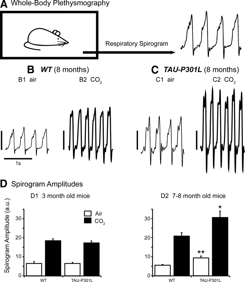
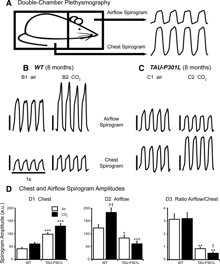
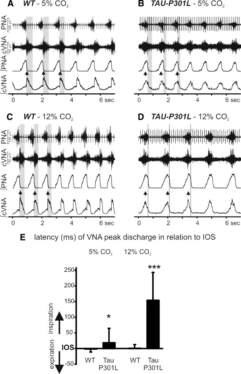
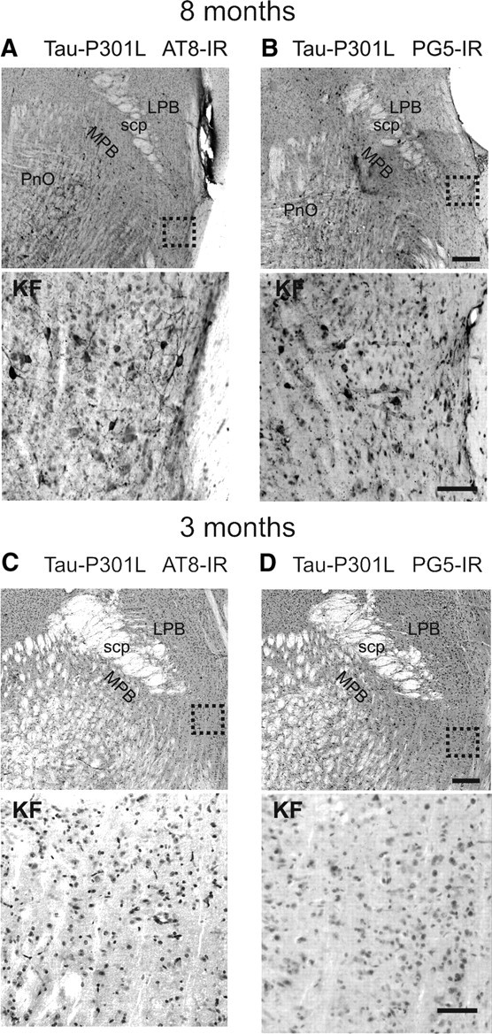
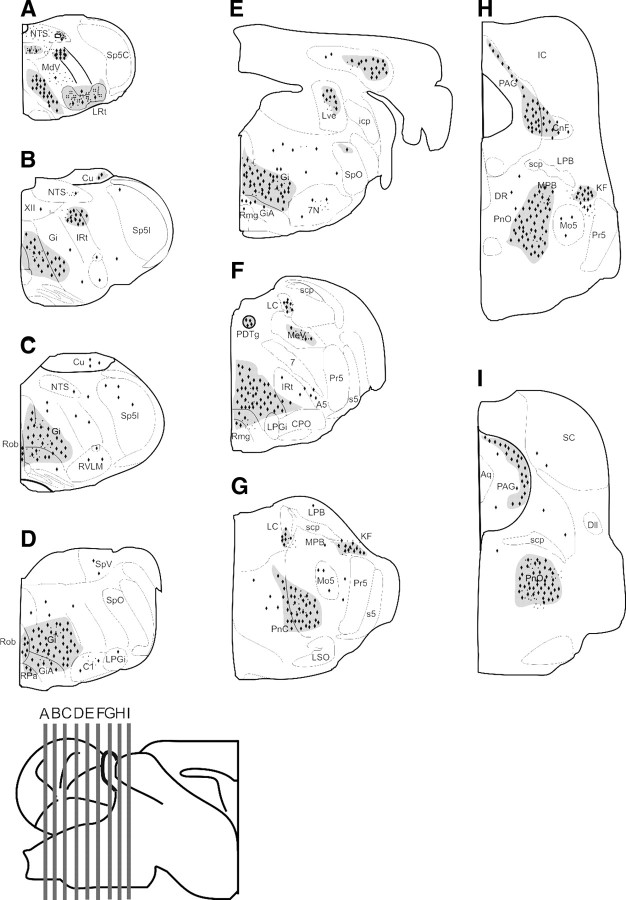
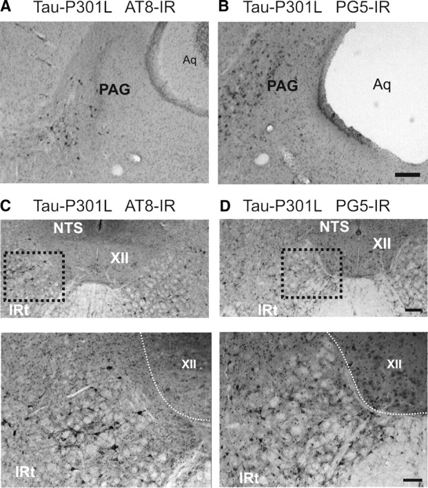
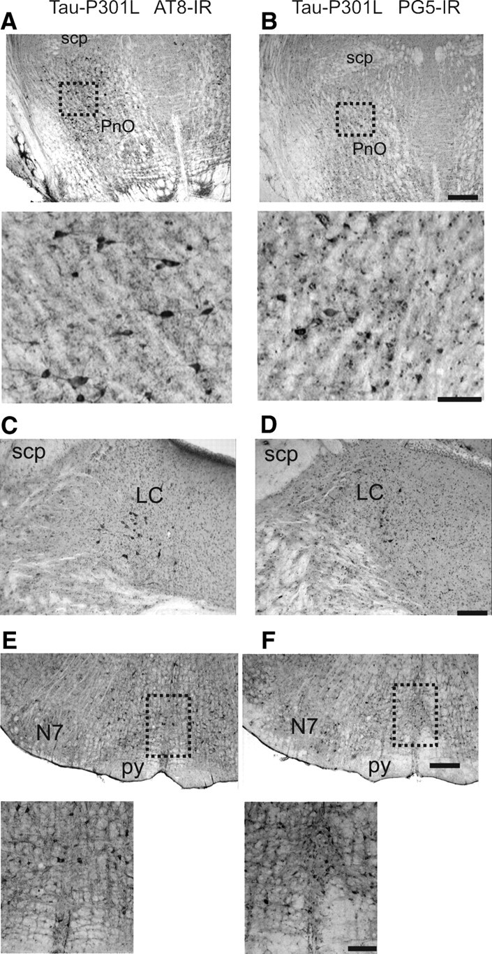
Tauopathy comprises hyperphosphorylation of the microtubule-associated protein tau, causing intracellular aggregation and accumulation as neurofibrillary tangles and neuropil treads. Some primary tauopathies are linked to mutations in the MAPT gene coding for protein tau, but most are sporadic with unknown causes. Also, in Alzheimer's disease, the most frequent secondary tauopathy, neither the cause nor the pathological mechanisms and repercussions are understood. Transgenic mice expressing mutant Tau-P301L suffer cognitive and motor defects and die prematurely from unknown causes. Here, in situ electrophysiology in symptomatic Tau-P301L mice (7-8 months of age) revealed reduced postinspiratory discharges of laryngeal motor outputs that control laryngeal constrictor muscles. Under high chemical drive (hypercapnia), postinspiratory discharge was nearly abolished, whereas laryngeal inspiratory discharge was increased disproportionally. The latter may suggest a shift of postinspiratory laryngeal constrictor activity into inspiration. In vivo double-chamber plethysmography of Tau-P301L mice showed significantly reduced respiratory airflow but significantly increased chest movements during baseline breathing, but particularly in hypercapnia, confirming a significant increase in inspiratory resistive load. Histological analysis demonstrated hyperphosphorylated tau in brainstem nuclei, directly or indirectly involved in upper airway motor control (i.e., the Kölliker-Fuse, periaqueductal gray, and intermediate reticular nuclei). In contrast, young Tau-P301L mice did not show breathing disorders or brainstem tauopathy. Consequently, in aging Tau-P301L mice, progressive upper airway dysfunction is linked to progressive tauopathy in identified neural circuits. Because patients with tauopathy suffer from upper airway dysfunction, the Tau-P301L mice can serve as an experimental model to study disease-specific synaptic dysfunction in well defined functional neural circuits.


Figures

Figure 1.
Figure 1.
Whole-body plethysmographic recordings in unrestrained, conscious WT and Tau-P301L mice. A, Schematic representation of the experimental setup used to record breathing in unrestrained, conscious mouse placed in a 200 ml recording chamber (to be compared with the experimental setup illustrated in Fig. 2A, in which the mouse is restrained). B, C, Individual traces show spirograms (inspiration upward) recorded from 8-month-old WT (B) and Tau-P301L (C) mice placed under control (B1, C1) (air; room air, 0.03% CO2) and hypercapnic (B2, C2) (CO2; 4% CO2 in air, 5 min) breathing conditions. SA values are expressed in arbitrary units (vertical calibration bars: 5 a.u. for B1 and C1; 10 a.u. for B2 and C2); note the increased SA under hypercapnic conditions in B2 and C2. D, The bars in the histograms show the mean SA (and SEM) from 6 WT and 6 Tau-P301L mice at 3 months of age (D1) and from 16 WT and 12 Tau-P301L mice at 7–8 months of age (D2). Note that the mean SA did not significantly differ in young WT versus Tau-P301L mice placed under identical breathing conditions (in D1; either air or CO2), whereas the mean SA was significantly larger in Tau-P301L versus WT old mice under air (D2; *p < 0.01) and CO2 (D2; **p < 0.05) breathing conditions. In all groups, hypercapnia significantly increased mean SA when compared with air.
Figure 2.
Figure 2.
Double-chamber plethysmographic recordings in conscious WT and Tau-P301L mice. A, Schematic representation of the experimental setup used for simultaneous recordings of chest and airflow spirograms in conscious but restrained mouse placed in a 50 ml recording chamber (to be compared with the experimental setup illustrated in Fig. 1A, in which the mouse is not restrained). B, C, Individual traces show chest and airflow spirograms (inspiration upward) recorded from 8-month-old WT (B) and Tau-P301L (C) mice placed under control (B1, C1) (air; room air, 0.03% CO2) and hypercapnic (B2, C2) (CO2; 4% CO2 in air, 5 min) breathing conditions. SA values are expressed in arbitrary units (vertical calibration bars, 50 a.u.). Note that hypercapnia increased both chest SA and airflow SA in the WT mouse (B), whereas hypercapnia increased chest SA but reduced airflow SA in the Tau-P301L mouse (C). D, The bars in the histograms show the mean (and SEM) values of chest SA (D1), airflow SA (D2), and ratio airflow versus chest SA (D3) obtained from double-plethysmographic recordings in 7- to 8-month-old WT (n = 6) and Tau-P301L (n = 6) mice placed under control (air, white bars) and hypercapnic (CO2, black bars) breathing conditions. The asterisk (*) indicated a significant difference between WT and Tau-P301L mice placed under identical breathing conditions (either air or CO2) (*p < 0.05; **p < 0.01; ***p < 0.001). The dagger (†) indicated a significant difference in control versus hypercapnic conditions between either paired WT or paired Tau-P301L mice (p < 0.05; ††p < 0.01). Note chest SA was significantly larger in Tau-P301L than WT mice under both breathing conditions (D1), whereas airflow SA was significantly smaller in Tau-P301L than WT mice under both breathing conditions (D2). D3 shows that the ratio airflow/chest SA was significantly lower in Tau-P301L than WT mice under air and was further lowered by hypercapnia.
Figure 3.
Figure 3.
In situ recordings of the respiratory motor pattern from WT and Tau-P301L mice. A–D, Recordings of inspiratory phrenic and inspiratory/postinspiratory vagal nerve activity (PNA; cVNA) of WT (A) and Tau-P301L mice (B) under control condition, 5% CO2 in perfusate and hypercapnic conditions 12% CO2 in perfusate (C, D). The gray-shaded areas highlight the reduced postinspiratory activity in Tau-P301L mice. Postinspiratory activity was virtually absent, whereas inspiratory-related cVNA discharge was disproportionally increased. For additional details and statistical analysis, see text. The double arrowheads in the integrated signals (A–D) illustrate cVNA peak discharge in coincidence with inspiratory PNA. Group data for the occurrence of cVNA peak discharge during the respiratory cycle are illustrated in E (*p < 0.05, ***p < 0.001). IOS, Inspiratory off-switch.
Figure 4.
Figure 4.
Tauopathy in the Kölliker–Fuse nucleus. The photomicrographs in A–D illustrate staining for phospho-epitopes AT8 and PG5 in the KF nucleus at 8 and 3 months of age. Scale bars: low magnification, 300 μm; high magnification, 50 μm. Abbreviations: LPB, Lateral parabrachial nuclei; MPB, medial parabrachial nucleus; PnO, pontine reticular nucleus, oral part; scp, superior cerebellar peduncle.
Figure 5.
Figure 5.
Relative intensity of tauopathy in brainstem and midbrain nuclei by immunohistochemistry with phospho-tau epitopes AT8 and PG5. Schematic rendering of the location of tauopathy [i.e., neurons containing NFTs within the caudal midbrain and ponto-medullary brainstem]. A–I, Sections from the caudal medulla oblongata to the caudal midbrain. The gray-shaded areas with black diamonds (NFTs) and points (AT8- and PG-5-IR staining of terminal-like structures) symbolize dense staining. 7, Facial nerve; 7N, facial nucleus; XII, hypoglossal nucleus; A5, A5 noradrenaline cells; Aq, aqueduct; C1, C1 adrenaline cells; Cu, cuneate nucleus; Dll, dorsal nucleus of the lateral lemniscus; PDTg, posterodorsal tegmental nucleus; DR, dorsal raphe nucleus; Gi, gigantocellular reticular nucleus; IC, inferior colliculus; IRt, intermediate reticular nucleus; KF, Kölliker–Fuse nucleus; LC, locus ceruleus; LPB, lateral parabrachial nuclei, LRt, spinal trigeminal nucleus, caudal part; LSO, lateral superior olive; Mo5, motor trigeminal nucleus; MPB, medial parabrachial nucleus; PAG, periaqueductal gray; PnC, pontine reticular nucleus, caudal part; PnO, pontine reticular nucleus, oral part; Pr5, principal trigeminal nucleus; RMg, raphe magnus nucleus; ROb, raphe obscurus nucleus; RPa, raphe pallidus nucleus; RVLM, rostral ventrolateral medulla oblongata; scp, superior cerebellar peduncle; SolC, nucleus of the solitary tract, commissural part; SolM, nucleus of the solitary tract, medial part; SolVL, nucleus of the solitary tract, ventrolateral part; Sp5C, spinal trigeminal nucleus, caudal part; Sp5I, spinal trigeminal nucleus, interpolar part; Sp5O, spinal trigeminal nucleus, oral part; LVe, lateral vestibular nucleus.
Figure 6.
Figure 6.
Tauopathy in lateral periaqueductal gray and intermediate reticular nucleus. The photomicrographs in A and B illustrate staining for phospho-epitopes AT8 and PG5 in the lateral periaqueductal gray (PAG) of Tau-P301L mouse. The photomicrographs in C and D illustrate staining in a Tau-P301L mouse for phospho-epitopes AT8 and PG5 in the intermediate reticular nucleus (IRt), which is located ventrolateral to the nucleus of the solitary tract (NTS) and lateral to hypoglossal nucleus (XII). Scale bars: low magnification, 300 μm; high magnification, 50 μm. Aq, Aqueduct.
Figure 7.
Figure 7.
Tauopathy in pontine reticular nucleus, locus ceruleus, and raphe obscurus. A and B show AT8 and PG5 immunoreactivity (IR) in the dorsal aspect of the oral part of pontine reticular nucleus (PnO). Scale bars: low magnification, 300 μm; high magnification, 100 μm. The photomicrographs in C and D illustrate staining for phospho-epitopes AT8 and PG5 in the locus ceruleus (LC) of Tau-P301L mouse. E and F show the same immunoreactivity (IR) in the raphe obscurus nucleus (ROb), mediolateral to the facial nucleus (7N). Scale bars: low magnification, 300 μm; high magnification, 100 μm. scp, Superior cerebellar peduncle; py, pyramidal tract.

Similar articles

Cited by

References

    1. Alheid GF, Milsom WK, McCrimmon DR. Pontine influences on breathing: an overview. Respir Physiol Neurobiol. 2004;143:105–114. - PubMed
    1. Andorfer CA, Davies P. PKA phosphorylations on tau: developmental studies in the mouse. Dev Neurosci. 2000;22:303–309. - PubMed
    1. Ballantyne D, Andrzejewski M, Mückenhoff K, Scheid P. Rhythms, synchrony and electrical coupling in the locus coeruleus. Respir Physiol Neurobiol. 2004;143:199–214. - PubMed
    1. Besnard S, Denise P, Cappelin B, Dutschmann M, Gestreau C. Stimulation of the rat medullary raphe nuclei induces differential responses in respiratory muscle activity. Respir Physiol Neurobiol. 2009;165:208–214. - PubMed
    1. Boekhoorn K, Terwel D, Biemans B, Borghgraef P, Wiegert O, Ramakers GJ, de Vos K, Krugers H, Tomiyama T, Mori H, Joels M, van Leuven F, Lucassen PJ. Improved long-term potentiation and memory in young tau-P301L transgenic mice before onset of hyperphosphorylation and tauopathy. J Neurosci. 2006;26:3514–3523. - PMC - PubMed

Publication types

MeSH terms

LinkOut - more resources