

Lack of functional pregnancy-associated plasma protein-A (PAPPA) compromises mouse ovarian steroidogenesis and female fertility
- PMID: 20130263
- PMCID: PMC2874498
- DOI: 10.1095/biolreprod.109.079517
Lack of functional pregnancy-associated plasma protein-A (PAPPA) compromises mouse ovarian steroidogenesis and female fertility
Abstract
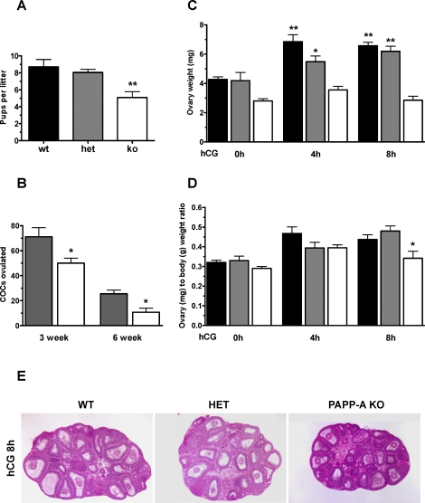
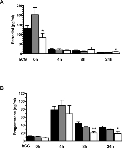
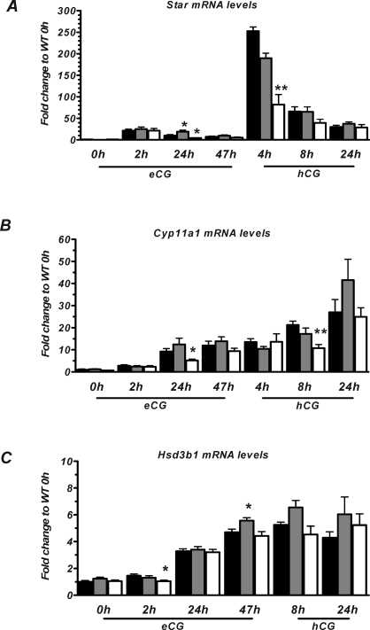
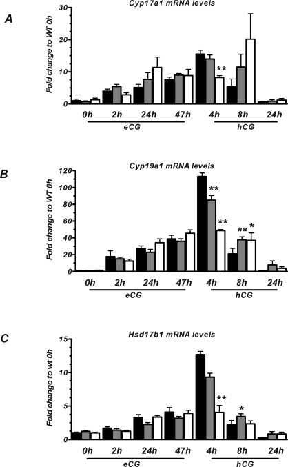
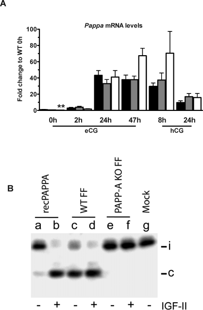
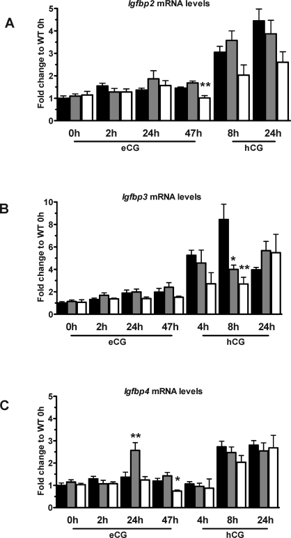
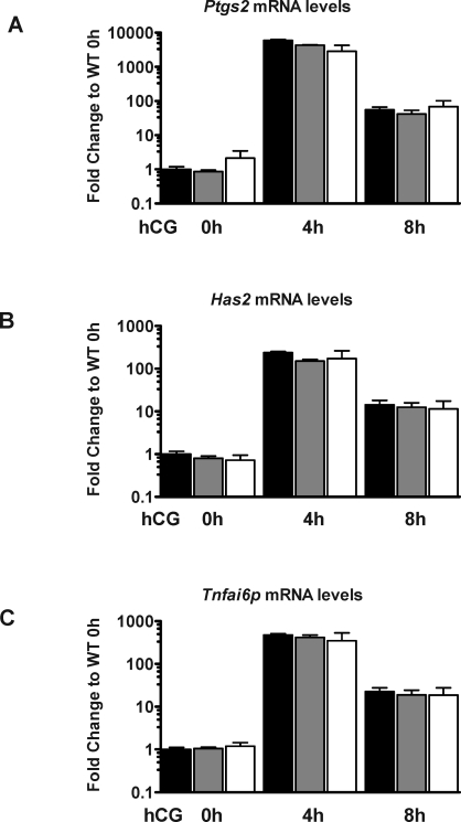
The insulin-like growth factor (IGF) system plays an important role in regulating ovarian follicular development and steroidogenesis. IGF binding proteins (IGFBP) mostly inhibit IGF actions, and IGFBP proteolysis is a major mechanism for regulating IGF bioavailability. Pregnancy-associated plasma protein-A (PAPPA) is a secreted metalloprotease responsible for cleavage of IGFBP4 in the ovary. The aim of this study was to investigate whether PAPPA plays a role in regulating ovarian functions and female fertility by comparing the reproductive phenotype of wild-type (WT) mice with mice heterozygous or homozygous for a targeted Pappa gene deletion (heterozygous and PAPP-A knockout [KO] mice, respectively). When mated with WT males, PAPP-A KO females demonstrated an overall reduction in average litter size. PAPP-A KO mice had a reduced number of ovulated oocytes, lower serum estradiol levels following equine chorionic gonadotropin administration, lower serum progesterone levels after human chorionic gonadotropin injection, and reduced expression of ovarian steroidogenic enzyme genes, compared to WT controls. In PAPP-A KO mice, inhibitory IGFBP2, IGFBP3, and IGFBP4 ovarian gene expression was reduced postgonadotropin stimulation, suggesting some compensation within the ovarian IGF system. Expression levels of follicle-stimulating hormone receptor, luteinizing hormone receptor, and genes required for cumulus expansion were not affected. Analysis of preovulatory follicular fluid showed complete loss of IGFBP4 proteolytic activity in PAPP-A KO mice, demonstrating no compensation for loss of PAPPA proteolytic activity by other IGFBP proteases in vivo in the mouse ovary. Taken together, these data demonstrate an important role of PAPPA in modulating ovarian function and female fertility by control of the bioavailability of ovarian IGF.
Figures
Similar articles
-
Depot-specific and GH-dependent regulation of IGF binding protein-4, pregnancy-associated plasma protein-A, and stanniocalcin-2 in murine adipose tissue.Growth Horm IGF Res. 2018 Apr;39:54-61. doi: 10.1016/j.ghir.2018.01.001. Epub 2018 Feb 3. Growth Horm IGF Res. 2018. PMID: 29398370
-
Insulin-like growth factor binding protein 4 expression parallels luteinizing hormone receptor expression and follicular luteinization in the primate ovary.Biol Reprod. 2003 Jul;69(1):22-9. doi: 10.1095/biolreprod.102.009191. Epub 2003 Jan 8. Biol Reprod. 2003. PMID: 12606427
-
Pregnancy-associated plasma protein-A (PAPP-A) in ovine, bovine, porcine, and equine ovarian follicles: involvement in IGF binding protein-4 proteolytic degradation and mRNA expression during follicular development.Endocrinology. 2001 Dec;142(12):5243-53. doi: 10.1210/endo.142.12.8517. Endocrinology. 2001. PMID: 11713222
-
Follicular development: the role of the follicular microenvironment in selection of the dominant follicle.Anim Reprod Sci. 2004 Jul;82-83:109-26. doi: 10.1016/j.anireprosci.2004.04.031. Anim Reprod Sci. 2004. PMID: 15271447 Review.
-
Insulin-like growth factor family in Graafian follicle development and function.J Soc Gynecol Investig. 2001 Jan-Feb;8(1 Suppl Proceedings):S26-9. doi: 10.1016/s1071-5576(00)00102-7. J Soc Gynecol Investig. 2001. PMID: 11223367 Review.
Cited by
-
Novel function of pregnancy-associated plasma protein A: promotes endometrium receptivity by up-regulating N-fucosylation.Sci Rep. 2017 Jul 13;7(1):5315. doi: 10.1038/s41598-017-04735-0. Sci Rep. 2017. PMID: 28706275 Free PMC article.
-
Proteolysis in Reproduction: Lessons From Gene-Modified Organism Studies.Front Endocrinol (Lausanne). 2022 May 4;13:876370. doi: 10.3389/fendo.2022.876370. eCollection 2022. Front Endocrinol (Lausanne). 2022. PMID: 35600599 Free PMC article. Review.
-
Insulin-Like Growth Factor Binding Proteins and IGFBP Proteases: A Dynamic System Regulating the Ovarian Folliculogenesis.Front Endocrinol (Lausanne). 2018 Mar 29;9:134. doi: 10.3389/fendo.2018.00134. eCollection 2018. Front Endocrinol (Lausanne). 2018. PMID: 29643837 Free PMC article. Review.
-
Epigenetic effects of endocrine-disrupting chemicals on female reproduction: an ovarian perspective.Front Neuroendocrinol. 2010 Oct;31(4):420-39. doi: 10.1016/j.yfrne.2010.06.003. Epub 2010 Jul 4. Front Neuroendocrinol. 2010. PMID: 20609371 Free PMC article. Review.
-
Insulin-like growth factor (IGF)-I and IGF-II contribute differentially to the phenotype of pregnancy associated plasma protein-A knock-out mice.Growth Horm IGF Res. 2011 Oct;21(5):243-7. doi: 10.1016/j.ghir.2011.06.002. Epub 2011 Jul 28. Growth Horm IGF Res. 2011. PMID: 21802327 Free PMC article.
References
-
- Kwintkiewicz J, Giudice LC.The interplay of insulin-like growth factors, gonadotropins, and endocrine disruptors in ovarian follicular development and function. Semin Reprod Med 2009; 27: 43–51. - PubMed
-
- Dupont J, Dunn SE, Barrett JC, LeRoith D.Microarray analysis and identification of novel molecules involved in insulin-like growth factor-1 receptor signaling and gene expression. Recent Prog Horm Res 2003; 58: 325–342. - PubMed
-
- Samani AA, Yakar S, LeRoith D, Brodt P.The role of the IGF system in cancer growth and metastasis: overview and recent insights. Endocr Rev 2007; 28: 20–47. - PubMed
-
- Boldt HB, Conover CA.Pregnancy-associated plasma protein-A (PAPP-A): a local regulator of IGF bioavailability through cleavage of IGFBPs. Growth Horm IGF Res 2007; 17: 10–18. - PubMed
Publication types
MeSH terms
Substances
Grants and funding
LinkOut - more resources
Full Text Sources
Medical
Molecular Biology Databases
Research Materials
Miscellaneous
