. 2010 May;82(5):948-57.
doi: 10.1095/biolreprod.109.079889. Epub 2010 Feb 3.

Absence of estrogen receptor alpha leads to physiological alterations in the mouse epididymis and consequent defects in sperm function

Affiliations

Absence of estrogen receptor alpha leads to physiological alterations in the mouse epididymis and consequent defects in sperm function

Avenel Joseph et al. Biol Reprod. 2010 May.

Abstract

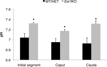
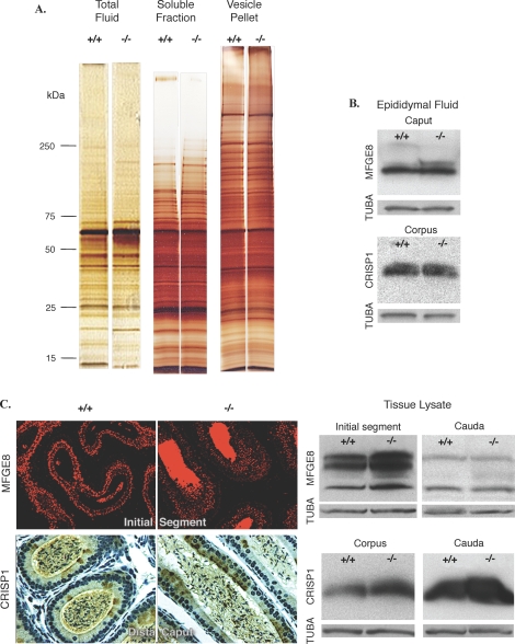
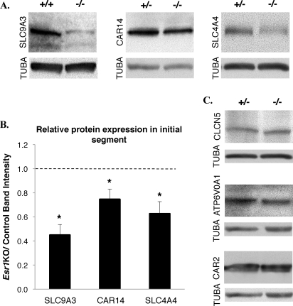
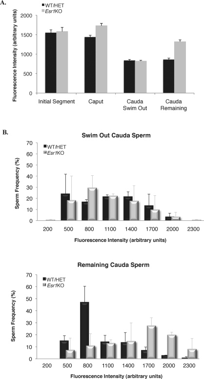
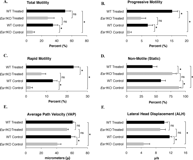
Male mice deficient in ESR1 (ERalpha) (Esr1KO mice) are infertile, and sperm recovered from the cauda epididymis exhibit reduced motility and fail to fertilize eggs in vitro. These effects on sperm appear to result from defective epididymal function and not a direct effect on spermatogenesis, as Esr1KO germ cells transplanted into wild-type testes yield normal offspring. We hypothesized that the previously described defect in efferent duct fluid reabsorption would lead to alterations in the epididymal fluid milieu, which would negatively impact sperm function. Analysis of the epididymal fluid revealed that the Esr1KO maintains a higher luminal pH throughout the epididymis, confirming an inability of the efferent ducts and/or epididymis to properly acidify the luminal contents. Subsequent studies showed that these abnormalities were not the result of global defects in epididymal function since protein secretion by the Esr1KO epididymis appeared normal as judged by SDS-PAGE of total secreted proteins and by immunoblotting of candidate secreted proteins. To gain insight into the basis of the aberrant fluid homeostasis in the Esr1KO epididymis, the expression of several enzymes and transporters known to be involved in acid/base regulation were analyzed. The levels of SLC9A3 (NHE3) as well as carbonic anhydrase XIV and SLC4A4 (NBC1) were all reduced in the proximal portion of the Esr1KO epididymis, while other components appeared unaffected, including other ion transporters and ATP6V0A1 (V-ATPase). The altered luminal milieu of the Esr1KO epididymis was shown to lead to a corresponding increase in the intracellular pH of Esr1KO sperm, relative to sperm from control animals. Since pH and bicarbonate ions are critical regulators of sperm cAMP levels and motility, we attempted to bypass the abnormal luminal and intracellular environment by supplementing sperm with exogenous cAMP. This treatment rescued all defective motility parameters, as assayed by CASA, further showing that motility defects are not intrinsic to the sperm but, rather, result from the abnormal epididymal milieu.


Figures

FIG. 1.
FIG. 1.
The Esr1KO epididymis fails to acidify the luminal fluid. Epididymides were isolated and subdissected, and the pH of the luminal contents was measured using an ex vivo assay. In control animals, the luminal pH decreases during epididymal transit, whereas it remains alkaline in the Esr1KO epididymis. In this regard, fluid isolated from the initial segment of Esr1KO animals was significantly more alkaline than controls (control: 7.04 ± 0.08, Esr1KO: 7.32 ± 0.02). This difference persists in the caput (control: 6.95 ± 0.07, Esr1KO: 7.17 ± 0.03) as well as in the cauda (control: 6.93 ± 0.11, Esr1KO: 7.31 ± 0.05). At least five independent samples of luminal fluid from each genotype and each region were assayed. Bars = SEM. WT, wild type; HET, heterozygote; *P ≤ 0.04.
FIG. 2.
FIG. 2.
Many epididymal proteins are expressed and localized normally in the Esr1KO epididymis. As a simple readout of overall epididymal function, total epididymal protein expression was compared between control and Esr1KO mice, as was the immunolocalization of MFGE8 and CRISP1, two epididymal secreted proteins important in sperm function. A) Total luminal fluid protein was separated by single-dimension electrophoresis and visualized by silver stain. At this resolution, the relative protein abundance and separation profiles appeared to be indistinguishable between genotypes. Furthermore, the protein composition of soluble and membrane-bound fractions were analyzed by SDS-PAGE and silver staining and shown to be similar between Esr1KO and control animals. B) Western blot analysis of MFGE8 and CRISP1 in epididymal fluid revealed similar levels in the Esr1KO and control caput and corpus, respectively. C) Immunolocalization of MFGE8 and CRISP1 in the Esr1KO epididymis was indistinguishable from that observed in control animals and similar to that previously reported [47, 52, 53]. Original magnification ×400. Furthermore, the relative abundance of CRISP and the known MFGE8 isoforms, as judged by Western blot analysis, was similar between genotypes. (A large bolus of unlabeled protein present in the epididymal fluid distorts the migration of the higher-molecular-weight MFGE8 isoform, which is easily seen in tissue lysates.) For all blots, α-tubulin, TUBA, serves as a loading control.
FIG. 3.
FIG. 3.
Reduced expression of cell-specific components involved in acid/base homeostasis. A) Western blot analysis of principle cell-specific regulatory proteins SLC9A3 (83 kDa), CAR14 (50 kDa), and SLC4A4 (130 kDa) showed reduced expression within the Esr1KO initial segment. α-tubulin, TUBA, serves as a loading control. B) Densitometric quantification of Western blots expressed as ratios of the control value revealed statistically significant reduction of SLC9A3, CAR14, and SLC4A4 in Esr1KO tissue. Histograms represent the mean ± SEM; *P ≤ 0.03. The dotted line represents equal levels of protein expression between Esr1KO and control animals. C) Other components involved in fluid regulation that are specifically expressed in narrow cells, such as ATP6V01 and CLCN5, or that are expressed by both principle and narrow cells, that is, CAR2, have similar levels of expression between genotypes. α-tubulin, TUBA, serves as a loading control.
FIG. 4.
FIG. 4.
Intracellular pH of Esr1KO sperm fails to acidify during epididymal transit. Sperm from the initial segment, caput, and cauda epididymis were loaded with BCECF, a pH-dependent fluorophore, and their fluorescence intensity was measured. A) Esr1KO and control sperm recovered from the initial segment have a similar fluorescent intensity. However, caput Esr1KO sperm show increased fluorescent intensity and pHi, relative to controls (P = 0.04). Cauda sperm were separated on the basis of motility, revealing that the overall mean fluorescent intensity of Esr1KO sperm that are sufficiently motile to swim out of the cauda is similar to that of control sperm, while the fluorescent intensity of the remaining nonmotile Esr1KO sperm is significantly increased. Presented are mean values ± SEM; *p ≤ 0.05. B) Frequency distributions of cauda sperm fluorescence intensity reveals that the relative pHi is similar between both control sperm populations and the Esr1KO swim-out sperm but is significantly more alkaline in the nonmotile Esr1KO cells.
FIG. 5.
FIG. 5.
Exogenous treatment with cAMP rescues Esr1KO sperm motility deficits. CASA measurements of caput sperm indicate that the raw numbers and relative percentages of overall sperm motility, as well as progressive and rapid motility, were significantly lower in the Esr1KO, whereas those sperm classified as static were drastically increased. The data also indicate that the path velocities of sperm (VAP) and amplitudes of their lateral head displacement (ALH) are both greatly reduced in the Esr1KO sperm. Treatment of Esr1KO sperm with a dp-cAMP cocktail significantly increased their total motility (A), progressive motility (B), and rapid motility (C) and decreased their static motility (D) to levels comparable to control cells. Similarly, the path velocities of sperm (VAP; E) and amplitudes of their lateral head displacement (ALH; F) were also increased to levels statistically similar to control cells following cAMP treatment. Although WT sperm generally showed stronger responses to exogenous cAMP treatment, the increase in Esr1KO total motility, ALH, and VAP induced by cAMP treatment were statistically similar to that seen in treated wild-type cells. *P ≤ 0.03; ns, not significant.

Similar articles

Cited by

References

    1. Eddy EM, Washburn TF, Bunch DO, Goulding EH, Gladen BC, Lubahn DB, Korach KS.Targeted disruption of the estrogen receptor gene in male mice causes alteration of spermatogenesis and infertility. Endocrinology 1996; 137: 4796–4805. - PubMed
    1. Lubahn DB, Moyer JS, Golding TS, Couse JF, Korach KS, Smithies O.Alteration of reproductive function but not prenatal sexual development after insertional disruption of the mouse estrogen receptor gene. Proc Natl Acad Sci U S A 1993; 90: 11162–11166. - PMC - PubMed
    1. Hess RA, Bunick D, Lee KH, Bahr J, Taylor JA, Korach KS, Lubahn DB.A role for oestrogens in the male reproductive system. Nature 1997; 390: 509–512. - PMC - PubMed
    1. Mahato D, Goulding EH, Korach KS, Eddy EM.Estrogen receptor-alpha is required by the supporting somatic cells for spermatogenesis. Mol Cell Endocrinol 2001; 178: 57–63. - PubMed
    1. Mahato D, Goulding EH, Korach KS, Eddy EM.Spermatogenic cells do not require estrogen receptor-alpha for development or function. Endocrinology 2000; 141: 1273–1276. - PubMed

Publication types