. 2010 Jul;51(7):3599-610.
doi: 10.1167/iovs.09-4797. Epub 2010 Feb 3.

Growth factor signaling in vitreous humor-induced lens fiber differentiation

Affiliations

Growth factor signaling in vitreous humor-induced lens fiber differentiation

Qian Wang et al. Invest Ophthalmol Vis Sci. 2010 Jul.

Abstract

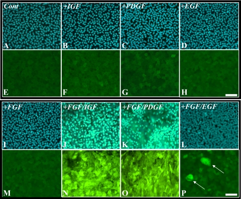
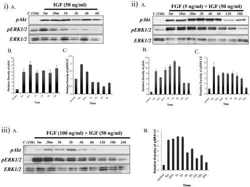
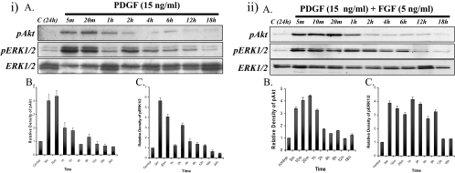
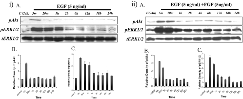
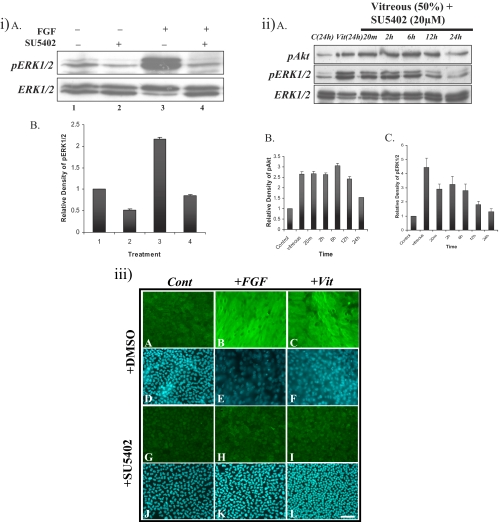
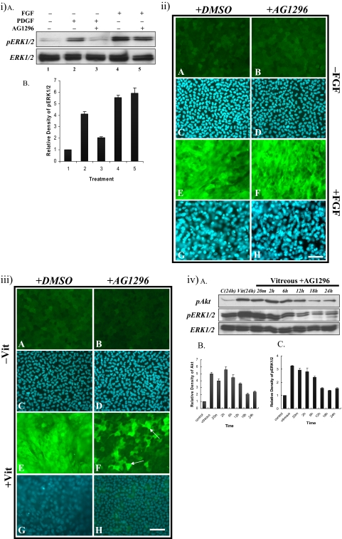
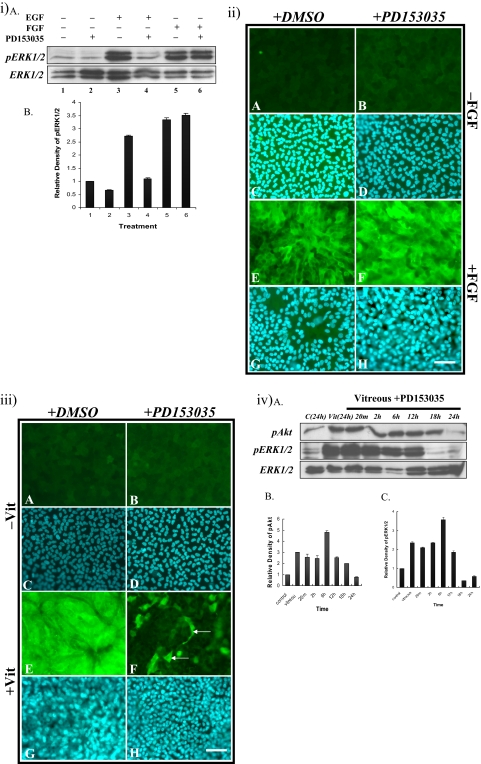
PURPOSE. Although some of the factors and signaling pathways that are involved in induction of fiber differentiation have been defined, such as FGF-mediated MAPK/ERK and PI3-K/Akt signaling, the factors in the vitreous that regulate this differentiation process in vivo have yet to be identified. The purpose of this study was to better understand the role of growth factors in vitreous that regulate this process by further characterizing the signaling pathways involved in lens fiber differentiation. METHODS. Rat lens epithelial explants were used to compare the ability of vitreous, IGF-1, PDGF-A, EGF, and FGF-2 to stimulate the phosphorylation of ERK1/2 and Akt leading to fiber differentiation, in the presence or absence of selective receptor tyrosine kinase (RTK) inhibitors. RESULTS. Similar to vitreous, FGF induced a sustained ERK1/2 signaling profile, unlike IGF, PDGF, and EGF, which induced a more transient (shorter) activation of ERK1/2. For Akt activation, IGF was the only factor that induced a profile similar to vitreous. IGF, PDGF, and EGF potentiated the effects of a low dose of FGF on lens fiber differentiation by extending the duration of ERK1/2 phosphorylation. In the presence of selective RTK inhibitors, although the sustained vitreous-induced ERK1/2 signaling profile and subsequent fiber differentiation was perturbed, the results also showed that, although prolonged ERK1/2 phosphorylation was necessary, it was not sufficient for fiber differentiation to proceed. CONCLUSIONS. These results are consistent with FGF's being the key growth factor involved in vitreous-induced signaling leading to lens fiber differentiation; however, they also indicate that other vitreal growth factors such as IGF may be involved in fine-tuning ERK1/2- and Akt-phosphorylation to the level that is necessary for initiation and/or maintenance of lens fiber differentiation in vivo.


Figures

Figure 1.
Figure 1.
Explants treated for 5 days with no growth factors (A, E) or a combination of different growth factors: 50 ng/mL IGF (B, F); 15 ng/mL PDGF (C, G); 5 ng/mL EGF (D, H); 5 ng/mL FGF (I, M), or a combination of a low dose of FGF (5 ng/mL) with IGF (FGF/IGF, J, N), PDGF (FGF/PDGF, K, O), or EGF (FGF/EGF, L, P), immunofluorescently labeled for β-crystallin (E–H, M–P) or counterstained with Hoechst dye (A–D, I–L). Only combinations of FGF with IGF (J, N), PDGF (K, O), or EGF (L, P, arrows) induced cell multilayering and accumulation of β-crystallin. Scale bar, 50 μm.
Figure 2.
Figure 2.
(i) Representative Western blots (iA) of explants cultured with no growth factor (control) or IGF from 5 minutes up to 6 hours, assayed for phosphorylated Akt (top), phosphorylated ERK1/2 (middle), or total ERK1/2 (bottom). Quantification of relative density of phosphorylated Akt (iB) and ERK1/2 (iC) showed the same trend as Western blot analysis. (ii) Representative Western blots (iiA) of explants cultured with no growth factor (control) or IGF+FGF from 5 minutes up to 18 hours, assayed for phosphorylated Akt (top), phosphorylated ERK1/2 (middle), and total ERK1/2 (bottom). Quantification of the relative density of phosphorylated Akt (iiB) and ERK1/2 (iiC) showed the same trend as the Western analysis. (iii) Explants treated with a high dose of FGF (100 ng/mL)+IGF (50 ng/mL). (iiiA) Explants cultured without growth factor (control) or with a combination of a high dose of FGF+IGF from 5 minutes up to 24 hours, assayed for phosphorylated Akt (top), phosphorylated ERK1/2 (middle), and total ERK1/2 (bottom). (iiiB) Quantification of the relative density of ERK1/2.
Figure 3.
Figure 3.
Explants treated with PDGF (15 ng/mL) or a low dose of FGF (5 ng/mL)+PDGF (15 ng/mL). (i) Representative Western blots of explants cultured without growth factor (control) or with PDGF (iA) from 5 minutes up to 24 hours, assayed for phosphorylated Akt (top), phosphorylated ERK1/2 (middle), and total ERK1/2 (bottom). Strong Akt phosphorylation was detected in 5 minutes and continued for up to 2 hours, followed by a return to basal levels. ERK1/2 phosphorylation was transiently activated within 5 minutes and peaked by 20 minutes. This level returned to basal level by 1 hour. Total ERK1/2 showed that the same amount of protein was loaded in each lane. Quantification of the relative density of phosphorylated Akt (iB) and ERK1/2 (iC) showed the same trend as the Western analysis. (ii) Representative Western blots of explants cultured without growth factor (control) or with PDGF+FGF (iiA) from 5 minutes up to 18 hours, assayed for phosphorylated Akt (top), phosphorylated ERK1/2 (middle), or total ERK1/2 (bottom). Quantification of the relative density of phosphorylated Akt (iiB) and ERK1/2 (iiC) showed the same trend as the Western analysis.
Figure 4.
Figure 4.
Explants treated with EGF (5 ng/mL) or a low dose of FGF (5 ng/mL)+EGF (5 ng/mL). (i) Representative Western blots of explants cultured without growth factor (control) or with EGF (iA) from 5 minutes up to 24 hours, assayed for phosphorylated Akt (top), phosphorylated ERK1/2 (middle), and total ERK1/2 (bottom). Quantification of the relative density of phosphorylated Akt (iB) and ERK1/2 (iC) showed the same trend as the Western analysis. (ii) Representative Western blots of explants cultured without growth factor (control) or with EGF+FGF (iiA) from 5 minutes up to 24 hours, assayed for phosphorylated Akt (top), phosphorylated ERK1/2 (middle), and total ERK1/2 (bottom). Quantification of the relative density of phosphorylated Akt (iiB) and ERK1/2 (iiC) showed the same trend as the Western analysis.
Figure 5.
Figure 5.
Effect of the FGFR inhibitor SU5402 on vitreous-induced lens fiber differentiation. (iA) Representative Western blots of explants cultured without growth factor (lanes 1 and 2) or with FGF (lanes 3 and 4) in the presence of DMSO (lanes 1 and 3) or 20 μM SU5402 (lanes 2 and 4) for 20 minutes. SU5402 completely blocked FGF-induced ERK1/2 phosphorylation. Quantification of the relative density of phosphorylated ERK1/2 (iB) showed the same trend as the Western analysis. (iiA) Representative Western blots of explants cultured without growth factor (control) or with 50% vitreous with 20 μM SU5402 from 20 minutes to 24 hours, assayed for phosphorylated Akt (top), phosphorylated ERK1/2 (middle), and ERK1/2 (bottom). Akt and ERK1/2 were phosphorylated within 20 minutes and maintained for up to 12 and 6 hours, respectively. Quantification of the relative density of phosphorylated Akt (iiB) and ERK1/2 (iiC) showed the same trend as the Western analysis. (iii) Representative micrographs of lens explants cultured with 20 μM SU5402. Explants were cultured without growth factors (A, D, G, J), with FGF (100 ng/mL; B, E, H, K), or with vitreous (50%; C, F, I, L), in the presence (G–L) or absence (A–F) of 20 μM SU5402 added 2 hours before FGF or vitreous treatment. The explants were assayed for β-crystallin accumulation after 5 days. Those cultured without SU5402 were treated with an equivalent volume of DMSO (A, D). When SU5402 was added 2 hours before FGF or vitreous treatment, lens fiber differentiation was blocked (H, I). Scale bar, 50 μm.
Figure 6.
Figure 6.
Effect of the IGFR inhibitor AG1204 on vitreous-induced lens fiber differentiation. (iA) Representative Western blots of explants cultured without (lanes 1 and 2) or with (10 ng/mL; lanes 3 and 4) IGF in the presence (lanes 2 and 4) or absence (lanes 1 and 3) of 5 μM AG1024 added 2 hours before growth factor treatment. AG1024 blocked IGF-induced ERK1/2 phosphorylation. (iB) Quantification of the relative density of phosphorylated ERK1/2 showed the same trend as the Western analysis. (iiA) Representative Western blots of explants cultured without (lanes 1 and 2) or with (lanes 3 and 4) FGF in the presence (lanes 2 and 4) or absence (lanes 1 and 3) of 5 μM AG1024 added 2 hours before growth factor treatment. AG1204 did not block FGF-induced ERK1/2 phosphorylation. (iiB) Quantification of the relative density of phosphorylated ERK1/2 that showed the same trend as the Western analysis. (iii) Representative micrographs of lens explants cultured without (A–D) or with (E–H) FGF, in the presence (B, D, F, H) or absence (A, C, E G) of 5 μM AG1024 added 2 hours before FGF treatment. AG1024 did not block FGF-induced lens fiber differentiation (F). (iv) Representative micrographs of lens explants cultured with vitreous and 5 μM AG1024. Explants were cultured without (A–D) or with (E–H) vitreous, in the presence (B, D, F, H, I, J) or absence (A, C, E, G) of 5 μM AG1024, added 2 hours before vitreous treatment. AG1024 reduced the accumulation of β-crystallin and cell elongation, but did not completely block the elongation. Some bare patches were observed over the explants (F, arrows), indicative of cell death/loss. Scale bar, (A–H) 50 μm; (I, J) 200 μm. (vA) Representative Western blots of explants cultured without or with vitreous in the presence or absence of 5 μM AG1024 added 2 hours before vitreous treatment, assayed for phosphorylated Akt (top), phosphorylated ERK1/2 (middle), and total ERK1/2 (bottom). Akt was phosphorylated within 20 minutes and maintained for up to 18 hours. The duration of ERK1/2 phosphorylation was reduced to 6 hours. Quantification of the relative density of phosphorylated Akt (vB) and ERK1/2 (vC) showed the same trend as the Western analysis.
Figure 7.
Figure 7.
Effect of PDGFR inhibitor, AG1296 on vitreous-induced lens fiber differentiation. (iA) Representative Western blots of explants cultured without growth factor (lane 1) with PDGF (15 ng/mL; lanes 2 and 3) or FGF (100 ng/mL; lanes 4 and 5) in the presence (lanes 3 and 5) or absence (lanes 2 and 4) of 1 mM AG1296, added 2 hours before growth factor treatment. AG1296 blocked PDGF-induced ERK1/2 phosphorylation, but had no effect on FGF-induced ERK1/2 phosphorylation. Quantification of the relative density of phosphorylated ERK1/2 (iB) showed the same trend as the Western analysis. (ii) Representative micrographs of lens explants cultured with 1 mM AG1296 and FGF (100 ng/mL). The explants were cultured without (A–D) or with (E–H) FGF, in the presence (B, D, F, H) or absence (A, C, E, G) of 1 mM AG1296 added 2 hours before FGF treatment. AG1296 did not block FGF-induced fiber differentiation (F). (iii) Representative micrographs of lens explants cultured with AG1296 and vitreous. Explants were cultured without (A–D) or with (E–H) vitreous, in the presence (B, D, F, H) or absence (A, C, E, G) of 1 mM AG1296 added 2 hours before vitreous treatment. AG1296 reduced vitreous-induced lens fiber differentiation. Some cells still expressed β-crystallin (F, arrows). Scale bar, 50 μm. (ivA) Representative Western blots of explants cultured without (control) or with vitreous in the presence or absence of 1 mM AG1296, assayed for phosphorylated Akt (top), phosphorylated ERK1/2 (middle), and total ERK1/2 (bottom). Duration of ERK1/2 phosphorylation was reduced to 6 hours. Quantification of the relative density of phosphorylated Akt (ivB) and ERK1/2 (ivC) showed the same trend as the Western analysis.
Figure 8.
Figure 8.
(iA) Representative Western blots of explants cultured without growth factor (lanes 1 and 2), with EGF (lanes 3 and 4), or with FGF (lanes 5 and 6) in the presence (lanes 2, 4, and 6) or absence (lanes 1, 3, and 5) of 50 nM PD153035, added 2 hours before growth factor treatment. Quantification of the relative density of (iB) phosphorylated ERK1/2 showed the same trend as the Western analysis. (ii) Representative micrographs of lens explants cultured without (A–D) or with (E–H) 100 ng/mL FGF, in the presence (B, D, F, H) or absence (A, C, E, G) of 50 nM PD153035 added 2 hours before FGF treatment. PD153035 did not block FGF-induced fiber differentiation (F). (iii) Representative micrographs of lens explants cultured without (A-D) or with (E–H) vitreous, in the presence (B, D, F, H) or absence (A, C, E, G) of 50 nM PD153035, added 2 hours before vitreous treatment. PD153035 reduced vitreous-induced lens fiber differentiation. Some cells still expressed β-crystallin (F, arrows). (iv) Western blot analysis of explants treated with vitreous and PD153035. (ivA) Representative Western blots of explants cultured without or with vitreous in the presence or absence of 50 nM of PD153035, added 2 hours before vitreous treatment, assayed for phosphorylated Akt (top), phosphorylated ERK1/2 (middle), or total ERK1/2 (bottom). Akt and ERK 1/2 were phosphorylated within 20 minutes and continued for up to 18 and 12 hours, respectively. Quantification of the relative density of phosphorylated Akt (ivB) and ERK1/2 (ivC) showed the same trend as the Western analysis. Scale bar, 50 μm.

Similar articles

Cited by

References

    1. Coulombre JL, Coulombre AJ. Lens development: fiber elongation and lens orientation. Science 1963;142:1489–1490 - PubMed
    1. McAvoy JW, Chamberlain CG. Growth factors in the eye. Prog Growth Factor Res 1990;2:29–43 - PubMed
    1. Beebe DC, Silver MH, Belcher KS, Van Wyk JJ, Svoboda ME, Zelenka PS. Lentropin, a protein that controls lens fiber formation, is related functionally and immunologically to the insulin-like growth factors. Proc Natl Acad Sci USA 1987;84:2327–2330 - PMC - PubMed
    1. Schulz MW, Chamberlain CG, de Iongh RU, McAvoy JW. Acidic and basic FGF in ocular media and lens: implications for lens polarity and growth patterns. Development 1993;118:117–126 - PubMed
    1. Cassidy L, Barry P, Shaw C, Duffy J, Kennedy S. Platelet derived growth factor and fibroblast growth factor basic levels in the vitreous of patients with vitreoretinal disorders. Br J Ophthalmol 1998;82:181–185 - PMC - PubMed

Publication types

MeSH terms

Substances