. 2010 Jun;51(6):3083-91.
doi: 10.1167/iovs.09-3838. Epub 2010 Feb 3.

Desiccating stress promotion of Th17 differentiation by ocular surface tissues through a dendritic cell-mediated pathway

Affiliations

Desiccating stress promotion of Th17 differentiation by ocular surface tissues through a dendritic cell-mediated pathway

Xiaofen Zheng et al. Invest Ophthalmol Vis Sci. 2010 Jun.

Abstract

Purpose: To explore the phenomenon that corneal and conjunctival tissues subjected to desiccating stress (DS) promote Th17 differentiation by stimulating the production of Th17-inducing cytokines through a dendritic cell (DC)-mediated pathway.

Methods: Experimental dry eye was created by subjecting C57BL/6 mice to desiccating environmental stress. Corneal and conjunctival explants from dry eye or control mice were cocultured with DCs for 24 hours before CD4(+) T cells were added for an additional 4 to 7 days. Expression of Th17-associated genes in the cornea, conjunctiva, DCs, and CD4(+) T cells was evaluated by real-time PCR. Cytokine concentrations in coculture supernatants were measured by immunobead assay. IL-17-producing T cells were identified by ELISPOT bioassay.

Results: Higher levels of IL-17A, TGF-beta1, TGF-beta2, IL-6, IL-23, and IL-1beta mRNA transcripts and TGF-beta1, IL-6, and IL-1beta protein were observed in corneal epithelium and conjunctiva from dry eye mice. DCs cocultured with epithelial explants from dry eye mice for 2 days produced higher levels of TGF-beta1, IL-6, IL-23, and IL-1beta mRNA transcripts and of TGF-beta1, IL-6, and IL-1beta protein. CD4(+) T cells cocultured with DCs and epithelial explants from dry eye mice expressed increased levels of IL-17A, IL-17F, IL-22, CCL-20, and retinoic acid receptor-related orphan receptor-gammat mRNA transcripts and increased IL-17A protein and number of IL-17-producing T cells (Th17 cells).

Conclusions: These findings demonstrate that DS creates an environment on the ocular surface that stimulates the production of Th17-inducing cytokines by corneal and conjunctival epithelia that promote Th17 differentiation through a dendritic cell-mediated pathway.


Figures

Figure 1.
Figure 1.
(A, arrows) Representative image of conjunctival section of nonstressed mice stained for CD11c+. Inset, dotted square: high magnification of CD11c+. Note dendritic cell appearance of stained cells. (B) Flow cytometry analysis of freshly isolated cells from the corneal (CN) and conjunctival (CJ) epithelia stained with CD11c-FITC conjugated antibody. Lymphocytes were gated based on characteristic light-scatter properties, and single lymphocytes were gated based on forward scatter height versus forward scatter area (FSC-A). Mean ± SD percentage of positive cells in three independent experiments is noted on the graph.
Figure 2.
Figure 2.
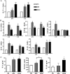
Stimulated production IL-17A– and Th17-inducing cytokines in the corneal and conjunctival epithelia of dry eye mice. (A) Real-time PCR data showing the relative expression (x-fold) of IL-17A in cornea (CN) and conjunctiva (CJ) in nonstressed controls mice (NS) and mice subjected to DS for 5 or 10 days (DS5, DS10, respectively; n = 5 per group). (B) Real-time PCR data showing the relative expression (x-fold) of Th17-inducing cytokines (IL-6, TGF-β1, TGF-β2, IL-23, IL-1β) in cornea (CN) and conjunctiva(CJ) in NS, DS5, and DS10 groups. (C) Concentrations of IL-6, TGF-β1, and IL-1β in the supernatants of 2-day cultured corneal and conjunctival explants obtained from NS, DS5, and DS10 groups measured by immunobead assay. Data are expressed as the mean ± SD of three separate experiments. *P < 0.05; **P < 0.01; ***P < 0.001; DS5 or DS10 versus NS group.
Figure 3.
Figure 3.
Stimulated Th17-inducing cytokines in the DCs cocultured with ocular surface tissue explants from nonstressed (NS) mice and mice subjected to DS (DS) for 10 days. (A) Real-time PCR data showing the relative expression (x-fold) of Th17-inducing cytokines (TGF-β1, TGF-β2, IL-6, IL-23, IL-1β, IL-1α) in DCs cultured for 2 days alone without corneal and conjunctival explants, or with corneal and conjunctival explants from a nonstressed control group (DC+NS) or eyes subjected to DS for 10 days (DC+DS). (B) Concentrations of IL-6, TGF-β1, and IL-1β in 2-day supernatants of DCs alone or DCs cocultured with corneal and conjunctival explants removed from NS eyes or eyes subjected to DS for 10 days, measured by immunobead assay. Data are expressed as mean ± SD of three separate experiments. *P < 0.05; **P < 0.01; ***P < 0.001.
Figure 4.
Figure 4.
Th17-induced Th17 differentiation of CD4+ T cells cocultured with DCs in the presence of cornea and conjunctival tissues subjected to DS. (A) Real-time PCR data showing the relative expression (x-fold) of Th17-expressed cytokines (IL-17A, IL-17F, IL-22, CCL-20) in CD4+ T cells cocultured with DCs for 1 day, 2 days, and 4 days in the T cell+DC group (absence of corneal and conjunctival explants) or DCs and corneal and conjunctival explants from a nonstressed control group (T cell+DC+NS) or from a 10-day DS group (T cell+DC+DS) (n = 4). (B, C) ELISPOT bioassay showing the numbers of IL-17 or IFN-γ–producing cells in T cell+DC group, T cell+DC+NS explant group, and T cell+DC+DS explant group. Cells (3 × 10⁵) were seeded per well. (D, E) Mean ± SD of three independent IL-17 and IFN-γ ELISPOTS showing IL-17 or IFN-γ–producing cells derived from CD4+ T cells cocultured with DCs for 7 days in the absence or presence of corneal and conjunctival explants removed from NS and 10-day DS mice, respectively. (F) IL-17 concentration in the supernatant of CD4+ T cells cocultured with DCs for 4 days in the absence or presence of corneal and conjunctival explants removed from NS and 10-day DS mice, respectively. (G) Real-time PCR data showing relative expression (x-fold) of the Th17 cell transcription factor-RORγt in CD4+ T cells cocultured with DCs for 1 day, 2 days, and 4 days in the T cell+DC group, T cell+DC+NS group, and T cell+DC+DS group (n = 4). Data are presented as mean ± SD of three or four independent experiments. *P < 0.05; **P < 0.01; ***P < 0.001.
Figure 5.
Figure 5.
Decreased levels of Th1- and Th2-associated factors in CD4+ T cells cocultured with DCs in the presence of corneal and conjunctival tissue subjected to DS. Real-time PCR data showing the relative expression (x-fold) of Th1-associated factors (IFN-γ, IL-12, IL-2, T-bet) and Th2-associated factors (IL-4, IL-13, GATA-3) in CD4+ T cells cocultured with DCs for 4 days in the T cell+DC group, T cell+DC+NS group, and T cell+DC+DS group (n = 4). Data are expressed as mean ± SD. *P < 0.05; **P < 0.01; ***P < 0.001. T cell+DC+DS or T cell+DC+NS group versus T cell+DC group.

References

    1. Mosmann TR, Coffman RL. TH1 and TH2 cells: different patterns of lymphokine secretion lead to different functional properties. Annu Rev Immunol 1989;7:145–173 - PubMed
    1. McKenzie BS, Kastelein RA, Cua DJ. Understanding the IL-23-IL-17 immune pathway. Trends Immunol 2006;27:17–23 - PubMed
    1. Steinman L. A brief history of T(H)17, the first major revision in the T(H)1/T(H)2 hypothesis of T cell-mediated tissue damage. Nat Med 2007;13:139–145 - PubMed
    1. Komiyama Y, Nakae S, Matsuki T, et al. IL-17 plays an important role in the development of experimental autoimmune encephalomyelitis. J Immunol 2006;177:566–573 - PubMed
    1. Afzali B, Lombardi G, Lechler RI, Lord GM. The role of T helper 17 (Th17) and regulatory T cells (Treg) in human organ transplantation and autoimmune disease. Clin Exp Immunol 2007;148:32–46 - PMC - PubMed

Publication types

MeSH terms