

Decreased retinal ganglion cell layer thickness in patients with type 1 diabetes
- PMID: 20130282
- PMCID: PMC2904016
- DOI: 10.1167/iovs.09-5041
Decreased retinal ganglion cell layer thickness in patients with type 1 diabetes
Abstract
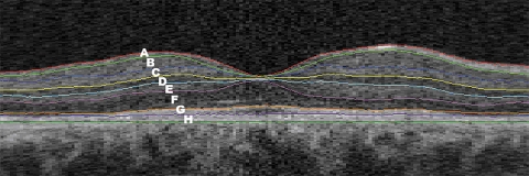
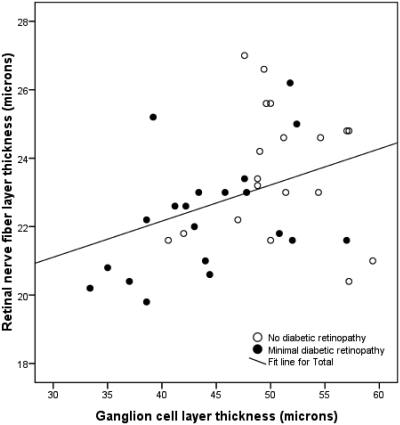
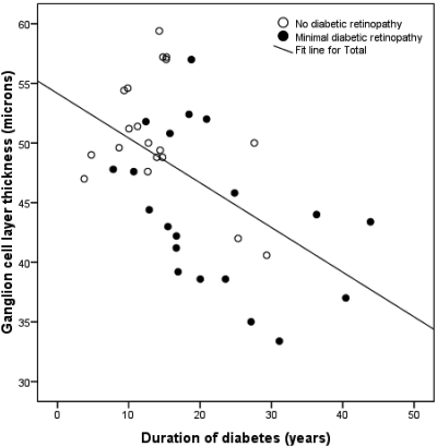
PURPOSE. To determine which retinal layers are most affected by diabetes and contribute to thinning of the inner retina and to investigate the relationship between retinal layer thickness (LT) and diabetes duration, diabetic retinopathy (DR) status, age, glycosylated hemoglobin (HbA1c), and the sex of the individual, in patients with type 1 diabetes who have no or minimal DR. METHODS. Mean LT was calculated for the individual retinal layers after automated segmentation of spectral domain-optical coherence tomography scans of patients with diabetes and compared with that in control subjects. Multiple linear regression analysis was used to determine the relationship between LT and HbA1c, age, sex, diabetes duration, and DR status. RESULTS. In patients with minimal DR, the mean ganglion cell layer (GCL) in the pericentral area was 5.1 mum thinner (95% confidence interval [CI], 1.1-9.1 mum), and in the peripheral macula, the mean retinal nerve fiber layer (RNFL) was 3.7 mum thinner (95% CI, 1.3-6.1 mum) than in the control subjects. There was a significant linear correlation (R = 0.53, P < 0.01) between GCL thickness and diabetes duration in the pooled group of patients. Multiple linear regression analysis (R = 0.62, P < 0.01) showed that DR status was the most important explanatory variable. CONCLUSIONS. This study demonstrates GCL thinning in the pericentral area and corresponding loss of RNFL thickness in the peripheral macula in patients with type 1 diabetes and no or minimal DR compared with control subjects. These results support the concept that diabetes has an early neurodegenerative effect on the retina, which occurs even though the vascular component of DR is minimal.
Figures
References
-
- Abu-El-Asrar AM, Dralands L, Missotten L, Al-Jadaan IA, Geboes K. Expression of apoptosis markers in the retinas of human subjects with diabetes. Invest Ophthalmol Vis Sci 2004;45:2760–2766 - PubMed
-
- Antonetti DA, Barber AJ, Bronson SK, et al. Diabetic retinopathy: seeing beyond glucose-induced microvascular disease. Diabetes 2006;55:2401–2411 - PubMed
-
- Barber AJ. A new view of diabetic retinopathy: a neurodegenerative disease of the eye. Prog Neuropsychopharmacol Biol Psychiatry 2003;27:283–290 - PubMed
-
- Barber AJ, Antonetti DA, Kern TS, et al. The Ins2Akita mouse as a model of early retinal complications in diabetes. Invest Ophthalmol Vis Sci 2005;46:2210–2218 - PubMed
Publication types
MeSH terms
Substances
Grants and funding
LinkOut - more resources
Full Text Sources
Other Literature Sources
Medical
