

Novel markers of left ventricular hypertrophy in uremia
- PMID: 20130393
- PMCID: PMC2924237
- DOI: 10.1159/000279768
Novel markers of left ventricular hypertrophy in uremia
Abstract
Aims: Left ventricular hypertrophy (LVH) is the most frequent cardiac complication in chronic renal disease. Previous studies implicate elevated serum phosphorus as a risk factor for LVH.
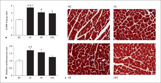
Methods: We treated 5/6 nephrectomized rats with enalapril or enalapril + sevelamer carbonate for 4 months to determine if sevelamer carbonate had an additional beneficial effect on the development of LVH and uremia-induced left ventricle (LV) remodeling.
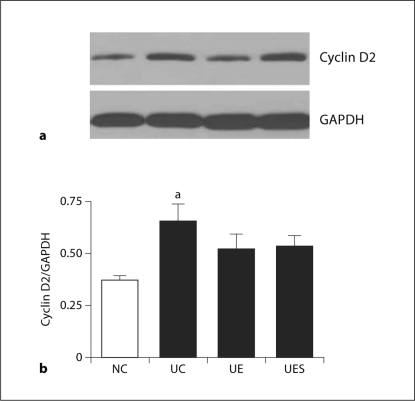
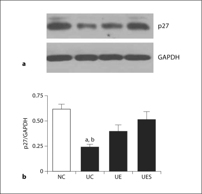
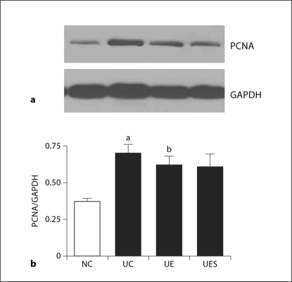
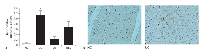
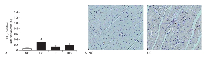
Results: Uremia increased LV weight and cardiomyocyte size. Enalapril and enalapril + sevelamer blunted the increase in left ventricular weight. Only enalapril + sevelamer diminished the increase in cardiomyocyte size. Uremia increased cyclin D2 and PCNA and decreased p27 protein expression in the heart. Enalapril + sevelamer diminished the decrease in p27 expression caused by uremia. Uremia increased Ki67-positive and phosphohistone H(3)-positive interstitial cells. This was not seen in cardiomyocytes. Multivariable regression analysis showed that increased phosphorus was an independent risk factor for both increased LV weight and cardiomyocyte size.
Conclusions: These data suggest left ventricular remodeling consists of cardiomyocyte hypertrophy and interstitial cell proliferation, but not cardiomyocyte proliferation. p27 and cyclin D2 may play important roles in the development of LVH. In addition, phosphorus can be an independent risk factor for the development of LVH.
2010 S. Karger AG, Basel.
Figures
Similar articles
-
Blockage of the renin-angiotensin system attenuates mortality but not vascular calcification in uremic rats: sevelamer carbonate prevents vascular calcification.Am J Nephrol. 2009;29(6):582-91. doi: 10.1159/000192844. Epub 2009 Jan 15. Am J Nephrol. 2009. PMID: 19145073
-
Cardiomyocyte loss in experimental renal failure: prevention by ramipril.Kidney Int. 2003 May;63(5):1708-13. doi: 10.1046/j.1523-1755.2003.00927.x. Kidney Int. 2003. PMID: 12675846
-
Angiotensin converting enzyme inhibition and dihydropyridine calcium channel blockade in the treatment of left ventricular hypertrophy in arterial hypertension.Minerva Cardioangiol. 2002 Jun;50(3):169-74. Minerva Cardioangiol. 2002. PMID: 12107398 Review.
-
Reversal of interstitial fibroblast hyperplasia via apoptosis in hypertensive rat heart with valsartan or enalapril.Cardiovasc Res. 2003 Mar;57(3):775-83. doi: 10.1016/s0008-6363(02)00789-7. Cardiovasc Res. 2003. PMID: 12618239
-
Angiotensin-converting enzyme inhibitors and effects on left ventricular hypertrophy.Blood Press Suppl. 1994;2:35-40. Blood Press Suppl. 1994. PMID: 8061844 Review.
Cited by
-
Adiponectin expression and the cardioprotective role of the vitamin D receptor activator paricalcitol and the angiotensin converting enzyme inhibitor enalapril in ApoE-deficient mice.Ther Adv Cardiovasc Dis. 2014 Dec;8(6):224-36. doi: 10.1177/1753944714542593. Epub 2014 Jul 18. Ther Adv Cardiovasc Dis. 2014. PMID: 25037058 Free PMC article.
-
Identification and analysis of a key long non-coding RNAs (lncRNAs)-associated module reveal functional lncRNAs in cardiac hypertrophy.J Cell Mol Med. 2018 Feb;22(2):892-903. doi: 10.1111/jcmm.13376. Epub 2017 Nov 20. J Cell Mol Med. 2018. PMID: 29154475 Free PMC article.
-
Cardiac and renal effects of atrasentan in combination with enalapril and paricalcitol in uremic rats.Kidney Blood Press Res. 2014;39(4):340-52. doi: 10.1159/000355811. Epub 2014 Sep 19. Kidney Blood Press Res. 2014. PMID: 25300759 Free PMC article.
-
Mineral and Bone Disorder and Its Association with Cardiovascular Parameters in Chinese Patients with Chronic Kidney Disease.Chin Med J (Engl). 2016 Oct 5;129(19):2275-80. doi: 10.4103/0366-6999.190678. Chin Med J (Engl). 2016. PMID: 27647184 Free PMC article.
-
Assessment of the left ventricular function in patients with uremia using layer-specific 2-dimensional speckle tracking echocardiography.Medicine (Baltimore). 2019 Mar;98(9):e14656. doi: 10.1097/MD.0000000000014656. Medicine (Baltimore). 2019. PMID: 30817588 Free PMC article.
References
-
- Foley RN, Parfrey PS, Harnett JD, et al. Clinical and echocardiographic disease in patients starting end-stage renal disease therapy. Kidney Int. 1995;47:186–192. - PubMed
-
- Silberberg JS, Barre PE, Prichard SS, et al. Impact of left ventricular hypertrophy on survival in end-stage renal disease. Kidney Int. 1989;36:286–290. - PubMed
-
- Levin A, Thompson CR, Ethier J, et al. Left ventricular mass index increase in early renal disease: impact of decline in hemoglobin. Am J Kidney Dis. 1999;34:125–134. - PubMed
-
- London GM. Cardiovascular disease in chronic renal failure: pathophysiologic aspects. Semin Dial. 2003;16:85–94. - PubMed
-
- Block GA, Hulbert-Shearon TE, Levin NW, et al. Association of serum phosphorus and calcium × phosphate product with mortality risk in chronic hemodialysis patients: a national study. Am J Kidney Dis. 1998;31:607–617. - PubMed
Publication types
MeSH terms
Substances
Grants and funding
LinkOut - more resources
Full Text Sources
Other Literature Sources
Miscellaneous
