Review
. 2010 Apr;77(8):662-8.
doi: 10.1038/ki.2009.559. Epub 2010 Feb 3.

Toward the development of podocyte-specific drugs

Affiliations
Review

Toward the development of podocyte-specific drugs

Jochen Reiser et al. Kidney Int. 2010 Apr.

Abstract

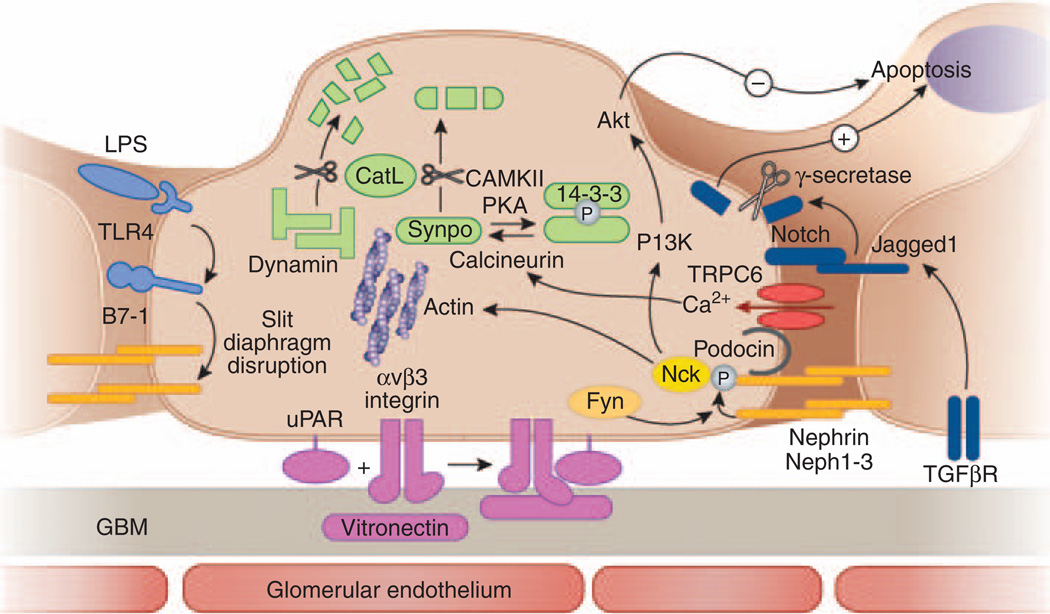
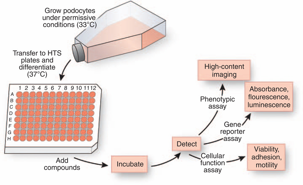
Most kidney diseases that ultimately lead to end-stage renal failure originate within the glomerulus and are associated with proteinuria. Treatment options are unspecific and offer partial cures at best because available therapies do not primarily treat glomerular cells but rather act systemically and thus cause many side effects. Most glomerulopathies directly stem from injury to podocytes, cells that have a key role in the maintenance of the glomerular filter. Thus, these cells constitute an obvious and promising target for the development of novel kidney-protective drugs. During the last decade, enormous advances have been made in the understanding of podocyte structure and function. A number of pathways that are altered during glomerular diseases may be targeted by novel small- and large-molecule drugs as well as biologicals that have been identified in nephrology and other areas of drug development. Cultured podocytes provide a valuable model for high-throughput drug screening assays. Furthermore, podocytes have been shown to possess many features that make them particularly good target cells for renal protection. This mini-review discusses some of the most recent promising data related to potential drug therapy for proteinuria and kidney disease through direct podocyte targeting.


Figures

Figure 1
Figure 1. Pathways involved in acquired glomerular diseases representing targets for podocyte-specific drugs
A schematic cross-section of a podocyte foot process with the corresponding cell body, the glomerular basement membrane, and the glomerular endothelium is shown. Pathways involved in podocyte injury that may be drug-targeted are depicted in different colors. Sustaining nephrin expression and phosphorylation (yellow) might contribute to both antiapoptotic signaling and actin polymerization. The B7-1 pathway (light blue) may be targeted by (1) toll-like receptor-4 antagonists or (2) blocking the binding of B7-1 to slit diaphragm structure proteins. Urokinase plasminogen-activator receptor (uPAR)-induced podocyte motility (violet) could be inhibited by (1) interfering with binding of uPAR to αvβ3 integrin, (2) inhibiting β3 integrin activation, or (3) inhibiting binding of αvβ3 integrin to vitronectin. The notch pathway (dark blue) can be targeted by (1) interfering with its upstream activation by blocking the TGF-β1 effect, (2) inhibiting γ-secretase, which is required for proteolytic receptor activation, or (3) interfering with target gene transcription. TRPC6 channels (red) may be targeted by (1) channel blockers or (2) inhibiting their expression. The CatL pathway (green) could be targeted by (1) specifically inhibiting CatL expression or activity, (2) shifting the equilibrium of synaptopodin toward the phosphorylated form by inhibiting calcineurin-mediated dephosphorylation or enhancing PKA or CAMKII-mediated phosphorylation, (3) protecting synaptopodin and dynamin by compounds that bind to the CatL cleavage site, or (4) delivering cleavage-resistant synaptopodin or dynamin mutants.
Figure 2
Figure 2. Phenotypic podocyte assays used for high-content screening (HCS)
Cultured podocytes can be used in HCS assays for automated detection of cellular morphology and stress fiber formation in a high-throughput environment. (a) An HCS captured image from a well of a 96-well plate with podocytes stained with phalloidin to detect actin-stress fibers. (b) Same image as in panel a stained with Cell Mask Blue (to stain cell nuclei and cytoplasm). (c) An overlay of images in panels a and b. (d) An overlay of automatically calculated cellular boundaries that can be used in measuring various cellular characteristics (such as cell number, cell size, and intensity of stress fibers per cell) on the raw image (a) from the 96-well plate.
Figure 3
Figure 3. Schematic of a workflow for a primary drug screen with cultured podocytes
Cultured podocytes are grown under permissive conditions at 33°C to allow for proliferation. Next, cells are transferred to 96-well HTS microtiter plates and allowed to differentiate under nonpermissive conditions at 37°C. Fully differentiated podocytes are incubated with chemical compound libraries, and the effect of compounds on cells is measured using a variety of cell-based readout assays. These detection assays include high-content imaging to detect changes in cellular phenotypes, measurement of homogenous changes in absorbance, fluorescence- or luminescence-based gene reporter assays as well as determination of changes in cell viability and cellular function, such as adhesion and motility.
Figure 4
Figure 4. Cyclo-RGDfV as an example of a small molecule targeting podocytes
(a) Chemical structure of the anti-integrin αvβ3 drug cyclo-RGDfV. (b) Three-dimensional crystal structure of the extracellular domains of integrin αvβ3 (shown as a ribbon model with αv-chain in blue and β3-chain in red) bound to cyclo-RGDfV (green, shown as a CPK model). Bound metal ions are shown as gray spheres. Models a and b are based on the study by Dechantsreiter et al. and Xiong et al., respectively. Cyclo-RGDfV has been successfully used to treat proteinuria in the lipopolysaccharide-mouse model.

Similar articles

Cited by

References

    1. Wiggins RC. The spectrum of podocytopathies: a unifying view of glomerular diseases. Kidney Int. 2007;71:1205–1214. - PubMed
    1. Reiser J, Kriz W, Kretzler M, et al. The glomerular slit diaphragm is a modified adherens junction. J Am Soc Nephrol. 2000;11:1–8. - PubMed
    1. Tryggvason K, Patrakka J, Wartiovaara J. Hereditary proteinuria syndromes and mechanisms of proteinuria. N Engl J Med. 2006;354:1387–1401. - PubMed
    1. Shankland SJ, Pippin JW, Reiser J, et al. Podocytes in culture: past, present, and future. Kidney Int. 2007;72:26–36. - PubMed
    1. Benzing T. The promise of well-being: stay in shape with N(i)ck. J Am Soc Nephrol. 2009;20:1425–1427. - PubMed

Publication types

MeSH terms