. 2010 Apr;77(8):736-42.
doi: 10.1038/ki.2009.550. Epub 2010 Feb 3.

Tamm-Horsfall protein and urinary exosome isolation

Affiliations

Tamm-Horsfall protein and urinary exosome isolation

Patricia Fernández-Llama et al. Kidney Int. 2010 Apr.

Abstract

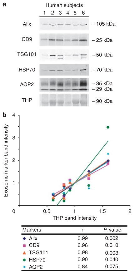
Urinary exosomes have been proposed as starting material for discovery of protein biomarkers of kidney disease. Current protocols for their isolation use a two-step differential centrifugation process. Due to their low density, exosomes are expected to remain in the low-speed (17,000 x g) supernatant and to sediment only when the sample is spun at high speed (200,000 x g). Analysis using western blot and electron microscopy found that urinary exosomes are also present in the low-speed pellet entrapped by polymeric Tamm-Horsfall protein, thus diminishing the procedure's reproducibility. Here we show that addition of dithiothreitol to the low-speed pellet disrupted the polymeric network, presumably by reduction of disulfide bonds linking the monomers. This modification shifted the exosomal proteins from the low- to the high-speed pellet. Also, by shifting the Tamm-Horsfall protein to the high-speed pellet, the use of dithiothreitol makes it feasible to use Tamm-Horsfall protein to normalize excretion rates of exosomal proteins in spot urines. We tested this by western blot, and found that there was a high degree of correlation between exosomal proteins and Tamm-Horsfall protein in the high-speed pellet. Since the yield of exosomes by differential centrifugation can be increased by chemical reduction, Tamm-Horsfall protein may be a suitable normalizing variable for urinary exosome studies when quantitative urine collections are not practical.


Conflict of interest statement

DISCLOSURE

All the authors declared no competing interests.

Figures

Figure 1
Figure 1. Sodium dodecyl sulfate-polyacrylamide gel electrophoresis (SDS—PAGE) of 17,000 and 200,000 × g pellets treated with and without dithiothreitol (DTT) and stained with Coomassie blue
Tamm-Horsfall protein (THP) is seen as a broad band around 92 kDa. The addition of DTT, a reducing agent, shifted the sedimentation of THP mainly from the low-speed pellet to the high-speed pellet. The procedure applied in this experiment involved incubation of the 17,000 × g pellet with DTT and a second 17,000 × g centrifugation after resuspension. The same percentages of the 17,000 and 200,000 × g pellet’s final volume were loaded on the gel.
Figure 2
Figure 2. Electron microscope images of the 17,000 × g pellets without (a and b) and with (c) dithiothreitol (DTT) treatment and the 200,000 × g pellet with DTT treatment (d)
Tamm-Horsfall protein (THP) formed long polymeric filaments that associated laterally to form ropelike structures (a, original magnification: × 5000 and b, depicting the dashed box in a with magnification × 40,000). The THP network contained small (20–100 nm) vesicles compatible with exosomes (b, arrowheads). Treatment with DTT disrupted the THP meshwork (c, original magnification: × 5000), leaving short convoluted THP oligomers (d, original magnification: × 20,000).
Figure 3
Figure 3. Immunoblots of the 17,000 and the 200,000 × g pellets with and without dithiothreitol (DTT) treatment probed against exosomal proteins Alix, TSG101, CD9, and AQP2
The same urine samples were processed fresh (a), stored at 4 °C for 3 days before processing (b), and stored at −80 °C for 3 days before processing (c).
Figure 4
Figure 4. Electron microscope images of the dithiothreitol (DTT)-treated 200,000 × g pellets from normal human subjects at × 20,000 magnification
This magnification was used to identify exosome-like particles based on the size (20–100 nm) and the shape (round) of the vesicles (arrowheads). Figures ac represent fields from different subjects.
Figure 5
Figure 5. Immunoblotting of 200,000 × g pellets from six normal human subjects probed against exosomal proteins Alix, CD9, TSG101, HSP70, and AQP2 as well as Tamm-Horsfall protein (THP)
(a) These immunoblots were loaded based on an equal number of exosome-like particles. (b) Scatter plot relating the band intensities of the exosomal proteins and THP.
Figure 6
Figure 6. Correlation of Tamm-Horsfall protein (THP) band intensities from Coomassie-stained gels and THP immunoblots of 200,000 × g urine pellets
A correlation between the amounts of THP deduced from densitometry of Coomassie-stained gels and THP immunoblots was very high (r = 0.99, linear regression analysis).
Figure 7
Figure 7. Current procedure for urinary exosome isolation
The new procedure adds the step of dithiothreitol (DTT) incubation of the 17,000 × g pellet to depolymerized the Tamm-Horsfall protein (THP) polymeric network followed by a second 17,000 × g spin to recover the exosomes that were entrapped by the THP network. The two 17,000 × g supernatants are pooled.

References

    1. Pisitkun T, Shen RF, Knepper MA. Identification and proteomic profiling of exosomes in human urine. Proc Natl Acad Sci USA. 2004;101:13368–13373. - PMC - PubMed
    1. Hogan MC, Manganelli L, Woollard JR, et al. Characterization of PKD protein-positive exosome-like vesicles. J Am Soc Nephrol. 2009;20:278–288. - PMC - PubMed
    1. Simpson RJ, Jensen SS, Lim JW. Proteomic profiling of exosomes: current perspectives. Proteomics. 2008;8:4083–4099. - PubMed
    1. Nilsson J, Skog J, Nordstrand A, et al. Prostate cancer-derived urine exosomes: a novel approach to biomarkers for prostate cancer. Br J Cancer. 2009;100:1603–1607. - PMC - PubMed
    1. Gonzales PA, Pisitkun T, Hoffert JD, et al. Large-scale proteomics and phosphoproteomics of urinary exosomes. J Am Soc Nephrol. 2009;20:363–379. - PMC - PubMed

Publication types