. 2010 Feb 4;463(7281):627-31.
doi: 10.1038/nature08728.

An aspartyl protease directs malaria effector proteins to the host cell

Affiliations

An aspartyl protease directs malaria effector proteins to the host cell

Justin A Boddey et al. Nature. .

Abstract

Plasmodium falciparum causes the virulent form of malaria and disease manifestations are linked to growth inside infected erythrocytes. To survive and evade host responses the parasite remodels the erythrocyte by exporting several hundred effector proteins beyond the surrounding parasitophorous vacuole membrane. A feature of exported proteins is a pentameric motif (RxLxE/Q/D) that is a substrate for an unknown protease. Here we show that the protein responsible for cleavage of this motif is plasmepsin V (PMV), an aspartic acid protease located in the endoplasmic reticulum. PMV cleavage reveals the export signal (xE/Q/D) at the amino terminus of cargo proteins. Expression of an identical mature protein with xQ at the N terminus generated by signal peptidase was not exported, demonstrating that PMV activity is essential and linked with other key export events. Identification of the protease responsible for export into erythrocytes provides a novel target for therapeutic intervention against this devastating disease.


Figures

Figure 1
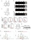
Figure 1. PEXEL processing is sensitive to HIV protease inhibitors
a, Structure of PfEMP3-GFP. PSS, signal sequence; RxLxQ PEXEL; residues 65-83 of PfEMP3. Arrow, PEXEL cleavage. b, PfEMP3-GFP processing with HIV-1 inhibitors. c, Densitometry of uncleaved bands in b. d, Structure of KAHRP-GFP. KSS, signal sequence; RxLxQ PEXEL, residues 59-96 of KAHRP. e, KAHRP-GFP PEXEL processing with inhibitors. f, Densitometry of uncleaved bands in e. g, Structure of ACPs-GFP. ACP SS, signal sequence-GFP. Arrow, signal sequence cleavage. h, ACPs-GFP signal sequence processing is not inhibited. ‘Cleaved’, signal sequence cleavage. i, Densitometry of the uncleaved region of immunoblot. (see Supplementary for details)
Figure 2
Figure 2. Plasmepsin V cleaves PEXEL
a, Plasmepsin V HA tagged (3D7-PMVHA). b, Plasmepsin V localises to ER. c, Inactive HA-tagged Plasmepsin V (3D7-PMVmutHA). d, Mutant Plasmepsin V (3D7-PMVmutHA) localises to ER. e, HA-tagged Plasmepsin V (ipPMVHA) with KAHRP peptides; unmutated, R>A, L>A, Q>A, RLQ>A. ipPMVmutHA (inactive) or ipPMIXHA (Plasmepsin IX) with unmutated peptide. f, Recovered enzyme. g, Recombinant GBP130 cleavage by Plasmepsin V. Last lanes are GBP130 mutant (RLE>A). U, uncleaved; C, cleaved. h, Identification of cleaved peptide GNGSGDSFDFRNKRTL. Identification of uncleaved RLQ>A mutant peptide. * refers to multiple H20 and NH3 losses. (see Supplementary for details)
Figure 3
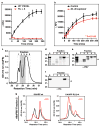
Figure 3. Activity of Plasmepsin V and cleavage of PEXEL
a, Plasmepsin V cleavage of fluorescent substrate with PEXEL (black line) and PEXEL RL>A mutant (red line). Data shown is one representative experiment done in triplicate. Data shown is the mean ± standard deviation. b, Lopinavir reduces Plasmepsin V activity (P=0.0145). Data shown is one representative experiment done in triplicate. Data shown is the mean ± standard deviation. c, N-terminal hexa-His fusion of Plasmepsin V (rHisPMV) from E. coli separated by HPLC. Elutions with activity indicated in grey. d, Starting material (S) and re-folded fractions were Coomassie stained. e, Starting material (S) and fractions probed with anti-Plasmepsin V antibodies. f, Starting material (S) and fractions probed with anti-hexaHis antibodies. g, HPLC of recombinant Plasmepsin V activity (rHisPMV) with KAHRP peptides. (see Supplementary for details)
Figure 4
Figure 4. Cleavage by Plasmepsin V is essential for export
a, Exported PfEMP3-GFP (top) whilst R>A mutant accumulated in ER (bottom). Right, fractionation showing full-length R>A mutant accumulates in membrane. b, Proteomic analysis of Plasmepsin V immuno-precipitated proteins. c, Structure of PfEMP3-GFP. PSS, signal sequence; RxLxQ PEXEL, residues 65-83 of PfEMP3. Arrow, PEXEL cleavage. d, Structure of PfEMP3xQ-GFP. Arrow, signal peptidase cleavage. e, N-terminal processing confirmed with α-GFP. f, Immuno-purified PfEMP3-GFP and PfEMP3xQ-GFP analysed by Coomassie and LC-MS/MS. g, Identification of N-terminal AcAQVLGNTR of PfEMP3-GFP after Plasmepsin V processing. h, Identification of AcAQVLGNTR at N-terminus of PfEMP3xQ-GFP after signal peptidase processing. i, Imaging of exported PfEMP3-GFP (top) and non-exported PfEMP3xQ-GFP (bottom). (see Supplementary for details)

Similar articles

Cited by

References

    1. Snow RW, et al. The global distribution of clinical episodes of Plasmodium falciparum malaria. Nature. 2005;434:214–217. - PMC - PubMed
    1. Miller LH, Good MF, Milon G. Malaria pathogenesis. Science. 1994;264:1878–1883. - PubMed
    1. Raventos Suarez C, Kaul DK, Macaluso F, Nagel RL. Membrane knobs are required for the microcirculatory obstruction induced by Plasmodium falciparum-infected erythrocytes. Proc Natl Acad Sci USA. 1985;82:3829–3833. - PMC - PubMed
    1. Barnwell JW, et al. A human 88-kD membrane glycoprotein (CD36) functions in vitro as a receptor for a cytoadherence ligand on Plasmodium falciparum-infected erythrocytes. J Clin Invest. 1989;84:765–772. - PMC - PubMed
    1. Marti M, et al. Targeting malaria virulence and remodeling proteins to the host erythrocyte. Science. 2004;306:1930–1933. - PubMed

Publication types

MeSH terms