. 2010 Feb 4;463(7281):632-6.
doi: 10.1038/nature08726.

Plasmepsin V licenses Plasmodium proteins for export into the host erythrocyte

Affiliations

Plasmepsin V licenses Plasmodium proteins for export into the host erythrocyte

Ilaria Russo et al. Nature. .

Abstract

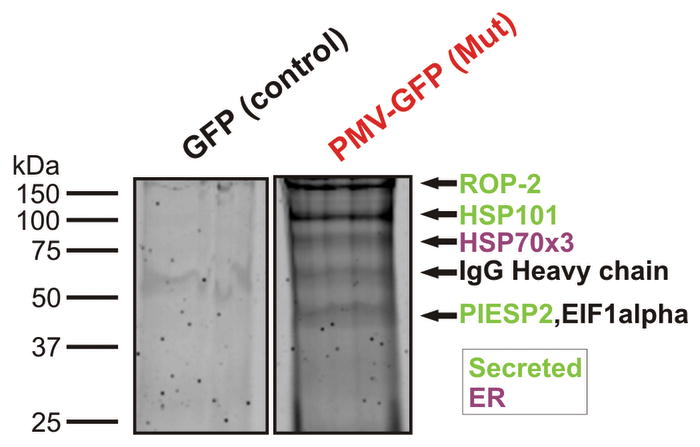
During their intraerythrocytic development, malaria parasites export hundreds of proteins to remodel their host cell. Nutrient acquisition, cytoadherence and antigenic variation are among the key virulence functions effected by this erythrocyte takeover. Proteins destined for export are synthesized in the endoplasmic reticulum (ER) and cleaved at a conserved (PEXEL) motif, which allows translocation into the host cell via an ATP-driven translocon called the PTEX complex. We report that plasmepsin V, an ER aspartic protease with distant homology to the mammalian processing enzyme BACE, recognizes the PEXEL motif and cleaves it at the correct site. This enzyme is essential for parasite viability and ER residence is essential for its function. We propose that plasmepsin V is the PEXEL protease and is an attractive enzyme for antimalarial drug development.


Conflict of interest statement

Competing financial interests: none.

Figures

Figure 1
Figure 1
The transmembrane region of plasmepsin V confers ER localization. a) Schematic of PM V and C-terminal integrants. SP: signal peptide; N-term.: N-terminal domain; Ct: C-terminal; TM: transmembrane region. b) Southern blots of full-length (DC6) or C-terminal tail deletion (EF2) integrant clones and transmembrane region deletion transfectants (ΔTM). Parental strain (3D7) and plasmids are shown for reference. c) Western blot using anti-PM V antibody. BiP served as loading control. d and e) Live fluorescence images of DC6 and EF2, respectively. Left to right: phase, GFP, DAPI, fluorescence merge and total merge. Bar, 2 μm. f) Localization of PM V fragments (defined in a) fused to yellow fluorescent protein (YFP). Expression was episomal, using the HSP86 promoter. Panel order as in d and e.
Figure 2
Figure 2
Plasmepsin V is essential for intraerythrocytic parasite viability. a) Active site allelic replacement scheme. Integration vectors possessing a synonymous (S) or non-synonymous (NS) mutation in the Asp 108 codon and a new TfiI restriction site just upstream were transfected into parental strain 3D7. Possible outcomes for upstream crossover (pre-mutation) and downstream crossover (post-mutation) are drawn. Integrants were selected and assessed by PCR. Primers used for amplification (241 and 247) are marked. b) Flow cytometry of parasites. Parasites were assessed for GFP expression, an indication of integration at the endogenous locus. Parental strain 3D7 (WT) is shown as control. c) Southern blot of transfected parasite pools. At left are expected positions of possible products of the BsrGI/TfiI digest mapped in c. d) Screening of integrants by PCR and restriction digest. Arrows indicate a doublet (predicted 787 and 748 bp) resulting from restriction fragmentation of product from recombinants that have crossed over upstream of the active site codon.
Figure 3
Figure 3
Episomal expression of wild-type and catalytic mutant PM V. a,b) Immunofluorescence of wild-type and 108 D to A (mutant) PM V expressing lines, respectively. Left to right: phase, GFP, DAPI, merge. Bar, 2 μm. c) Fluorescence intensity of 52 transfected early trophozoites was measured. Mean relative fluorescence units were 82,559 and 32,059 for wild-type and mutant-transfected parasites, respectively. d) Western blot. BiP serves as a loading control. e) Mutant PM V-expressing parasites encased in erythrocyte ghosts. f) Growth curves. Asynchronous cultures episomally expressing wild-type (green) or mutant (red) PM V were monitored by flow cytometry for four days in triplicate (error bars indicate standard deviation). Mutant growth rate is reduced by 49.2+/−1.6 %. g) Double-infected erythrocyte containing a parasite expressing high levels of mutant PM V. h and i) Western blots of HRPII processing in parasites episomally expressing mutant or wild-type PM V. h: top - anti-HRPII; middle panels - anti-PM V; bottom - anti-BiP. Contrast and brightness enhanced slightly over the entire panel to bring out features seen on the original film. i: top - anti-HRPII; middle panels - antibodies against non-exported secretory proteins dipeptidyl peptidase I (DPAP1) and plasmepsin II (PMII); bottom - anti-BiP. j) Flow cytometry. Wild-type (green) and mutant (red) PM V-expressing parasites were fixed, treated with tetanolysin (5 min, 20 units/ml) to selectively permeabilize the erythrocyte compartment and exported RESA was quantified by flow cytometry with specific antibody. Control without primary antibody is shown below. Results are representative of 4 experiments. k) HRP II staining was inconsistent by the fixation procedure used in j, so HRPII erythrocyte fluorescence intensity (80 cells) was quantified. Mean relative fluorescence intensity per μm2 was 2671 and 1179 for wild-type and mutant, respectively.
Figure 4
Figure 4
Plasmepsin V activity. a) Substrate cleavage. PM V was isolated from parental (3D7) and PM V-GFP fusion (DC6) clones using anti-GFP (yellow and red, respectively) or anti-PM V (blue and green, respectively). A similar isolation without Ab (protein A) served as a control for non-specific binding (purple and cyan, respectively). Purified enzyme was incubated at pH 6.5 for 35 min with fluorogenic peptide (Anaspec) corresponding to the PEXEL motif for HRPII (DABCYL-LNKRLLHETQ-EDANS). Error bars indicate standard deviation of three activity curves. b) PM V specificity for PEXEL motif. Processing of fluorogenic peptides containing the PEXEL motifs of HRPII (blue) or PfEMP2 (red, DABCYL-RYVRILSETE-EDANS) are shown. Left - wild-type peptide (WT); middle - L to A mutant peptide; right - R to A mutant peptide. Inset: peptide sequences with mutated residues in black. Error bars as in a. c) pH dependence in Tris-Malate buffer. Two separate experiments are shown in different colors. Error bars as in a. d) Activity of active-site mutant PM V compared to wild type. PMV-GFP was isolated from 3D7 (untransfected mock isolation), wild-type- or mutant-PM V-GFP-transfected parasites using anti-GFP for purification. Activity on HRPII-PEXEL peptide is normalized to PM V protein content. Error bars as in a. e) Cleavage of pro-HRPII by isolated enzyme from d. p: pro-form; m: mature protein.
Figure 5
Figure 5
Analysis of substrate cleavage. PM V was incubated with HRPII (a) or PfEMP2 (c) PEXEL peptides (DABCYL-LNKRLLHETQ-EDANS and DABCYL-RYVRILSETE-EDANS, respectively). Cleavage products were separated on a C18 column by reverse-phase HPLC. Back to front: incubation for 0, 2 and 16h. S, substrate peak. b,d) isolated products and substrates from a and c were analyzed by MALDI-TOF mass spectrometry. Ion peaks and sodium adducts are labeled. HRPII peptide product masses: calculated 762.51 and 1008.25; detected 762.32 and 1007.80. PfEMP2 peptide product masses: calculated 1071.30 and 713.72; detected 1071.31 and 713.23. Ions corresponding to alternative peptide bond cleavage were not detected.
Figure 6
Figure 6
Proteomic analysis of PM V-associated proteins. Shown is a Coomassie-stained gel of an anti-GFP pulldown for the episomal GFP-expressing control strain (GFP) and the mutant PM V-GFP-expressing strain (Mut). Bands were excised, trypsinized and analyzed by MS-MS. The same analysis was also performed on the wild-type PM V-GFP strain, and on the parental 3D7 strain but using anti-PM V antibody. Shown are proteins for which at least four peptides were identified from the anti-PM V pulldown and three or more peptides from at least one of the two anti-GFP pulldowns of episomal PM V-expressing parasites. All proteins identified are tabulated in Supplementary Table 1. None of the proteins were detected in the GFP control pulldown.

Similar articles

Cited by

References

    1. Sargeant TJ, et al. Lineage-specific expansion of proteins exported to erythrocytes in malaria parasites. Genome Biol. 2006;7:R12. - PMC - PubMed
    1. van Ooij C, et al. The malaria secretome: from algorithms to essential function in blood stage infection. PLoS Pathogens. 2008;4:e1000084. - PMC - PubMed
    1. Haldar K, Mohandas N. Erythrocyte remodeling by malaria parasites. Current Opinion Hematol. 2007;14:203–209. - PubMed
    1. Maier AG, Cooke BM, Cowman AF, Tilley L. Malaria parasite proteins that remodel the host erythrocyte. Nature Reviews. 2009;7:341–354. - PubMed
    1. Hiller NL, et al. A host-targeting signal in virulence proteins reveals a secretome in malarial infection. Science. 2004;306:1934–1937. - PubMed

Publication types

MeSH terms