Review
. 2010 Feb;88(1):77-88.
doi: 10.1139/o09-159.

Comparison of the multiple oligomeric structures observed for the Rvb1 and Rvb2 proteins

Affiliations
Review

Comparison of the multiple oligomeric structures observed for the Rvb1 and Rvb2 proteins

Kevin L Y Cheung et al. Biochem Cell Biol. 2010 Feb.

Abstract

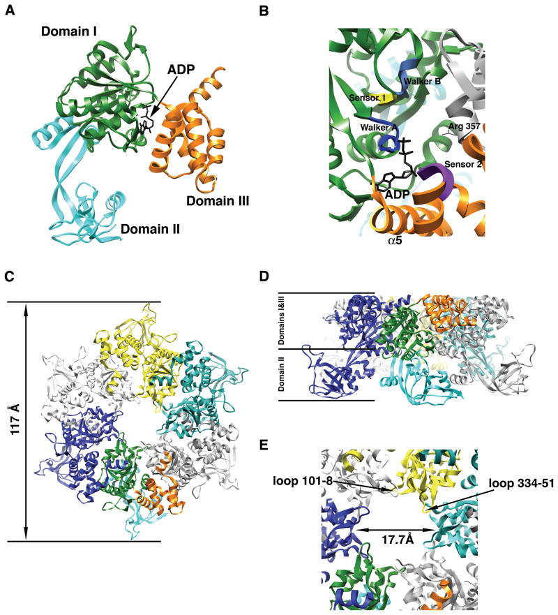
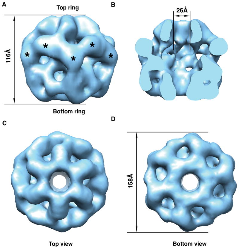
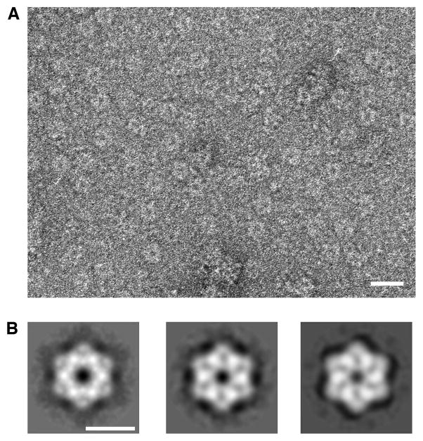
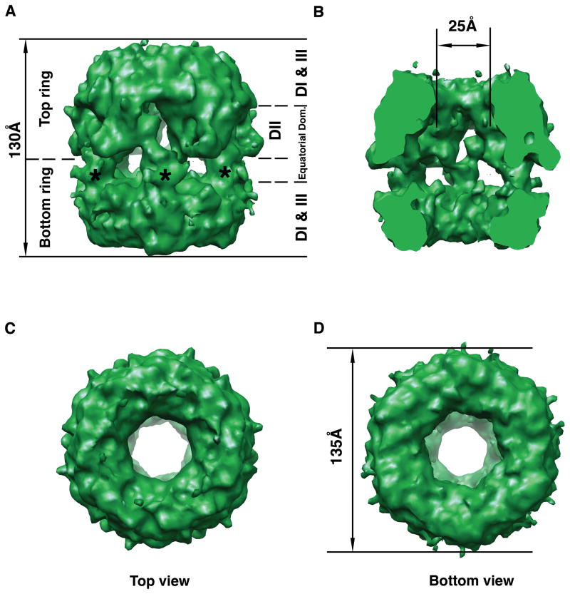
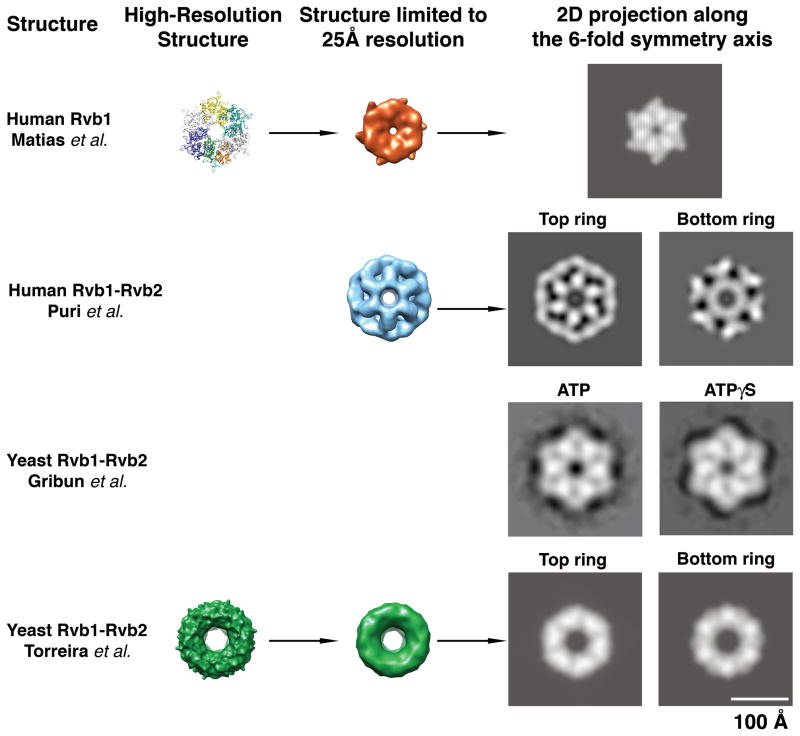
The Rvb1 and Rvb2 proteins are 2 members of the AAA+ family, involved in roles as diverse as chromatin remodeling, transcription, small nucleolar RNA maturation, cellular transformation, signaling of apoptosis and mitosis. These proteins are capable of playing a role in such diverse cellular activities because they are components of different macromolecular assemblies. In the last few years, there has been a number of groups reporting on the structure of purified Rvbs. The reported results have been rather controversial, because there are significant differences observed among the published structures in spite of the high degree of homology among these proteins. Surprisingly, contradictions are observed not only between structures representing the Rvb proteins from different species, but also between protein structures from the same species. This review describes the available Rvb structures from different species and also makes a comparative analysis of them. Finally, we identify some aspects of these structural studies worth pursuing in additional investigations to ensure that the reported structures reflect physiologically relevant conformations of the Rvb1-Rvb2 complex.


Figures

Figure 1
Figure 1. X-ray structure of the human Rvb1 protein
(A) Ribbon representation of the monomer of human Rvb1 in complex with an ADP molecule. Each domain is represented in a different color. (B) Closed up view of the nucleotide-binding site in the human Rvb1 structure. The elements of the AAA+ module important for nucleotide binding and hydrolysis are represented in different colors and labeled. Domains within the monomer are color coded as in Fig. 1A and the neighboring subunit contributing the “arginine finger” (Arg-357) is colored in grey. (C) Top-view of the ribbon representation of the hexameric ring formed by the human Rvb1 protein in the crystal structure. Each monomer is represented in a different color except the monomer at the bottom that follows the same color code as Fig. 1A for the domains. (D) Side-view of the ribbon representation of the human Rvb1 hexamer. The location of domains I, II and III from the Rvb1 monomers in the hexamer is indicated. Each monomer is represented in a different color and the monomer at the front has its domains colored as in Fig. 1A. (E) Close-up view of the central pore in the human Rvb1 hexamer. The diameter of the pore is indicated and also the two loops within one monomer that are predicted to mediate the binding of ssDNA.
Figure 2
Figure 2. Three-dimensional structure of the human Rvb1/Rvb2 complex obtained from negative staining electron micrographs
(A) Surface rendering representations of the side view of the structure. Top and bottom rings are defined arbitrarily. Asterisks indicate the putative location of the DII domains. (B) Surface representation of the inside of the complex. The front half of the structure has been removed to appreciate the cavity and the channel going through the structure. (C) & (D) Surface representations of the 3D structure showing the top and bottom view, respectively of the dodecameric structure.
Figure 3
Figure 3. Two-dimensional structure of the yeast Rvb1/Rvb2 complex
(A) Electron micrographs of negatively stained ring-shaped particles obtained upon incubation of the Rvb1 and Rvb2 proteins in the presence of ADP. Scale bar represents 200Å. (B) Two-dimensional averages of the yeast Rvb1/Rvb2 complex in the presence of ADP (left panel), ATP (center panel) and ATPγS (right panel). Scale bar represents 100Å.
Figure 4
Figure 4. Cryo-electron microscopy 3D structure of the yeast Rvb1/Rvb2 complex
(A) Side view of the three-dimensional structure of the Rvb1/Rvb2 complex. Top and bottom rings are indicated as well as the location of the DI, DII, DIII and equatorial domains of the monomers assembles within the dodecamer. The asterisks indicate the projected densities from the bottom ring into the equatorial domain. (B) The structure has been cut open to show the internal chamber, and the channel going through the structure. (C) Top view of the cryo-EM map. (D) Bottom view of the 3D EM map of the complex. The diameter of the structure is indicated.
Figure 5
Figure 5. Comparison of the Rvb structures
To compare the available Rvb structures, high-resolution models such as human Rvb1 and yeast Rvb1/Rvb2 complex were first limited to a resolution of 25Å by applying a Gaussian low-pass filter. A two-dimensional projection along the 6-fold symmetry axis was calculated from the limited resolution 3D structures. In the case of the double-ring EM structure of the human and yeast Rvb1/Rvb2 complexes independent projections were calculated for the top (“Top”) and bottom (“Bottom”) rings (as defined in Fig 2 and 4). The structures are represented at scale thus; their relative size is comparable. In the case of the yeast Rvb1/Rvb2 complex from Gribun et al. (Gribun et al. 2008) the images represent 2D averages of the Rvb1/Rvb2 complex in ATP (left panel) and ATPγS (right panel) obtained upon averaging several hundred 2D projections of these complexes.

References

    1. Bakshi R, Mehta AK, Sharma R, Maiti S, Pasha S, Brahmachari V. Characterization of a human SWI2/SNF2 like protein hINO80: demonstration of catalytic and DNA binding activity. Biochem Biophys Res Commun. 2006;339(1):313–320. - PubMed
    1. Bauer A, Chauvet S, Huber O, Usseglio F, Rothbacher U, Aragnol D, Kemler R, Pradel J. Pontin52 and reptin52 function as antagonistic regulators of beta-catenin signalling activity. EMBO J. 2000;19(22):6121–6130. - PMC - PubMed
    1. Davey MJ, Indiani C, O’Donnell M. Reconstitution of the Mcm2-7p heterohexamer, subunit arrangement, and ATP site architecture. J Biol Chem. 2003;278(7):4491–4499. - PubMed
    1. Fletcher RJ, Bishop BE, Leon RP, Sclafani RA, Ogata CM, Chen XS. The structure and function of MCM from archaeal M. Thermoautotrophicum. Nat Struct Biol. 2003;10(3):160–167. - PubMed
    1. Fuchs M, Gerber J, Drapkin R, Sif S, Ikura T, Ogryzko V, Lane WS, Nakatani Y, Livingston DM. The p400 complex is an essential E1A transformation target. Cell. 2001;106(3):297–307. - PubMed

Publication types

MeSH terms

LinkOut - more resources