

Chemical synthesis, characterization, and biocompatibility study of hydroxyapatite/chitosan phosphate nanocomposite for bone tissue engineering applications
- PMID: 20130797
- PMCID: PMC2814093
- DOI: 10.1155/2009/512417
Chemical synthesis, characterization, and biocompatibility study of hydroxyapatite/chitosan phosphate nanocomposite for bone tissue engineering applications
Abstract
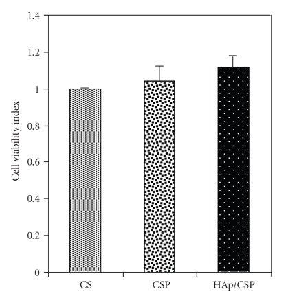
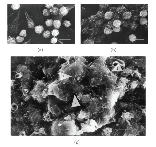
A novel bioanalogue hydroxyapatite (HAp)/chitosan phosphate (CSP) nanocomposite has been synthesized by a solution-based chemical methodology with varying HAp contents from 10 to 60% (w/w). The interfacial bonding interaction between HAp and CSP has been investigated through Fourier transform infrared absorption spectra (FTIR) and x-ray diffraction (XRD). The surface morphology of the composite and the homogeneous dispersion of nanoparticles in the polymer matrix have been investigated through scanning electron microscopy (SEM) and transmission electron microscopy (TEM), respectively. The mechanical properties of the composite are found to be improved significantly with increase in nanoparticle contents. Cytotoxicity test using murine L929 fibroblast confirms that the nanocomposite is cytocompatible. Primary murine osteoblast cell culture study proves that the nanocomposite is osteocompatible and highly in vitro osteogenic. The use of CSP promotes the homogeneous distribution of particles in the polymer matrix through its pendant phosphate groups along with particle-polymer interfacial interactions. The prepared HAp/CSP nanocomposite with uniform microstructure may be used in bone tissue engineering applications.
Figures


Similar articles
-
Synthesis and characterization of a novel chitosan/montmorillonite/hydroxyapatite nanocomposite for bone tissue engineering.Biomed Mater. 2008 Sep;3(3):034122. doi: 10.1088/1748-6041/3/3/034122. Epub 2008 Sep 3. Biomed Mater. 2008. PMID: 18765898
-
Hydroxyapatite-TiO(2)-based nanocomposites synthesized in supercritical CO(2) for bone tissue engineering: physical and mechanical properties.ACS Appl Mater Interfaces. 2014 Oct 8;6(19):16918-31. doi: 10.1021/am5044888. Epub 2014 Sep 23. ACS Appl Mater Interfaces. 2014. PMID: 25184699
-
Glycol chitosan/nanohydroxyapatite biocomposites for potential bone tissue engineering and regenerative medicine.Int J Biol Macromol. 2016 Dec;93(Pt B):1465-1478. doi: 10.1016/j.ijbiomac.2016.04.030. Epub 2016 Apr 13. Int J Biol Macromol. 2016. PMID: 27086294
-
Preparation of novel bioactive nano-calcium phosphate-hydrogel composites.Sci Technol Adv Mater. 2010 Feb 22;11(1):014103. doi: 10.1088/1468-6996/11/1/014103. eCollection 2010 Feb. Sci Technol Adv Mater. 2010. PMID: 27877318 Free PMC article. Review.
-
Hydroxylapatite nanoparticles: fabrication methods and medical applications.Sci Technol Adv Mater. 2012 Dec 28;13(6):064103. doi: 10.1088/1468-6996/13/6/064103. eCollection 2012 Dec. Sci Technol Adv Mater. 2012. PMID: 27877527 Free PMC article. Review.
Cited by
-
Calcium Phosphate Bioceramics: A Review of Their History, Structure, Properties, Coating Technologies and Biomedical Applications.Materials (Basel). 2017 Mar 24;10(4):334. doi: 10.3390/ma10040334. Materials (Basel). 2017. PMID: 28772697 Free PMC article. Review.
-
Zn and Ag Doping on Hydroxyapatite: Influence on the Adhesion Strength of High-Molecular Polymer Polycaprolactone.Molecules. 2022 Mar 16;27(6):1928. doi: 10.3390/molecules27061928. Molecules. 2022. PMID: 35335291 Free PMC article.
-
A porous medium-chain-length poly(3-hydroxyalkanoates)/hydroxyapatite composite as scaffold for bone tissue engineering.Eng Life Sci. 2016 Oct 24;17(4):420-429. doi: 10.1002/elsc.201600084. eCollection 2017 Apr. Eng Life Sci. 2016. PMID: 32624787 Free PMC article.
-
Synthesis and in vitro characteristics of biogenic-derived hydroxyapatite for bone remodeling applications.Bioprocess Biosyst Eng. 2024 Jan;47(1):23-37. doi: 10.1007/s00449-023-02940-y. Epub 2023 Nov 12. Bioprocess Biosyst Eng. 2024. PMID: 37952238
-
Biological and Physico-Chemical Properties of Composite Layers Based on Magnesium-Doped Hydroxyapatite in Chitosan Matrix.Micromachines (Basel). 2022 Sep 22;13(10):1574. doi: 10.3390/mi13101574. Micromachines (Basel). 2022. PMID: 36295927 Free PMC article.
References
-
- Hench LL. Bioceramics. Journal of the American Ceramic Society. 1998;81:1705–1728.
-
- Shikinami Y, Okuno M. Bioresorbable devices made of forged composites of hydroxyapatite (HA) particles and poly-L-lactide (PLLA)—part I. Basic characteristics. Biomaterials. 1999;20(9):859–877. - PubMed
-
- Wang M, Joseph R, Bonfield W. Hydroxyapatite-polyethylene composites for bone substitution: effects of ceramic particle size and morphology. Biomaterials. 1998;19(24):2357–2366. - PubMed
-
- Fang L, Leng Y, Gao P. Processing of hydroxyapatite reinforced ultrahigh molecular weight polyethylene for biomedical applications. Biomaterials. 2005;26(17):3471–3478. - PubMed
-
- Huang J, Di Silvio L, Wang M, Tanner KE, Bonfield W. In vitro mechanical and biological assessment of hydroxyapatite-reinforced polyethylene composite. Journal of Materials Science: Materials in Medicine. 1997;8(12):775–779. - PubMed
LinkOut - more resources
Full Text Sources
